Our Services
Others
- DFPP
- Shockwave
- IV Drip


“This literature review integrates recent work on the links between the degeneration of internal endosomal waste and motor transport networks and the resulting catastrophic clogging of the axon and nutrient deprivation in neurodegenerative diseases.”
Efficient intracellular logistics over long distances and a robust waste clearance machinery are essential for the structural integrity and survival of highly polarized neurons. Neurons are special in that their axons can be up to a meter long, so they face unique challenges to transport important nutrients and to remove cellular waste over large subcellular distances. Recent literature shows that the endosomal-lysosomal network and the microtubule motor machinery operate as a functional unit. Disruptions of this integrated network lead to endosomal-lysosomal collapse, organelle motility arrest and focal axonal traffic jams. In this review, published evidence is compiled to explain how defects in early endosomal sorting, loss of lysosomal acidification and impaired motor-adaptor coupling block degradative vesicle transport along the axon. We discuss how aberrant accumulation of undegraded autophagic vacuoles and endolysosomes leads to focal axonal swellings, clogging of nutrients and local metabolic starvation in Alzheimer’s disease, Parkinson’s disease, amyotrophic lateral sclerosis, Huntington’s disease and lysosomal storage disorders. This review also discusses recent findings of lysosomal vesicle hitchhiking of messenger RNA granules, and provides evidence that disruption of anterograde mRNA delivery compromises axonal mitochondrial integrity and local protein synthesis. Finally, the emerging therapeutic approaches to restore lysosomal pH and to stabilize the motor‐adaptor interactions to relieve neuroaxonal dystrophy are discussed.
In the nervous system, neurons are among the most morphologically complex and metabolically demanding cells. These cells have specialized axons that can extend as long as a meter from the neuronal soma and need constant internal logistics to support synaptic transmission, organelle renewal, and structural survival(Berth & Lloyd, 2023; Ferguson, 2018). Macromolecules and structural components synthesized in the cell body are transported to the distal axon terminals, and damaged proteins, worn-out organelles, and endocytosed signaling complexes generated at presynaptic regions are shuttled back to the soma for proteolysis. For this bidirectional long-distance movement, anterograde transport is powered by plus-end-directed kinesin motors, and retrograde transport by minus-end-directed cytoplasmic dynein-dynactin complexes on polarized microtubule tracks(Berth & Lloyd, 2023).
The endosomal-lysosomal network is an essential component of neuronal longevity, serving as the general cell’s hub for waste disposal, macromolecular recycling, and nutrient sensing(Lee, Sato, & Nixon, 2011). Under physiological conditions, cytosolic and extracellular substrates are delivered to early endosomes by endocytosis, phagocytosis and autophagy that mature into late endosomes and amphisomes prior to fusion with degradative lysosomes(Malik, Maddison, Smith, & Peters, 2019). In highly compartmentalized neurons this clearance pathway is physically linked to axonal transport. Mature, enzyme-rich lysosomes are concentrated near the cell body but soma-derived degradative lysosomes are actively transported anterogradely into distal axons to provide local digestive capacity(Farfel-Becker et al., 2019). Simultaneously, autophagosomes formed in the distal axon terminal fuse with late endosomes to recruit dynein motors to initiate processive retrograde transport toward the soma for ultimate degradation(Roney, Cheng, & Sheng, 2022).
As discussed in our previous review, misfolded toxic protein seeds can co-opt the machinery for long-distance transport in neurons during cell-to-cell propagation and network-level disease progression. Building upon these previous findings on mechanisms of extracellular dissemination, this literature review provides a more detailed description of the intracellular processes that regulate waste breakdown and organelle trafficking along the axon(Kimura, Noda, & Yoshimori, 2007). The breakdown of the endosomal-lysosomal network severely affects organelle biogenesis, membrane sorting and vesicular transport. Endosomal-lysosomal dysfunction and axonal transport defects create a vicious feed-forward loop; stalled transport blocks delivery of digestive enzymes to distal axon regions, while impaired proteolysis initiates cargo detachment and organelle immobilization along the axon. This review summarizes and synthesizes the current research on the molecular mechanisms underlying endosomal–lysosomal network collapse and transport deficits that converge to cause axonal clogging, nutrient starvation, and progressive neurodegeneration.
To understand how the endosomal-lysosomal network collapses in disease, we must first review the molecular mechanics that govern the trafficking of these organelles along the axon in physiology(Yang, Ma, Lian, Xu, & Cao, 2023). Microtubules inside the axonal shaft have a consistent structural polarity with the fast-growing plus ends oriented toward the axon terminal and the minus ends anchored toward the soma. The major class of plus-end-directed kinesin motors, such as kinesin-1 (KIF5) and kinesin-3 (KIF1A) family motors, couple ATP hydrolysis to processive stepping toward the distal axon terminal. Conversely, cytoplasmic dynein mediates minus-end-directed retrograde transport toward the cell body together with the dynactin complex and cargo-specific adaptors(Zhang et al., 2021).
The recruitment of motor proteins to endolysosomal membranes is coordinated by specialized multiprotein complexes and small GTPases. Anterograde transport of soma-derived lysosomes into the axon depends on the BLOC-one-related complex (BORC), an octameric complex associated with the lysosomal membrane(Keren-Kaplan & Bonifacino, 2021). BORC recruits and activates the small GTPase Arl8, which subsequently interacts with its effector protein SKIP (PLEKHM2). Activated SKIP binds directly to kinesin light chains and couples lysosomes to kinesin-1 motors to enter the axon. KIF1A and Arl8 coordinate to drive plus-end organelle motility along axonal microtubules. Late endosomes and autolysosomes recruit dynein-dynactin via specific adaptors such as Rab7-RILP, Snapin and JIP3 on retrograde pathway(De Pace et al., 2024; Roney et al., 2022).
Along this physical transport pathway organelle maturation occurs dynamically. Autophagosomes produced at distal terminals are initially devoid of retrograde motility and acid hydrolases. Heterotypic fusion of autophagosomes with Rab7-positive late endosomes generates amphisomes, making autophagosomes transport competent. The motor-adaptor sharing mechanism transfers these preloaded dynein-Snapin complexes from late endosomes to the newly forming amphisomes, allowing processive retrograde transport to the soma(Jia, Guardia, Pu, Chen, & Bonifacino, 2017). Retrogradely moving amphisomes encounter soma-derived degradative lysosomes moving anterogradely and sequential fusion events lead to decreased lumenal pH and activation of cathepsins B, D, L and glucocerebrosidase(Kimura et al., 2007).
Bidirectional transport of degradative organelles and autophagic intermediates (Figure 1) is achieved by a cooperative relay between BORC-driven kinesin anterograde motors and Snapin- or RILP-mediated dynein retrograde complexes along polarized axonal microtubules. Such motor-adaptor interactions in normal functioning allow neurons to sustain local degradation capacity at distal tips while continuously clearing autophagic substrates toward the soma(Lee et al., 2011).
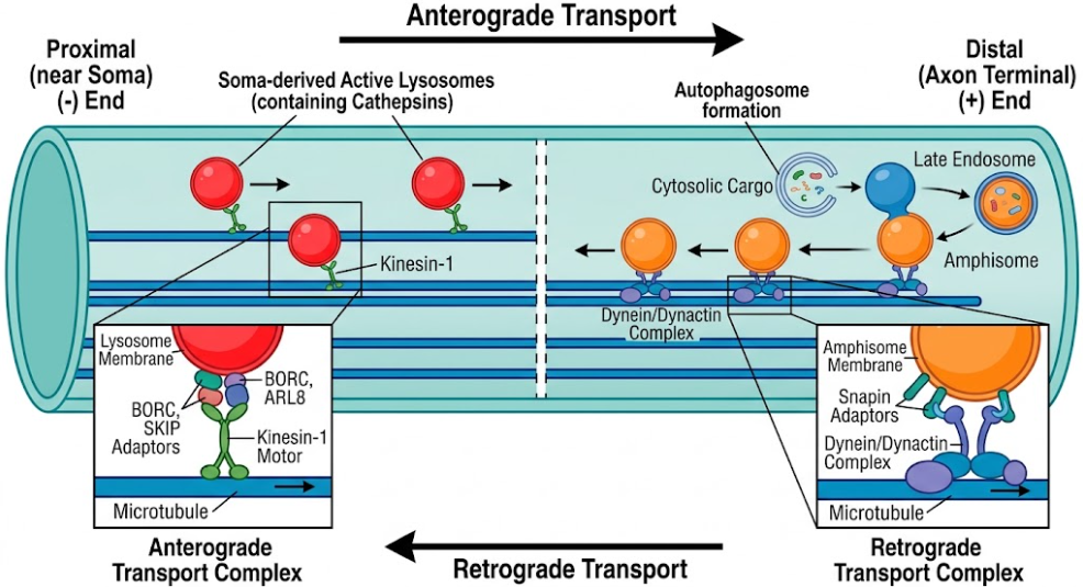
Figure 1. Molecular Mechanisms of Axonal Transport. Kinesin-1-mediated transport of soma-derived lysosomes (+ end) and Dynein/Dynactin-mediated return of amphisomes (- end) with key adaptors.
Genetic alterations, toxic protein interactions and metabolic insults in neurological disorders collapse the endosomal-lysosomal network by disrupting lumenal acidification, endosomal sorting and membrane trafficking(Almeida, Bahr, & Kinsey, 2020). At the heart of this vulnerable network lies the vacuolar-type H+ ATPase (v-ATPase), a multi-subunit proton pump that acidifies the lysosomal lumen to pH 4.5–5.0, thus providing the acidic milieu required for cathepsin activation and hydrolase maturation(Kim, Cho, & Jung, 2025). In familial Alzheimer’s disease, loss-of-function mutations of Presenilin 1 (PSEN1) inhibit ER-to-lysosome trafficking of the v-ATPase V0a1 subunit. Consequently, lysosomal acidification is impaired, leading to loss of intralumenal protease activity and accumulation of undegraded autophagic substrates. Furthermore, the v-ATPase subunits directly interact with amyloid-beta (Aβ) peptides and hyperphosphorylated tau, which inhibit the proton pumping activity and lead to localized acidification deficits(Kim et al., 2025).
Disruptions in early endosomal sorting and retromer recycling also speed up network breakdown(Malik et al., 2019). The retromer complex is composed of VPS35, VPS26 and VPS29 and it is responsible for coordinating retrograde transport of membrane receptors and processing enzymes from early endosomes to the trans-Golgi network or plasma membrane. Pathogenic mutations in VPS35 linked to Parkinson’s disease and deficiencies of retromer associated proteins in Alzheimer’s disease disturb retromer assembly and cargo sorting(Almeida et al., 2020). This leads to abnormal retention and accumulation of amyloid precursor protein (APP) and β-site APP cleaving enzyme 1 (BACE1) in enlarged early endosomes, followed by hyper-amyloidogenic processing and endosomal swelling(Paumier & Gowrishankar, 2024).
Mutations in genes for the endocytic machinery also affect clathrin-coated vesicle uncoating and early endosome maturation, including Synaptojanin 1 (SYNJ1), Auxilin-1 (DNAJC6), and RME-8 (DNAJC13). Hyperactive LRRK2 kinase activity in models of Parkinson’s disease leads to hyperphosphorylation of Rab GTPases including Rab8a and Rab10, which disrupts Golgi-to-lysosome trafficking and causes Golgi fragmentation(Kim et al., 2025). In lysosomal storage diseases such as Niemann-Pick disease type C, NPC1 mutations result in cholesterol accumulation within lysosomal membranes, which aberrantly traps Arl8 and kinesin-1, blocking their release and anterograde exit of lysosomes into the axon(Roney et al., 2021).
As shown in Figure 2, failure of v-ATPase assembly and thus lysosomal acidification, combined with retromer sorting defects and Rab GTPase dysregulation, causes massive enlargement of endolysosomal compartments containing undegraded protein aggregates and processing enzymes. The failure of intralumenal proteolysis is a major trigger of organelle immobilization and network collapse(Kimura et al., 2007).
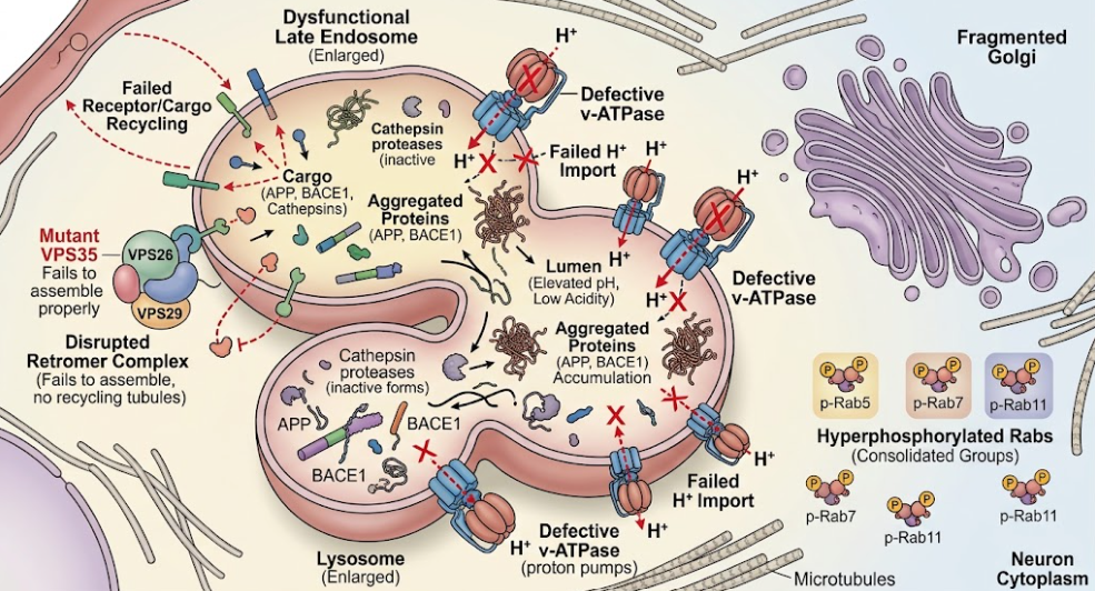
Figure 2. Dysfunction of Lysosomal Endo Network. Diagram of the collapse of the endosomal-lysosomal network in a neuron. It focuses on defective v-ATPase proton pumps, defective acidification, disrupted retromer complexes and the subsequent accumulation of aggregated proteins such as APP and BACE1 within enlarged late endosomes and lysosomes.
Impaired lysosomal proteolysis contributes most to deficits in axonal transport of degradative organelles, leading to focal organelle accumulations called axonal traffic jams or dystrophic neurites(Farfel-Becker et al., 2019). Studies show that inhibition of lysosomal protease activity by cysteine protease inhibitors (e.g., leupeptin or E64), aspartic protease inhibitors (e.g., pepstatin) or the v-ATPase inhibitor bafilomycin A1 substantially slows or stops the movement of LC3-positive autophagosomes, LAMP1-positive lysosomes and Rab7-positive late endosomes. Live-cell imaging revealed a dramatic increase in stationary vesicles containing cathepsins and a decrease in retrograde velocity(Lee et al., 2011).
Importantly, transport arrest due to proteolysis failure is cargo-specific and not a consequence of a global collapse of microtubules. Organelles that do not contain cathepsins, such as AcGFP-RhoB-positive early endosomes and mitochondria, continue to move normally in the axon, passing through and out of focal swellings unimpeded(Cai et al., 2010). In addition, non-vesicular structural elements such as neurofilaments do not build up in these swellings, distinguishing inhibition of lysosomal proteolysis from global transport blockades induced by microtubule severing. Mechanistically, failure of intralumenal proteolysis leads to dynein motors detachment from late endosomes and autolysosomes, making them stationary and promoting their coalescence into focal axonal dystrophies(Lee et al., 2011).
These dystrophic axonal swellings accumulate undegraded autophagic vacuoles, multilamellar structures, ubiquitin, hyperphosphorylated neurofilaments, APP and BACE1, reminiscent of the neuropathological hallmark of neuritic dystrophy surrounding amyloid plaques in Alzheimer’s disease(Yuan & Grutzendler, 2016). Experiments show that this clogging is reversible: Rab7 vesicle motility is restored by removal of the proteolytic block, autophagic substrates are cleared and axonal swellings are resolved. However, toxic protein species actively exacerbate transport failure in chronic disease conditions. Soluble amyloid-β 1-42 oligomers associate with amphisomes in distal axons and directly interact with dynein intermediate chain (DIC). This interaction antagonizes dynein-Snapin coupling and prevents dynein recruitment, thereby immobilizing autophagic vacuoles at presynaptic terminals(Tammineni, Ye, Feng, Aikal, & Cai, 2017). Similarly, mutant SOD1 in amyotrophic lateral sclerosis is associated with DIC to disrupt dynein loading to late endosomes, promoting axonal autophagic stress in motor neurons. In Huntington’s disease, polyglutamine expanded huntingtin impedes the HAP1-dynein interaction and blocks retrograde transport of autophagosomes(Xie et al., 2015).
Selective detachment of dynein motors from protease-deficient autolysosomes (as in Figure 3) leads to focal accumulation of dynein motors in dystrophic axonal swellings. These swellings represent physical traffic jams that inhibit retrograde organelle clearance, but permit passage of non-cathepsin cargos such as mitochondria, creating local nutrient and waste bottlenecks along the axon(Paumier & Gowrishankar, 2024).
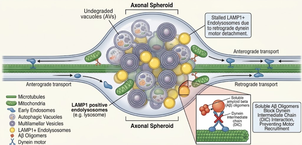
Figure 3. Mechanisms of focal axonal dystrophy in neurodegenerative disease. Scheme of the cell cascade where retrograde transport defects, often associated with factors such as Aβ, lead to cargo build-up (e.g. LAMP1+ endolysosomes and AVs) and focal axonal spheroid formation.
Endosomal-lysosomal collapse affects more than waste clearance, directly compromising inter-organellar communication and local axonal translation. Lysosome-related vesicles have recently been shown to act as mobile platforms for the transport of messenger RNA (mRNA) granules in a process termed RNA granule hitchhiking(De Pace et al., 2024). Annexin A11 and other RNA-binding proteins function as molecular tethers that connect non-translating mRNA granules to the surface of anterogradely moving lysosomal vesicles. This mechanism serves to transport nuclear-encoded mRNAs, especially those encoding ribosomal proteins and mitochondrial oxidative phosphorylation (OXPHOS) subunits, to distal axonal regions for local translation(De Pace et al., 2024).
For example, anterograde lysosomal transport is impaired by knockout of BORC subunits (BORCS5 or BORCS7) and lysosome-related vesicles are blocked from entering the axon. Thus, axonal mRNA pools for ribosomal components (RPS7, RPS27A and RPL24) and mitochondrial respiratory chain subunits (Complex I-V and MICOS complex) are strongly reduced(De Pace et al., 2024). This depletion leads to a local shutdown of axonal protein synthesis which leads to loss of mitochondrial membrane potential, increased reactive oxygen species (ROS) production, deformed cristae, mitochondrial fragmentation and eventual axonal degeneration. Restoring lysosome-kinesin coupling through artificial adaptors rescues axonal mRNA levels, protein translation and mitochondrial function, thus validating that lysosomal transport is directly necessary for axonal metabolic homeostasis(Lee et al., 2011).
Furthermore, endosomal-lysosomal collapse results in a severe disruption of crosstalk with endoplasmic reticulum (ER), Golgi apparatus and mitochondria. Protrudin, TMEM106B and VPS13C regulate ER-lysosome membrane contact sites and control lysosome positioning, lipid exchange and lysosome membrane repair through the PITT (phosphoinositide-initiated membrane tethering and lipid transport) pathway(Kim et al., 2025; Yang et al., 2023). TMEM106B or Protrudin mutations impair ER-endolysosome contact dynamics in frontotemporal lobar degeneration and hereditary spastic paraplegia, leading to accumulation of enlarged, hypocidic lysosomal intermediates at the axon initial segment. Moreover, Golgi fragmentation due to α-synuclein aggregation in Parkinson’s disease or mutant huntingtin in Huntington’s disease disrupts sorting of lysosomal enzymes through the cation-independent mannose-6-phosphate receptor (M6PR) pathway, leading to a deficiency of essential hydrolases in distal lysosomes and contributing to axonal transport deficits(De Pace et al., 2024; Kim et al., 2025; Roney et al., 2022).
The evidence presented in this literature review demonstrates that endosomal-lysosomal network collapse and impaired axonal transport are a primary driver of early disease pathogenesis, and not a passive secondary consequence(Farfel-Becker et al., 2019). This tight coupling between organelle degradation and microtubule motility reveals an architectural vulnerability of polarized neurons. Axons span long physical distances and therefore the local accumulation of undegraded autophagic vacuoles rapidly progresses to focal dystrophies that disrupt organelle quality control, cellular signaling and nutrient distribution(Kimura et al., 2007).
Recent literature has highlighted an important insight, that is the organelle selectivity of transport deficits arising from the failure of lysosomal proteolysis. The loss of lysosomal enzyme activity specifically prevents the movement of degradation vesicles containing cathepsins by uncoupling dynein, rather than a non-specific shutdown of all axonal transport(Lee et al., 2011). This selective mechanism explains the enrichment of dystrophic neurites in Alzheimer’s disease with autophagic vacuoles and lysosomal intermediates, but also the initial bypassing of mitochondria and non-degradative endosomes from these swellings. However, as axonal swellings increase, inter-organellar crosstalk breaks down, as evidenced by loss of RNA granule hitchhiking, leading to secondary mitochondrial crisis, localized ATP starvation and metabolic failure in the axon(De Pace et al., 2024).
As discussed in our previous review, extracellular propagation of misfolded protein seeds leads to the spread of pathology throughout neural networks. The data presented here show that traffic jams of internal organelles induce localized metabolic susceptibilities that make axons highly susceptible to toxic seed uptake and structural breakdown(Lee et al., 2011; Roney et al., 2022). These mechanistic insights offer promising avenues for therapeutic intervention to restore axonostasis. Small-molecule v-ATPase activators (e.g. C381 or EN6) or acidic nanoparticles (aNPs) promote lysosomal acidification, restore proteolytic efficiency and alleviate axonal dystrophy in rodent models. TFEB activation promotes autophagic flux by increasing lysosomal biogenesis(Kim et al., 2025). Stabilizing motor-adaptor interactions, for example by over-expressing Snapin to reestablish dynein recruitment, or targeting hyperactive LRRK2 kinase activity to stop motor tug-of-war, is a powerful way to rescue retrograde autophagic flux(Tammineni et al., 2017). Restoration of the continuous bidirectional flow of degradative organelles and metabolic cargo along the axon holds great potential to stop the progression of neurodegeneration before irreversible neuronal loss happens(Lee et al., 2011).
To summarize, disruption of the endosomal-lysosomal network and associated defects in axonal transport is a common pathogenic mechanism in neurological diseases. Failure of lysosomal proteolysis, whether due to genetic changes in v-ATPase subunits, retromer components, or pathogenic protein oligomers, causes selective immobilization of degradative vesicles and dystrophic axonal swellings. This organelle congestion disrupts important cellular processes such as the clearance of autophagic substrates, the dynamics of inter-organellar contact sites and mRNA transport, which are all necessary for local mitochondrial maintenance. Future studies targeting the exact molecular triggers of motor-adaptor detachment and specific therapies to restore lysosomal acidification and axonal trafficking will be critical in developing effective disease-modifying treatments for neurodegenerative diseases.
Almeida, M. F., Bahr, B. A., & Kinsey, S. T. (2020). Endosomal-lysosomal dysfunction in metabolic diseases and Alzheimer’s disease. Int Rev Neurobiol, 154, 303-324. doi:10.1016/bs.irn.2020.02.012
Berth, S. H., & Lloyd, T. E. (2023). Disruption of axonal transport in neurodegeneration. J Clin Invest, 133(11). doi:10.1172/jci168554
Cai, Q., Lu, L., Tian, J. H., Zhu, Y. B., Qiao, H., & Sheng, Z. H. (2010). Snapin-regulated late endosomal transport is critical for efficient autophagy-lysosomal function in neurons. Neuron, 68(1), 73-86. doi:10.1016/j.neuron.2010.09.022
De Pace, R., Ghosh, S., Ryan, V. H., Sohn, M., Jarnik, M., Rezvan Sangsari, P., . . . Bonifacino, J. S. (2024). Messenger RNA transport on lysosomal vesicles maintains axonal mitochondrial homeostasis and prevents axonal degeneration. Nat Neurosci, 27(6), 1087-1102. doi:10.1038/s41593-024-01619-1
Farfel-Becker, T., Roney, J. C., Cheng, X. T., Li, S., Cuddy, S. R., & Sheng, Z. H. (2019). Neuronal Soma-Derived Degradative Lysosomes Are Continuously Delivered to Distal Axons to Maintain Local Degradation Capacity. Cell Rep, 28(1), 51-64.e54. doi:10.1016/j.celrep.2019.06.013
Ferguson, S. M. (2018). Axonal transport and maturation of lysosomes. Curr Opin Neurobiol, 51, 45-51. doi:10.1016/j.conb.2018.02.020
Jia, R., Guardia, C. M., Pu, J., Chen, Y., & Bonifacino, J. S. (2017). BORC coordinates encounter and fusion of lysosomes with autophagosomes. Autophagy, 13(10), 1648-1663. doi:10.1080/15548627.2017.1343768
Keren-Kaplan, T., & Bonifacino, J. S. (2021). ARL8 Relieves SKIP Autoinhibition to Enable Coupling of Lysosomes to Kinesin-1. Curr Biol, 31(3), 540-554.e545. doi:10.1016/j.cub.2020.10.071
Kim, S. H., Cho, Y. S., & Jung, Y. K. (2025). Failure of lysosomal acidification and endomembrane network in neurodegeneration. Exp Mol Med, 57(11), 2418-2428. doi:10.1038/s12276-025-01579-x
Kimura, S., Noda, T., & Yoshimori, T. (2007). Dissection of the autophagosome maturation process by a novel reporter protein, tandem fluorescent-tagged LC3. Autophagy, 3(5), 452-460. doi:10.4161/auto.4451
Lee, S., Sato, Y., & Nixon, R. A. (2011). Lysosomal proteolysis inhibition selectively disrupts axonal transport of degradative organelles and causes an Alzheimer’s-like axonal dystrophy. J Neurosci, 31(21), 7817-7830. doi:10.1523/jneurosci.6412-10.2011
Malik, B. R., Maddison, D. C., Smith, G. A., & Peters, O. M. (2019). Autophagic and endo-lysosomal dysfunction in neurodegenerative disease. Mol Brain, 12(1), 100. doi:10.1186/s13041-019-0504-x
Paumier, J. M., & Gowrishankar, S. (2024). Disruptions in axonal lysosome transport and its contribution to neurological disease. Curr Opin Cell Biol, 89, 102382. doi:10.1016/j.ceb.2024.102382
Roney, J. C., Cheng, X. T., & Sheng, Z. H. (2022). Neuronal endolysosomal transport and lysosomal functionality in maintaining axonostasis. J Cell Biol, 221(3). doi:10.1083/jcb.202111077
Roney, J. C., Li, S., Farfel-Becker, T., Huang, N., Sun, T., Xie, Y., . . . Sheng, Z. H. (2021). Lipid-mediated motor-adaptor sequestration impairs axonal lysosome delivery leading to autophagic stress and dystrophy in Niemann-Pick type C. Dev Cell, 56(10), 1452-1468.e1458. doi:10.1016/j.devcel.2021.03.032
Tammineni, P., Ye, X., Feng, T., Aikal, D., & Cai, Q. (2017). Impaired retrograde transport of axonal autophagosomes contributes to autophagic stress in Alzheimer’s disease neurons. Elife, 6. doi:10.7554/eLife.21776
Xie, Y., Zhou, B., Lin, M. Y., Wang, S., Foust, K. D., & Sheng, Z. H. (2015). Endolysosomal Deficits Augment Mitochondria Pathology in Spinal Motor Neurons of Asymptomatic fALS Mice. Neuron, 87(2), 355-370. doi:10.1016/j.neuron.2015.06.026
Yang, X., Ma, Z., Lian, P., Xu, Y., & Cao, X. (2023). Common mechanisms underlying axonal transport deficits in neurodegenerative diseases: a mini review. Front Mol Neurosci, 16, 1172197. doi:10.3389/fnmol.2023.1172197
Yuan, P., & Grutzendler, J. (2016). Attenuation of β-Amyloid Deposition and Neurotoxicity by Chemogenetic Modulation of Neural Activity. J Neurosci, 36(2), 632-641. doi:10.1523/jneurosci.2531-15.2016
Zhang, H., Wei, W., Zhao, M., Ma, L., Jiang, X., Pei, H., . . . Li, H. (2021). Interaction between Aβ and Tau in the Pathogenesis of Alzheimer’s Disease. Int J Biol Sci, 17(9), 2181-2192. doi:10.7150/ijbs.57078