Our Services
Others
- DFPP
- Shockwave
- IV Drip


“This review focuses on the role of Paneth cell failure in the synthesis and activation of α-defensins (HD5 and HD6) in the crypt of Lieberkühn and its impact on chronic dysbiosis and the initiation of ileal Crohn’s disease.”
The human small intestinal epithelium is one of the largest and most delicate barriers of the body that is under constant attack by trillions of microbes living in the gut lumen. At the mucosal interface the host depends upon secretion of a specialized chemical shield to maintain homeostasis and prevent pathogenic invasion. Paneth cells are specialized, long-lived secretory intestinal epithelial cells located at the base of the crypts of Lieberkühn and are the major source of this antimicrobial armamentarium. Most strikingly, they secrete constitutively human α-defensin 5 (HD5) and human α-defensin 6 (HD6), highly concentrated in the crypt microenvironment. Recent scientific breakthroughs have demonstrated a profound paradigm shift, showing that a deficiency in these Paneth cell-derived defensins is the primary initiating factor in the pathogenesis of ileal Crohn’s disease. In ileal Crohn’s disease, the antimicrobial shielding gradient collapses because of a specific failure to produce or process functional HD5 and HD6 rather than a generalized inflammatory response. We systematically review the current literature on Paneth cell biology and describe the molecular basis of HD5 and HD6 activation and the dual and distinct mechanisms by which these two peptides act to kill pathogens directly and entrap bacteria. We also describe the complex genetic and environmental pathways that converge to impair HD5 and HD6 production including NOD2 mutations, variants in the TCF4 promoter, autophagy defects mediated by ATG16L1 and endoplasmic reticulum stress induced by XBP1. Finally, we discuss how the breakdown of this shield leads to microbial encroachment, shifts the mucosal immune balance toward pro-inflammatory Th17-skewing and establishes the persistent chronic inflammation characteristic of this disease, providing new therapeutic avenues thru defensin-mimetic supplementations and engineered probiotics.
One of the most difficult tasks facing the human mucosal immune system is to establish a balanced relationship with the enormous and complex gut microbiota. (Salzman, 2010; Schroeder et al., 2015) The small intestine is covered by a single layer of epithelial cells which creates an extensive, folded surface area of approximately 400 square meters.(Elphick & Mahida, 2005) This large surface area is designed to optimize nutrient and water absorption but at the same time creates a vulnerable interface for potential microbial encroachment and systemic translocation.(Salzman, 2010) This epithelial monolayer is constantly renewed every three to five days by pluri-potent intestinal stem cells (ISCs) at the crypt base. Protection of such a regenerative niche is essential, since damage or colonization would jeopardize epithelial integrity and thus barrier function.(Erkert, Haag, & Becker, 2026)
The small bowel has an immediate, germline-coded system of defense called innate mucosal immunity to protect this critical stem cell niche. Innate defense mechanisms are immediate responses to microbial challenges and involve mechanical, cellular and chemical barriers.(Elphick & Mahida, 2005) Paneth cells are a highly specialized, long-lived secretory epithelial lineage first described by Josef Paneth in 1888 at the center of the chemical barrier of the small intestine. Paneth cells are found solely at the bottom of the small intestinal crypts and are interspersed between Lgr5+ ISCs. They have a developed network of endoplasmic reticulum and large apical granules .(Erkert et al., 2026) These granules are packed with a variety of host defense proteins and peptides, which are rapidly degranulated into the crypt lumen following cholinergic stimulation or direct sensing of bacterial ligands.(Salzman, 2010)
Paneth cells secrete several microbicidal proteins including lysozyme and secretory phospholipase A2 (sPLA2), but the most abundant and biologically relevant components of their secretory arsenal are the enteric α-defensins: human α-defensin 5 (HD5) and human α-defensin 6 (HD6).(Ehmann et al., 2019) These cationic peptides constitute ~70% of the total repertoire of antimicrobial peptides (AMPs) produced by Paneth cells and are important factors in shaping the local microbial ecology.(Erkert et al., 2026) There is a wealth of clinical and genetic data collected over the last two decades showing that ileal Crohn’s disease is caused by a primary defect in Paneth cell production and secretion of these α-defensins.(J. Wehkamp & Stange, 2020) This literature review will break down the molecular mechanisms of this defensin shield, review the genetic and environmental pathways that lead to its collapse, and follow how the breakdown of this antimicrobial barrier serves as the initiating spark for the chronic, non-resolving mucosal inflammation that defines Crohn’s disease.
Paneth cells are located in a strategic position in the small intestinal architecture to create a unique decreasing gradient of antimicrobial peptides diffusing from the crypt base up toward the intestinal lumen.(Erkert et al., 2026) Paneth cells and their defensins increase in abundance gradually by three- to seven-fold from the proximal small intestine to the distal terminal ileum, reflecting the increasing density of the microbial population along the gastrointestinal tract, from about 103 bacteria per gram in the duodenum to more than 107 in the ileum.(Kennedy & Chang, 2020)
HD5 and HD6, the major α-defensins, are small (~4 kDa) cationic, arginine-rich peptides with six conserved cysteine residues that form three intramolecular disulfide bonds.(Salzman, 2010) These defensins are produced as inactive pre-pro-peptides to protect the host epithelial cell from premature cytotoxicity. During synthesis, the signal peptide is cleaved off resulting in a pro-peptide, which is stored in the dense secretory granules of Paneth cells.(Wilson et al., 1999) In humans, these pro-peptides are processed into active, mature and microbicidal forms upon or after degranulation into the extracellular space. This crucial activation step is mediated by luminal trypsin, which cleaves the pro-region and releases the active mature peptide.(Valore & Ganz, 1992)
Once secreted and activated, the concentration of α-defensins in the immediate vicinity of the crypt base is estimated to reach staggering physiological levels of 15 to 100 mg/ml. This concentration in the microenvironment is at least 1,000-fold higher than the minimal bactericidal concentrations needed to kill most pathogens in vitro.(Elphick & Mahida, 2005; Salzman, 2010) This high concentration ensures that the regenerative stem cell niche is completely sterile, protecting the Lgr5+ stem cells from direct contact with the heavy bacterial load of the lumen. Beyond this localized sterilizing role, defensins diffuse into the overlying mucus layer, where they create a “zone of exclusion” that prevents both commensal and pathogenic bacteria from approaching the epithelial monolayer, thereby establishing a state of immunological détente at the host-microbe interface.(Salzman, 2010)
HD5 and HD6 are co-expressed and stored in the same Paneth cell granules, yet they are functionally distinct and utilize radically different physical mechanisms to neutralize microbial threats. Human defensin 5 (HD5) functions mainly by direct bactericidal killing.(Courth et al., 2015) Mature HD5 is highly basic and cationic, which confers a net positive charge that allows the peptide to electrostatically interact with the negatively charged, anionic cell envelope components of bacteria, such as lipopolysaccharide in Gram-negative bacteria and lipoteichoic acid in Gram-positive bacteria.(Bel et al., 2017) Following this initial electrostatic binding, the amphipathic nature of HD5 allows it to insert, and disrupt, the bacterial membrane. Ultrastructural studies using transmission electron microscopy revealed that HD5 treatment induces detachment of the inner membrane, detachment of the plasma membrane and formation of vesicular structures around the bacterial envelope, leading to loss of cytoplasmic integrity and bactericidal death.(Ehmann et al., 2019)
Most impressive is the recent discovery of a complex post-translational “fragment clustering” mechanism that significantly boosts the antimicrobial versatility of HD5. Oxidized HD5 is reduced upon exposure to the proteolytic environment of the gut, especially natural duodenal fluid, either spontaneously by the low redox potential of the gut or enzymatically by the thioredoxin (TRX) system secreted by Paneth cells. The linear peptide structure of HD5 is vulnerable to proteolytic cleavage by duodenal proteases upon reduction of the disulfide bonds. This proteolytic digestion does not inactivate the peptide but rather cleaves HD5 into a massive cluster of up to 8000 unique, active antimicrobial fragments.(Ehmann et al., 2019) The N-terminal fragment HD5(1–9) has been found to be highly bio-active and versatile with potent bactericidal activity against both Gram-positive and Gram-negative bacteria. Importantly, these fragments kill pathogens but selectively spare key beneficial commensal species such as Akkermansia muciniphila, offering a fine-tuning tool to modulate the composition of the gut microbiota.(Salzman, 2010)
Conversely, human defensin 6 (HD6) is remarkably protease resistant and works primarily thru the formation of physical traps rather than thru direct chemical killing. Under physiological conditions, HD6 creates a complex and highly organized network of extracellular “nanonets”.(Salzman, 2010) These nanonets are assembled independently of the local redox state and are completely resistant to proteolytic digestion by human duodenal fluid as the peptide structure is protected within the assembled net. As bacteria migrate toward the crypt bottom, they are physically trapped within the HD6 nanonets, thereby impeding their motility, inhibiting their adherence to the epithelial membrane, and aborting further invasion. There was no direct bactericidal activity of HD6 under standard conditions.(Schroeder et al., 2015) However, reduction of HD6 in the luminal microenvironment triggers a conformational change exposing a direct killing activity specific for certain commensal anaerobic bacteria. This reduction occurs by an “all-or-nothing” single step mechanism and involves no partially reduced intermediates.(Ehmann et al., 2019) The linear, reduced HD6 maintains its extracellular nanonet trapping structures and leads to cytoplasmic disintegration of target bacteria under reducing conditions. This “Retiarius-like” mechanism, combining nanonet entrapment and localized environment-dependent bactericidal activity, broadens the host’s defense capabilities at the crypt interface. As shown in Figure 1., human α-defensin 5 (HD5) directly disrupts bacterial outer membranes and degrades into active peptide fragments, while human α-defensin 6 (HD6) acts as a physical trap by self-assembling into stable nanonets to catch and immobilize pathogens under physiological conditions.(Schroeder et al., 2015)
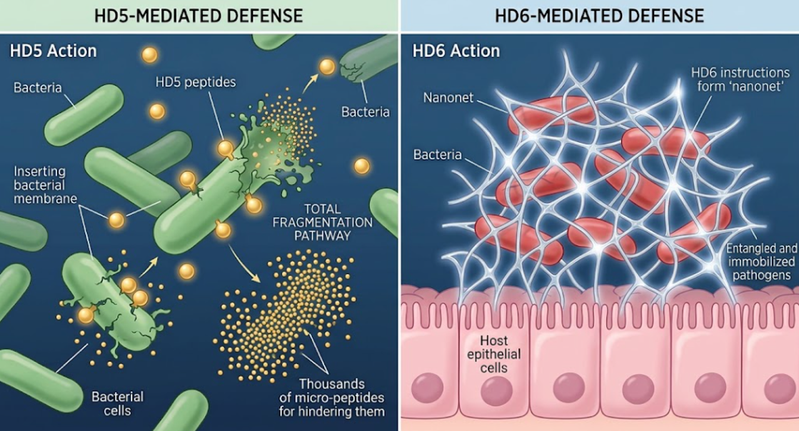
Figure 1. Dual defense weapons of Paneth cell defensins. Membrane-disrupting activity of HD5 (left) and nanonet-forming defense of HD6 (right)
The defensive shield of the mucosal barrier is gravely compromised in ileal Crohn’s disease.(Jan Wehkamp et al., 2005) Remarkably, this barrier failure is marked by a selective and profound decrease in the mRNA expression and peptide concentration of both HD5 and HD6, while other major Paneth cell products such as lysozyme and sPLA2 remain unchanged or are even increased. Importantly, this specific defensin deficiency is independent of tissue inflammation and is not observed in ulcerative colitis, colonic Crohn’s disease, or pouchitis. This important finding demonstrates that defensin failure is a primary or inciting cause of ileal Crohn’s disease pathology, not a secondary consequence of chronic tissue injury.(Salzman, 2010; Jan Wehkamp et al., 2005)
This defensin failure has been linked to specific molecular pathways that control Paneth cell differentiation, protein folding and exocytosis by pioneering genetic and transcriptomic analyzes.(Tan, Zeng, & Zhi, 2015)
The NOD2/CARD15 gene is the major genetic risk factor in ileal CD. NOD2 is an intracellular pattern recognition receptor in Paneth cells that detects bacterial muramyl dipeptide (MDP). In healthy conditions, MDP sensing by NOD2 activates downstream NF-κB signaling, driving α-defensins expression in the Paneth cell lineage.(Santaolalla, Fukata, & Abreu, 2011)
Loss-of-function mutations in the C-terminal leucine-rich repeat region of NOD2, especially the SNP13 (1007fs) frameshift mutation, abolish MDP sensing in Crohn’s disease.(Stappenbeck & McGovern, 2017) Thus, patients with the NOD2 SNP13 mutation have a nine-fold reduction in ileal HD5 mRNA and peptide levels compared with healthy controls. In Crohn’s patients with wild-type NOD2, there is a three-fold reduction in HD5, indicating that other upstream factors are also involved in silencing the defensin genes. In addition, epigenetic studies revealed that the DEFA5 gene is hypermethylated in Crohn’s patients, which leads to permanent silencing of its expression regardless of tissue inflammation.(Brabec et al., 2023)
The differentiation of Paneth cells from Lgr5+ stem cells is driven by the Wnt/β-catenin signaling pathway, and the transcription factor TCF-4 (TCF7L2) is the master regulator of DEFA5 and DEFA6 transcription. Overall TCF-4 expression is significantly reduced in patients with ileal Crohn’s disease and specific single nucleotide polymorphisms (SNPs) within the TCF-4 promoter region are strongly associated with early onset ileal Crohn’s disease and a concurrent reduction in α-defensins. Murine Tcf-4 deletion results in lack of mature Paneth cells.(Salzman, 2010; Schroeder et al., 2015)
The cellular autophagy pathway regulates granule packing and exocytosis in Paneth cells. The autophagy gene ATG16L1 and in particular the ATG16L1 T300A variant is mutated leading to Paneth cells being vulnerable to metabolic stress and diminished antibacterial defense.
ATG16L1 T300A variant–carrying Paneth cells exhibit dymorphic, diffuse, disordered, or absent lysozyme-positive granules and fragmented endoplasmic reticulum membranes. This exocytic block prevents the proper exocytosis of defensins into the crypt lumen, resulting in their premature intracellular degradation (crinophagy). LRRK2 mutations are analogous to the disruption of cargo sorting and aberrant autophagic degradation of defensins and lysozyme.(Erkert et al., 2026; Salzman, 2010)
Paneth cells have a colossal secretory output and are very sensitive to Endoplasmic Reticulum (ER) stress, relying on the transcription factor X-box-binding protein 1 (XBP1) to orchestrate the unfolded protein response (UPR).(Kaser et al., 2008) Complete failure of UPR, spontaneous endoplasmic reticulum stress, cellular apoptosis, and loss of the Paneth cell population results from the deletion of XBP1 in intestinal epithelial cells. Importantly, the combination of autophagy-deficient and ER stress-susceptible genotypes in mice results in severe, spontaneous transmural ileitis that recapitulates human Crohn’s disease, establishing Paneth cell dysfunction as a primary site of origin for intestinal inflammation.(Chen et al., 2026; Kaser et al., 2008) Genetic and transcriptomic alterations, including NOD2 mutations, TCF4 transcription factor deficiency, ATG16L1 autophagy defects and XBP1-driven endoplasmic reticulum stress, converge inside the Paneth cell as shown in the Figure 2. to block production and orderly secretion of HD5 and HD6, leading to a total collapse of the protective chemical barrier.(Chen et al., 2026)
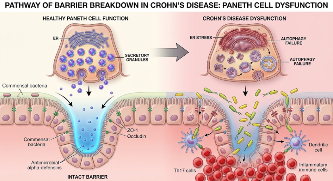
Figure 2. The Shield Break and Crohn’s Disease. Healthy Paneth cells (left) create secretory granules and secrete α-defensins to maintain an intact barrier and manage commensal bacteria. Paneth cell ER stress and autophagy failure drive the transition to Crohn’s dysfunction (right, large gray arrow). This leads to a decreased secretion of α-defensin, which enables the overpopulation of commensal bacteria, breach of the intestinal barrier, and an inflammatory immune cell response (including Th17 cells).
The physiological result of this multi-pathway defensin failure is the complete collapse of the chemical shield protecting the mucosa of the small intestine. In healthy conditions, the high concentration of HD5 and HD6 forms an impenetrable chemical barrier that repels bacteria from the crypt base and keeps the inner mucus layer sterile. The antimicrobial gradient in this chemical barrier “fades away” as defensin expression drops.(Ehmann et al., 2019; Salzman, 2010)
This loss of the defensin shield allows bacterial species (commensal and pathogenic) to get close and come into direct physical contact with the small bowel mucosa. In healthy subjects the mucosa of the small intestine is completely free of adherent bacteria. However, in ileal Crohn’s disease the mucosal surface is covered with heavy, adherent biofilms of Escherichia coli and other luminal micro-organisms.(Sartor, 2001) These bacteria, no longer shielded by the epithelial monolayer, breach the tight junctions, translocate across the epithelial barrier, and infiltrate the lamina propria.(Shen, Huang, Yao, & Jin, 2022)
The trauma of physical breach and the ensuing bacterial translocation causes a profound breakdown of immunological tolerance resulting in a state of severe ileal dysbiosis. 16S rRNA gene sequencing of mucosal tissue from defensin-deficient hosts shows drastically decreased overall microbial diversity.(Jan Wehkamp et al., 2005) There is a substantial loss of protective, anti-inflammatory anaerobes of the Firmicutes and Bacteroidetes phyla and a concomitant overgrowth of potentially pathogenic, pro-inflammatory pathobionts, such as Proteobacteria.(Chen et al., 2026; Schroeder et al., 2015)
Adherent bacteria and translocation products activate lamina propria macrophages and Adherent bacteria and products of translocation induce lamina propria macrophages and dendritic cells to produce pro-inflammatory cytokines (IL-6, IL-1β, TNF-α). This local dysbiosis skews the mucosal T cell responses toward pro-inflammatory Th17 cells at the expense of regulatory T cells (Tregs) resulting in a self-perpetuating inflammatory cascade. This immune cascade underpins the progressive tissue destruction, transmural injury and stricture formation that characterizes clinically ileal Crohn’s disease.(Schroeder et al., 2015; Jan Wehkamp et al., 2005)
The recognition of ileal Crohn’s disease as a primary deficiency syndrome of the defensins is a paradigm shift in gastroenterology that offers new therapeutic approaches restoring the gut’s innate chemical shield rather than merely suppressing downstream adaptive inflammation. Current mainstay pharmacologic therapies, such as corticosteroids and monoclonal anti-TNF antibodies, have important systemic side effects and do not correct the underlying mucosal barrier defect, and they have high relapse rates and eventual need for surgical bowel resection.(Chung, Jung, Keum, Kim, & Jon, 2020)
Among the most promising translational strategies is direct supplementation with exogenous α-defensins or development of synthetic defensin mimetics.(Palrasu et al., 2025) Orally administered recombinant HD5 was able to restore dysbiosis, prevent disruption of tight junctions (restoring ZO-1 and occludin localization) and inhibit mucosal release of pro-inflammatory cytokines in preclinical murine models of experimentally induced colitis and ethanol induced barrier breakdown . The synthetic defensin mimetic Brilacidin is currently being evaluated in clinical studies (phase 2 clinical trials) for inflammatory bowel disease.(Dong, Yang, Wang, Jiang, & Zhang, 2021) Brilacidin has demonstrated the induction of clinical remission with excellent safety and tolerability in patients.(Erkert et al., 2026)
Another strategy is to use genetically engineered probiotics to deliver mature defensins directly to the ileal mucosa. One example is a genetically engineered strain of Lactococcus lactis engineered to continuously secrete mature HD5. It was shown to significantly reduce mucosal damage, inhibit the pro-inflammatory NF-κB pathway and significantly reduce the severity of experimental colitis in mice.(Ehmann et al., 2019) This engineered biotherapeutic circumvents the primary challenge of peptide stability and delivery by releasing the biologically active α-defensins at the crypt interface where they are most needed.(Erkert et al., 2026)
Moreover, supporting Paneth cell homeostasis is a highly viable supportive strategy. Zinc supplementation has been shown to protect Paneth cells from TNF-α-induced necroptosis, stabilize lysozyme and provide the critical cofactor needed for the metalloproteinase MMP7 to carry out the proteolytic maturation of defensins. (Erkert et al., 2026; Geiser, Venken, De Lisle, & Andrews, 2012) Restoring the physical and functional integrity of the Paneth cell defensin shield is the frontier for customized, barrier-centric therapies for Crohn’s disease.
The classical paradigm of ileal Crohn’s disease as a primary autoimmune disorder has been replaced by a more sophisticated, barrier-centric understanding focusing on the biology of the Paneth cell. To summarize, the inability of these specialized cells to produce and process the human α-defensins HD5 and HD6 weakens the mucosal chemical shield leading to collapse of the antimicrobial gradient in the crypt of Lieberkühn. With this protective shield removed, the host small intestinal mucosa becomes exposed to bacterial invasion, translocation, and marked dysbiosis. This local microbial dysbiosis stimulates a mucosal immune response shifted toward a pro-inflammatory Th17 phenotype, which drives the chronic inflammation typical of the disease. Reestablishing this collapsed defensin shield with engineered probiotics, synthetic mimetics like Brilacidin, or supportive Paneth cell therapies is a highly promising, paradigm-shifting frontier for Crohn’s disease management, offering patients hope for true mucosal healing and long-term remission.
Bel, S., Pendse, M., Wang, Y., Li, Y., Ruhn, K. A., Hassell, B., . . . Hooper, L. V. (2017). Paneth cells secrete lysozyme via secretory autophagy during bacterial infection of the intestine. Science, 357(6355), 1047-1052. doi:10.1126/science.aal4677
Brabec, T., Vobořil, M., Schierová, D., Valter, E., Šplíchalová, I., Dobeš, J., . . . Filipp, D. (2023). IL-17-driven induction of Paneth cell antimicrobial functions protects the host from microbiota dysbiosis and inflammation in the ileum. Mucosal Immunology, 16(4), 373-385. doi:https://doi.org/10.1016/j.mucimm.2023.01.005
Chen, M., Lu, Z., Zhang, T., Li, G., Zheng, Q., & Zhang, T. (2026). Combined treatment with naringin and osthole ameliorates colitis through microbiota-amino acid metabolism and the JNK pathway. Nat Prod Bioprospect, 16(1), 31. doi:10.1007/s13659-025-00582-z
Chung, C. H., Jung, W., Keum, H., Kim, T. W., & Jon, S. (2020). Nanoparticles Derived from the Natural Antioxidant Rosmarinic Acid Ameliorate Acute Inflammatory Bowel Disease. ACS Nano, 14(6), 6887-6896. doi:10.1021/acsnano.0c01018
Courth, L. F., Ostaff, M. J., Mailänder-Sánchez, D., Malek, N. P., Stange, E. F., & Wehkamp, J. (2015). Crohn’s disease-derived monocytes fail to induce Paneth cell defensins. Proc Natl Acad Sci U S A, 112(45), 14000-14005. doi:10.1073/pnas.1510084112
Dong, L., Yang, H., Wang, Z., Jiang, N., & Zhang, A. (2021). Antimicrobial peptide CC34 attenuates intestinal inflammation via downregulation of the NF-κB signaling pathway. 3 Biotech, 11(9), 397. doi:10.1007/s13205-021-02948-9
Ehmann, D., Wendler, J., Koeninger, L., Larsen, I. S., Klag, T., Berger, J., . . . Wehkamp, J. (2019). Paneth cell α-defensins HD-5 and HD-6 display differential degradation into active antimicrobial fragments. Proc Natl Acad Sci U S A, 116(9), 3746-3751. doi:10.1073/pnas.1817376116
Elphick, D. A., & Mahida, Y. R. (2005). Paneth cells: their role in innate immunity and inflammatory disease. Gut, 54(12), 1802-1809. doi:10.1136/gut.2005.068601
Erkert, L., Haag, L.-M., & Becker, C. (2026). Paneth cells as orchestrators of epithelial barrier defense and emerging therapeutic targets in inflammatory bowel disease. Seminars in Immunopathology, 48(1), 1. doi:10.1007/s00281-026-01068-x
Geiser, J., Venken, K. J., De Lisle, R. C., & Andrews, G. K. (2012). A mouse model of acrodermatitis enteropathica: loss of intestine zinc transporter ZIP4 (Slc39a4) disrupts the stem cell niche and intestine integrity. PLoS Genet, 8(6), e1002766. doi:10.1371/journal.pgen.1002766
Kaser, A., Lee, A. H., Franke, A., Glickman, J. N., Zeissig, S., Tilg, H., . . . Blumberg, R. S. (2008). XBP1 links ER stress to intestinal inflammation and confers genetic risk for human inflammatory bowel disease. Cell, 134(5), 743-756. doi:10.1016/j.cell.2008.07.021
Kennedy, M. S., & Chang, E. B. (2020). The microbiome: Composition and locations. Prog Mol Biol Transl Sci, 176, 1-42. doi:10.1016/bs.pmbts.2020.08.013
Palrasu, M., Kakar, K., Marudamuthu, A., Hamida, H., Thada, S., Zhong, Y., . . . Nagarkatti, P. (2025). AhR Activation Transcriptionally Induces Anti-Microbial Peptide Alpha-Defensin 1 Leading to Reversal of Gut Microbiota Dysbiosis and Colitis. Gut Microbes, 17(1), 2460538. doi:10.1080/19490976.2025.2460538
Salzman, N. H. (2010). Paneth cell defensins and the regulation of the microbiome: détente at mucosal surfaces. Gut Microbes, 1(6), 401-406. doi:10.4161/gmic.1.6.14076
Santaolalla, R., Fukata, M., & Abreu, M. T. (2011). Innate immunity in the small intestine. Curr Opin Gastroenterol, 27(2), 125-131. doi:10.1097/MOG.0b013e3283438dea
Sartor, R. B. (2001). Intestinal microflora in human and experimental inflammatory bowel disease. Curr Opin Gastroenterol, 17(4), 324-330. doi:10.1097/00001574-200107000-00005
Schroeder, B. O., Ehmann, D., Precht, J. C., Castillo, P. A., Küchler, R., Berger, J., . . . Wehkamp, J. (2015). Paneth cell α-defensin 6 (HD-6) is an antimicrobial peptide. Mucosal Immunology, 8(3), 661-671. doi:10.1038/mi.2014.100
Shen, Q., Huang, Z., Yao, J., & Jin, Y. (2022). Extracellular vesicles-mediated interaction within intestinal microenvironment in inflammatory bowel disease. J Adv Res, 37, 221-233. doi:10.1016/j.jare.2021.07.002
Stappenbeck, T. S., & McGovern, D. P. B. (2017). Paneth Cell Alterations in the Development and Phenotype of Crohn’s Disease. Gastroenterology, 152(2), 322-326. doi:10.1053/j.gastro.2016.10.003
Tan, G., Zeng, B., & Zhi, F. C. (2015). Regulation of human enteric α-defensins by NOD2 in the Paneth cell lineage. Eur J Cell Biol, 94(1), 60-66. doi:10.1016/j.ejcb.2014.10.007
Valore, E. V., & Ganz, T. (1992). Posttranslational processing of defensins in immature human myeloid cells. Blood, 79(6), 1538-1544.
Wehkamp, J., Salzman, N. H., Porter, E., Nuding, S., Weichenthal, M., Petras, R. E., . . . Bevins, C. L. (2005). Reduced Paneth cell α-defensins in ileal Crohn’s disease. Proceedings of the National Academy of Sciences, 102(50), 18129-18134. doi:doi:10.1073/pnas.0505256102
Wehkamp, J., & Stange, E. F. (2020). An Update Review on the Paneth Cell as Key to Ileal Crohn’s Disease. Front Immunol, 11, 646. doi:10.3389/fimmu.2020.00646
Wilson, C. L., Ouellette, A. J., Satchell, D. P., Ayabe, T., López-Boado, Y. S., Stratman, J. L., . . . Parks, W. C. (1999). Regulation of intestinal alpha-defensin activation by the metalloproteinase matrilysin in innate host defense. Science, 286(5437), 113-117. doi:10.1126/science.286.5437.113