Our Services
Others
- DFPP
- Shockwave
- IV Drip


“Discover the secrets of skin regeneration and how our body rebuilds blood flow to turn stubborn, non-healing wounds into healthy skin again.”
Cutaneous wound healing is a highly orchestrated physiological process that consists of four overlapping but distinct phases: hemostasis, inflammation, proliferation, and remodeling. In the early stages of healing, the emphasis is on immediate defense and the removal of cellular debris, whereas the subsequent proliferative phase is responsible for the physical reconstruction of the damaged tissue barrier. In this review we describe the complex cellular and molecular networks which control the proliferative phase, focusing on the three hallmarks of tissue regeneration: reepithelialization, granulation tissue synthesis and sprouting angiogenesis. In this framework, we study spatiotemporal activation, migration and division of keratinocytes and dermal fibroblasts under the influence of epidermal growth factor (EGF) and transforming growth factor-β (TGF-β) cascades. Furthermore, we review the molecular machinery of neovascularization, highlighting the important role played by the vascular endothelial growth factor (VEGF) secretory axis, its interactions with VEGFR-2, and the recruitment of bone marrow-derived endothelial progenitor cells (BMD EPCs). Finally, we discuss the pathological changes that characterize non-healing chronic wounds, including the inhibitory mechanism of the pseudo-receptor BAMBI (BMP and Activin Membrane-Bound Inhibitor) on canonical TGF-β signaling, microRNA dysregulation, and the clinical implications of autologous platelet concentrates.
The clinical success of cutaneous tissue repair is dependent on the precise spatiotemporal regulation of sequential cellular events. The immediate response to a mechanical breach involves rapid neuroimmune modulated vasoconstriction, platelet activation and formation of a provisional fibrin-rich clot that sets the spatial boundaries of the wound, as discussed in our previous review on the spatiotemporal dynamics of hemostasis and acute inflammation. This temporary scaffold serves as a reservoir of early growth factors (e.g. PDGF, TGF-β and EGF) and recruits neutrophilic and monocytic cohorts to sterilize the wound bed.(Phool, Mohd, Mayur, Himanshu, & Neetu, 2026) However, as we discussed in our previous review, these pro-inflammatory cascades are very detrimental when they persist for long periods of time, resulting in an overabundance of reactive oxygen species (ROS) and destructive matrix metalloproteinases (MMPs)(He et al., 2025) that degrade the fibrin matrix in an indiscriminate manner, halting repair in a self-perpetuating inflammatory loop. (Werdin, Tenenhaus, & Rennekampff, 2008)
Hence, a timely transition from the inflammatory to the proliferative phase is an absolute prerequisite for successful scarless regeneration.(Landén, Li, & Ståhle, 2016) Hemostasis and inflammation are concerned with defense and debridement. The proliferative phase (usually days 4 to 21 post-injury) is devoted to reestablishing the physical and functional architecture of the skin.(Morbidelli, Genah, & Cialdai, 2021) This phase is characterized by rapid proliferation, migration and differentiation of resident parenchymal cells: keratinocytes migrate from the wound margins to perform re-epithelialization; resident dermal fibroblasts invade the provisional clot to synthesize a collagenous granulation tissue; endothelial cells undergo sprouting angiogenesis to reestablish the vascular network.(Chitturi et al., 2015; Iwami et al., 2000) hese processes are not single events but are controlled by complex paracrine feedback and bi-directional biochemical and mechanical communications between the cells and the surrounding extracellular matrix (ECM).(Jin et al., 2025) Here we detail these cellular and molecular dynamics and discuss the specific signaling pathways that control the reconstruction of vascularized granulation tissue.
1. How New Skin Cells Move to Cover Wound Surface?
Re-epithelialization is the restoration of a functional physical barrier between the internal tissue and the hostile external environment, and is mediated by the coordinated migration and proliferation of epidermal keratinocytes.(Crowe, Doetschman, & Greenhalgh, 2000; Lau, Paus, Tiede, Day, & Bayat, 2009) This is initiated by a dramatic shift in phenotype of keratinocytes in the basal layer of the wound edge and epithelial stem cells in adjacent hair follicles or sweat glands. Within hours of tissue injury, mechanical tension, electrical gradients, and local biochemical signals activate these cells.(Xu, Zhang, Yan, Wang, & Guo, 2021)
Keratinocytes at the wound edge must loosen their attachments to neighboring cells and to the underlying basement membrane to be able to move laterally over the wound bed.(Xu et al., 2021) This process is a partial epithelial-mesenchymal transition (partial EMT) with loss of apical-basal polarity and acquisition of front-to-rear migratory polarity with active cytoplasmic pseudopodia. To achieve this mobility, the cells must destroy their desmosomes between cells and hemidesmosomes between cells and matrix.(Abba, Patil, Leupold, & Allgayer, 2016; Nunan et al., 2015)
Regulation of intercellular desmosomal remodeling by protein kinase C (PKC) activation. Wound growth factors stimulate PKC to phosphorylate desmosomal proteins, shifting desmosomes from a highly adhesive, calcium-independent phenotype to a weakly adhesive, calcium-dependent state.(Ahluwalia & Tarnawski, 2012) At the same time, the transcription factor Slug (an important regulator of EMT) induces desmosome rupture, increasing keratinocyte detachment.(C. Wang et al., 2015) For hemidesmosome disassembly, basal keratinocytes (which constitutively express integrin, to anchor the cellular cytoskeleton to laminin-5 in the intact basement membrane) undergo a major integrin switch.(Veith, Henderson, Spencer, Sligar, & Baker, 2019) PKC-dependent phosphorylation induces internal trafficking and downregulation of α6β4 integrin during migration Concurrently, migrating keratinocytes upregulate expression of α3β1 and αvβ5 integrins, which bind with high affinity to the precursor form of laminin-5 deposited in the provisional wound stroma.(Ahluwalia & Tarnawski, 2012) The switch of this integrin from static adhesion to dynamic traction allows for collective cell migration. Active keratinocytes express specific keratins such as K6, K16 and K17 to support this migratory phenotype and improve viscoelastic properties of the migrating epithelial tongue.(Altaany, Yang, & Wang, 2013; Monti et al., 2018)
The leading-edge keratinocytes move laterally as a subset of basal keratinocytes located directly behind the moving front actively divides mitotically to replenish the cellular pool and propel the epithelial sheet forward.(Phool et al., 2026) The migration and proliferation of these cells occur in two steps and are tightly controlled by a sophisticated network of growth factors, such as Epidermal Growth Factor (EGF), Heparin-Binding EGF-like Growth Factor (HB-EGF) and Transforming Growth Factor-alpha (TGF-α). The ligands are bound to the Epidermal Growth Factor Receptor (EGFR/ErbB1) which is a transmembrane receptor tyrosine kinase on the surface of the keratinocyte cell.(Barrientos, Stojadinovic, Golinko, Brem, & Tomic-Canic, 2008) Binding of ligand to EGFR leads to homodimerization and autophosphorylation of its intracellular tyrosine kinase domain. serves as a docking site for adaptor proteins (e.g., Shc and Grb2) which activate two major intracellular cascades:(Jacinto, Martinez-Arias, & Martin, 2001; Witte & Barbul, 2002)
1. EGFR-MEK-ERK Pathway: Activation of the small GTPase Ras leads to the recruitment of the serine/threonine kinase Raf-1, which phosphorylates and activates Mitogen-Activated Protein Kinase (MEK1/2). MEK then phosphorylates Extracellular Signal Regulated Kinase (ERK1/2).(Lee et al., 2018) The phosphorylated ERK1/2 translocate to the nucleus and activate transcription factors (e.g. c-Fos and c-Jun, which form the AP-1 complex) which induce transcription of genes required for cell cycle progression and G1-to-S phase transition.(Huang et al., 2015)
2. The EGFR-PI3K-Akt Pathway: Binding of EGFR causes recruitment of Phosphoinositide 3-Kinase (PI3K), which converts PIP2 to PIP3 on the inner leaflet of the cell membrane.(Kim, Shin, & Kim, 2018) PIP3 serves as a membrane docking site for Phosphoinositide-Dependent Kinase-1 (PDK1) and Akt (Protein Kinase B).(Phool et al., 2026) PDK1 phosphorylates Akt which then phosphorylates and inactivates downstream pro-apoptotic proteins (e.g BAD) Akt also activates the mammalian target of rapamycin (mTOR) complex. This cascade supports protein synthesis, metabolic adaptation, and keratinocyte survival in the stressful hypoxic environment of the wound.(J. Y. Li et al., 2019)
Migrating keratinocytes release matrix metalloproteinases, notably MMP-1 (interstitial collagenase) and MMP-9 (gelatinase B), which degrade and rearrange the physical barriers of the provisional matrix to allow for cell migration.(Landén et al., 2016) The proliferation is inhibited by contact inhibition signals when re-epithelialization is complete and the opposing epithelial fronts meet and the keratinocytes resume their normal differentiation program to reconstitute the stratified epidermis. The migrating epithelial tongue is guided by desmosome remodeling and hemidesmosome disassembly, which is led by an integrin switch from α6β4 to α3β1, and the EGFR-MEK-ERK and EGFR-PI3K-Akt activation cascades promote lateral migration and proliferation, as shown in the Figure 1. (Ramirez, Patel, & Pastar, 2014)
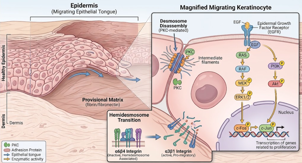
Figure 1. Desmosome disassembly and activation of EGFR-MEK-ERK cascade in migrating keratinocytes.
During re-epithelialisation the wound bed is populated by dermal fibroblasts that migrate from the adjacent uninjured dermis into the provisional fibrin-fibronectin clot. Sharp gradients of Platelet-Derived Growth Factor (PDGF) and Transforming Growth Factor-beta (TGF-β) secreted during early hemostasis and inflammation promote this migration.(Darby, Laverdet, Bonté, & Desmoulière, 2014)
Single-cell transcriptomic technologies have recently shown that dermal fibroblasts are not a uniform population, but possess considerable lineage heterogeneity and phenotypic plasticity.(Bao et al., 2009) The major functional fibroblast lineages are known to be at work in wound healing:
1. Engrailed-1 lineage-positive fibroblasts (CD26+/EN1+): These cells are predominantly found in the reticular dermis. They quickly invade the wound stroma upon activation and are the main cellular effectors of fibrotic scarring, secreting large quantities of dense, highly aligned Type I collagen.(Jin et al., 2025; Morbidelli et al., 2021)
2. Engrailed-1 lineage-negative fibroblasts (EN1−): This subset of fibroblasts is situated in the papillary dermis and correlates strongly with regeneration outcomes. EN1- fibroblasts do not form dense scar tissue but lay down a loose, disorganized matrix that favors hair follicle neogenesis and restoration of normal dermal pattern.(Kaplansky, Durnova, Burkovskaya, & Vorotnikova, 1991; Mascharak et al., 2021)
Once in the provisional matrix, activated fibroblasts proliferate and release proteolytic enzymes (MMP-1, MMP-13, MMP-2, etc.) to degrade the fibrin-rich clot and make space. Instead, they secrete a loose hydrated matrix of fibronectin, hyaluronic acid, glycosaminoglycans and immature Type III collagen comprising the provisional granulation tissue.(J. Li, Chen, & Kirsner, 2007) Normal adult skin is made up of approximately 80% Type I collagen and 10% Type III collagen. Early granulation tissue is primarily composed of embryo-associated Type III collagen (approximately 30%) which provides a flexible framework for cell migration and capillary ingrowth. Fibroblasts replace Type III collagen with Type I collagen in healing, increasing the tensile strength of the forming scar to about 80 % of pre-wounding strength.(Schultz & Wysocki, 2009)
A subset of activated fibroblasts switches to contractile myofibroblasts dramatically in response to sustained exposure to Transforming Growth Factor-beta 1 (TGF-β1). This transition is characterized by de novo expression of alpha-smooth muscle actin (α-SMA) that is assembled into stress fibers that insert into focal adhesions and form mechanical connections with the extracellular fibronectin scaffold. Myofibroblasts apply strong physical contractile forces that draw the wound edges together, thus accelerating wound closure.(Morbidelli et al., 2021; Phool et al., 2026)
The canonical TGF-β/Smad signaling pathway controls the molecular mechanism of myofibroblast differentiation and collagen deposition. Active TGF-β1 interacts with the transmembrane serine/threonine kinase receptor TGF-β Receptor Type II (TGF-βRII), which is constitutively active. Such binding results in recruitment, association and phosphorylation of TGF-β Receptor Type I (TGF-βRI/ALK5).(Landén et al., 2016; J. Li, Zhang, & Kirsner, 2003)
The activated TGF-βRI kinase domain directly phosphorylates receptor-regulated Smad proteins, especially Smad2 and Smad3. After phosphorylation, Smad2/3 detach from the receptor and associate with the common mediator Smad4 to create a heterotrimeric complex (Smad2/3-Smad4 complex). This complex translocates into the nucleus and binds to specific Smad binding elements (SBE) in the promoter regions of target genes and cooperates with co-activators (such as p300/CBP) to promote the transcription of genes encoding α-SMA, Type I collagen (COL1A1), Type III collagen (COL3A1) and Connective Tissue Growth Factor (CTGF). After the wound is closed, myofibroblasts must undergo apoptosis. Pathological persistence of myofibroblasts leads to hypertrophic scarring or keloids. As shown in the Figure 2., canonical TGF-β/Smad signaling cascade induces transcription of COL1A1, COL3A1, and α-SMA, resulting in transformation of resident fibroblasts into contractile, collagen-producing myofibroblasts.(Landén et al., 2016; J. Li et al., 1999)
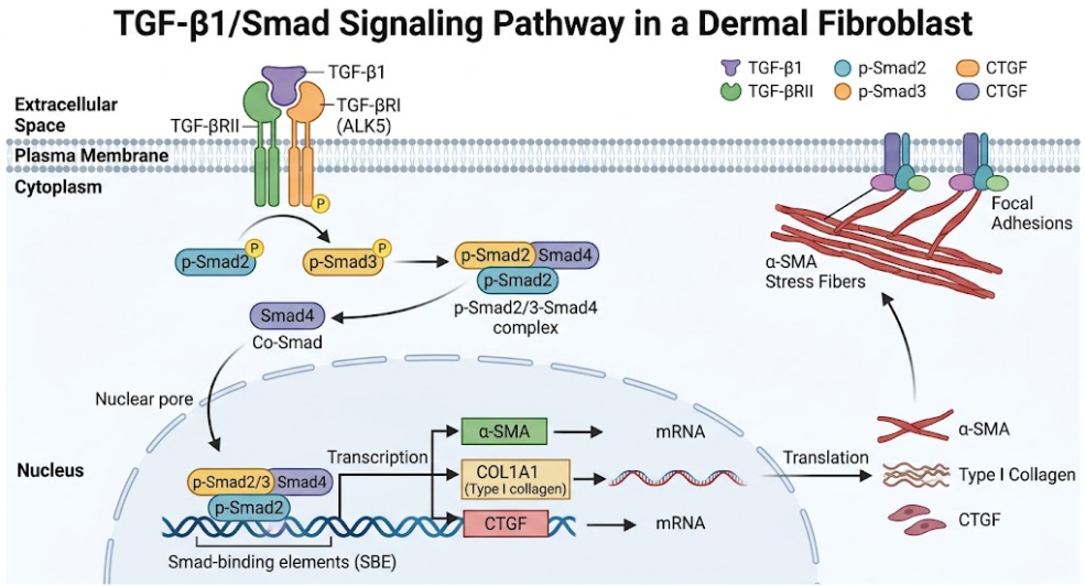
Figure 2. Canonical TGF-β/Smad pathway and myofibroblast phenotypic switch
The metabolic demand of hyperactive proliferating keratinocytes and fibroblasts requires rapid reestablishment of the vascular network. Neovascularization occurs by sprouting angiogenesis (new capillary budding from pre-existing vessels) and vasculogenesis (de novo recruitment of bone marrow derived progenitor cells).(Hunt, 1988; Santoro & Gaudino, 2005)
Sprouting angiogenesis is induced by local angiogenic signals, mainly VEGF-A released by parenchymal cells in the hypoxic wound stroma. When a VEGF gradient is detected, a single endothelial cell (EC) from an existing capillary is chosen to become the leading tip cell. The tip cell is highly polarized and migratory with many filopodia that sense the environment and migrate toward the VEGF gradient.(Jin et al., 2025; J. Li et al., 2003; Morbidelli et al., 2021)
The Notch/DLL4 lateral inhibition pathway controls the choice of the tip cell and the suppression of the tip phenotype in neighboring cells, which then become stalk cells that proliferate and form the vascular lumen. Binding of VEGF-A to VEGFR-2 on the selected tip cell upregulates expression of the transmembrane ligand Delta-like ligand 4 (DLL4).(Welch-Reardon et al., 2014) DLL4 binds to Notch receptors on adjacent stalk cells, triggering proteolytic cleavage of Notch by gamma-secretase and release of the Notch intracellular domain (NICD).(Y. Wang et al., 2023) NICD translocate to the nucleus to repress VEGFR-2 transcription and upregulate VEGFR-1 (a decoy receptor). Therefore, stalk cells become temporarily unresponsive to VEGF-A, so that only one tip cell is located at the tip of the capillary sprout, preventing unorganized, chaotic vascular growth. As illustrated in the Figure 3., the choice of leading endothelial tip cell and proliferating stalk cells is governed by a finely tuned Notch/DLL4 lateral inhibition loop in a gradient of tissue-derived VEGF-A. (Y. Wang et al., 2023; Welch-Reardon et al., 2014)
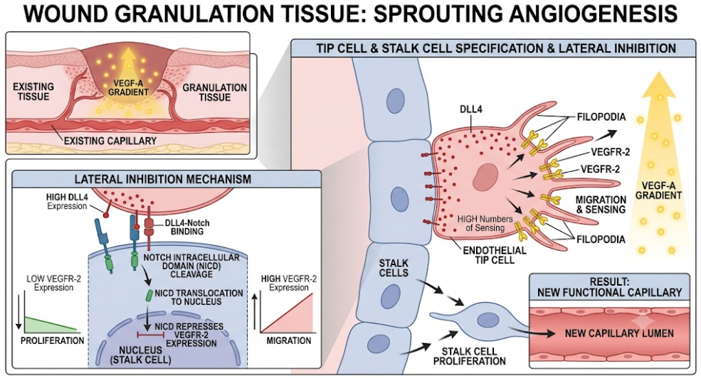
Figure 3. Insertion: Notch/DLL4-mediated lateral inhibition and sprouting capillary sprout
To traverse the dense provisional wound matrix, the leading tip cell has to degrade the surrounding extracellular matrix. Tip-cell VEGFR-2 activation results in secretion of matrix metalloproteinase (including MMP-2 and MMP-9) and plasminogen activators (uPA, tPA). These proteases degrade the capillary basement membrane (mainly Type IV collagen and laminins).(Morbidelli et al., 2021; Phool et al., 2026)
This active phase of migration is associated with a transient integrin switch, in which the static laminin-binding integrins are down-regulated and αvβ3 and α5β1 integrins are up-regulated. The αvβ3 integrin binds to provisional matrix proteins (fibronectin, fibrin and vitronectin) providing the traction needed for migration. In addition, αvβ3 physically associates with MMP-2 on the endothelial cell membrane, localizing the proteolytic activity right at the leading edge of the migrating sprout.(J. Li et al., 2003; Phool et al., 2026)
Beside sprouting from local vessels, post-natal vasculogenesis, which is driven by bone marrow-derived endothelial progenitor cells (BMD EPCs), also supports neovascularization. Local tissue hypoxia and injury trigger activation of systemic signaling cascades that induce mobilization of progenitor cells from the bone marrow to the circulation.(Velazquez, 2007) These circulating progenitor cells are recruited to the wound bed specifically by a steep gradient of stromal cell-derived factor-1 (SDF-1/CXCL12) secreted by hypoxic wound-edge cells.(Landén et al., 2016; Velazquez, 2007)
The homing of BMD EPCs to the wound stroma is thru CXCL12/CXCR4 receptor axis. BMD EPCs are physically incorporated into the new capillary sprouts when recruited into the granulation tissue, differentiate into mature endothelial cells and secrete high levels of pro-angiogenic paracrine factors to accelerate neovascularization.(Phool et al., 2026)
Once formed the immature, leaky capillary loops need to be matured and stabilized. This process involves a recruitment of mural cells (pericytes for capillaries, smooth muscle cells for larger vessels) Endothelial cells secrete Platelet-Derived Growth Factor-BB (PDGF-BB) which binds to PDGFR- on resident pericytes initiating their migration and recruitment to the outer surface of the endothelial tubes. (An et al., 2021)
At the same time pericytes secrete Angiopoietin-1 (Ang-1) that binds to the Tie-2 receptor on endothelial cells. Such activation favors the synthesis of a new basement membrane built up of laminin-8 and laminin-10 which reinforces cell-cell junctions and makes the newly formed microvasculature stable, functional and resistant to vascular leakage.(J. Li et al., 2003)
The proliferative phase is orchestrated by spatiotemporal regulation of the Vascular Endothelial Growth Factor (VEGF) secretory axis. Under normal conditions, healthy skin has low levels of VEGF at baseline. However, mechanical trauma disrupts the microvasculature and creates a highly hypoxic wound microenvironment.(Jin et al., 2025; Phool et al., 2026; Velazquez, 2007)
he VEGF secretory axis is centered around the transcription factor Hypoxia-Inducible Factor-1 (HIF-1), a heterodimer of a β subunit and a highly oxygen sensitive subunit (HIF-1α). In normoxia, prolyl hydroxylase domain (PHD) enzymes continuously hydroxylate HIF-1α at proline residues. This hydroxylation enables HIF-1α to be recognized by the von Hippel-Lindau (VHL) E3 ubiquitin ligase leading to its rapid degradation by the proteasome.(Landén et al., 2016)
The lack of molecular oxygen (a required co-substrate for PHD) causes prolyl hydroxylases to be inactivated under hypoxic conditions. Thus, HIF-1α is not degraded but accumulates in the cytoplasm and translocates to the nucleus, where it heterodimerizes with HIF-1β. The active HIF-1 complex binds the Hypoxia Response Elements (HRE) on the promoter regions of the target genes to induce the massive transcription of the proangiogenic factors, mainly VEGF-A.(Morbidelli et al., 2021)
The VEGF-A gene undergoes alternative splicing to produce several different isoforms, the major ones being VEGF121, VEGF165 and VEGF189.(J. Li et al., 2003) These isoforms have differential affinities to extracellular matrix heparin sulfate proteoglycans (HSPGs):
• VEGF121: Does not contain the heparin-binding domain, and is a very soluble, freely diffusible peptide that creates long-range chemotactic gradients.
• VEGF165: Possesses a highly basic heparin-binding domain and is tightly sequestered in the local extracellular matrix, giving rise to a localized high concentration angiogenic cue.
• VEGF189: Intermediate solubility and heparin-binding. This is the most physiologically active isoform in wound healing.
Different cellular players within the wound bed including activated keratinocytes, dermal fibroblasts and polarized M2 macrophages produce and secrete VEGF-A. Interestingly, although M1 macrophages dominate the early inflammatory phase, they mainly secrete pro-inflammatory cytokines (IL-1β, TNF-α) that promote single-cell migration of vascular cells.(Phool et al., 2026) Alternatively activated (M2) macrophages induce an orchestrated secretome enriched in VEGF165, basic FGF and active sphingolipids (including sphingosine-1-phosphate, S1P) that synergistically promote endothelial sprouting and capillary tube formation.(Beyer, Koch, Lee, Jung, & Blocki, 2018)
evere clinical complications are caused by pathological deviations in the cellular and molecular machinery of the proliferative phase. In chronic non-healing wounds, such as diabetic foot ulcers and venous stasis ulcers, the microenvironment is unable to shift from sustained inflammation to cellular proliferation. As depicted in the Figure 4., chronic non-healing wounds are marked by persistent M1 inflammation, excessive MMP-9 degradation of VEGF-A, and upregulation of the BAMBI pseudo-receptor that blocks regenerative TGF-β signaling.(Sen et al., 2009)
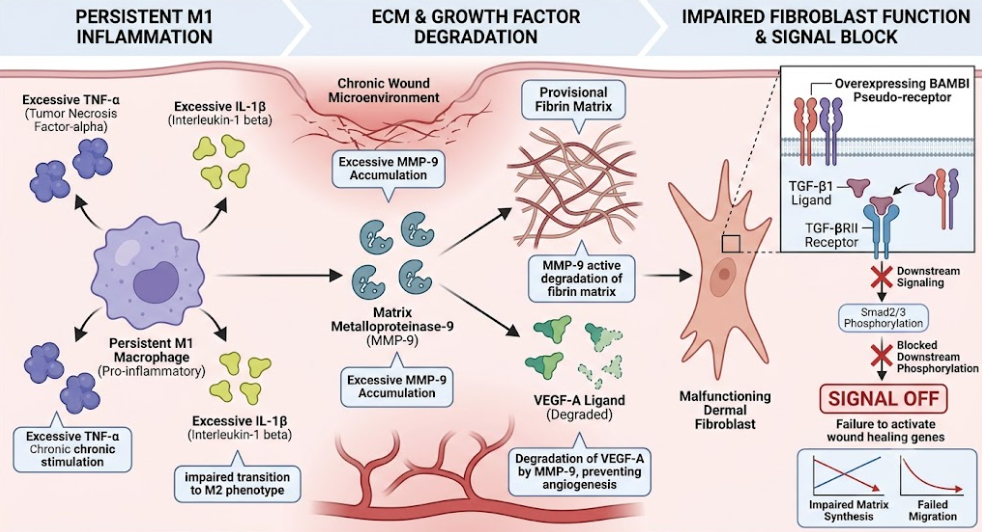
Figure 4. Molecular checkpoints and dysregulation in the chronic wound microenvironment
Chronic diabetic wounds are marked by persistent hyperglycemia and biofilm-producing bacterial infections that arrest macrophages in a persistent, pro-inflammatory M1 state. These cells secrete an excess of MMP-9 and serine proteases. The protease-rich environment that results in the degradation of the provisional fibrin scaffold indiscriminately cleaves essential growth factors such as VEGF-A and PDGF-BB, rendering them inactive. The proteolytic destruction inhibits the formation of stable capillary sprouts leading to severe hypoxia and tissue necrosis.(Landén et al., 2016; Phool et al., 2026)
Molecular analyzes have recently identified the BMP and Activin Membrane-Bound Inhibitor (BAMBI) as a key checkpoint in the pathogenesis of chronic wounds. BAMBI is a transmembrane glycoprotein whose extracellular domain shows extensive sequence similarity with the TGF-β Receptor Type I (TGF-βRI/ALK5). BAMBI, however, completely lacks the intracellular serine/threonine kinase domain for downstream signal transduction.(Ehnert et al., 2023)
BAMBI is markedly increased (>10-fold) in chronic wound tissue compared with healthy healing wounds. BAMBI forms homodimers with TGF-βRI and heterodimers with TGF-βRII. It competes with active TGF-β receptors for ligand binding. BAMBI does not have a kinase domain and therefore functions in a dominant-negative manner as a decoy receptor and totally prevents Smad2 and Smad3 phosphorylation. Therefore, the transcription of regenerative genes such as COL1A1 and CTGF is completely suppressed downstream, despite the high levels of active TGF-β1 ligands in the tissues of chronic wounds. This blockade of the molecule blocks fibroblast to myofibroblast differentiation and granulation tissue formation.(Zhang et al., 2020)
Cellular proliferation is disturbed by the epigenetic dysregulation by non-coding RNAs as well as by pseudo-receptors.(Landén et al., 2016) Several microRNAs (miRNAs) are negative regulators of the proliferative phase:
• miR-198: In healthy wounds, a molecular switch turns off miR-198, allowing translation of Follistatin-like 1 (FSTL1) to promote keratinocyte migration. Pathological overexpression of miR-198 in chronic diabetic ulcers silences FSTL1 and blocks re-epithelialization.
• miR-210: miR-210 is induced by severe hypoxia and inhibits keratinocyte proliferation and delays wound closure by targeting the cell-cycle regulator E2F3.
• miR-200b: Upregulation of miR-200b in diabetic wounds by high TNF-α directly silences the expression of VEGFR-2 in endothelial cells and inhibits sprouting angiogenesis.(Landén et al., 2016)
Clinical strategies have been to bypass these checkpoints either by delivering stabilized growth factors or with the use of autologous biomaterials. Recombinant PDGF-BB (becaplermin gel) is FDA-approved for diabetic ulcers, but its short half-life in protease-rich wound stroma limits its clinical efficacy.(Jin et al., 2025; Morbidelli et al., 2021)
To overcome this autologous platelet concentrates are widely used, in particular Platelet-Rich Plasma (PRP) gel. PRP gel is activated with calcium chloride to form a stable autologous fibrin scaffold, providing a sustained-release reservoir of active PDGF, TGF-β, and VEGF. PRP gel induces vigorous neovascularization, vascular regression and myofibroblast differentiation as demonstrated by clinical and histological assessment. In contrast, the second generation of Platelet-Rich Fibrin (PRF) is not appropriate for deep surgical or tissue reconstruction, because of its high leukocyte component, which causes excessive local inflammation, resulting in impaired myofibroblast generation and compromised structural remodeling.(Choi, Kim, & Park, 2022)
Functional cutaneous tissue reconstruction is a spatiotemporal masterpiece that relies on an accurate transition from an inflammatory environment to a proliferative, regenerative state. As illustrated by this review, the successful repair of tissue depends on the tight interplay between keratinocyte re-epithelialization, fibroblast-to-myofibroblast differentiation and sprouting angiogenesis.(Landén et al., 2016) These cellular events are mediated by different molecular pathways such as the EGFR signaling pathway in epithelial cells, the canonical TGF-β/Smad complex in dermal fibroblasts and Notch/DLL4 lateral inhibition in sprouting capillaries.(Jin et al., 2025)
But in chronic non-healing wounds, these spatiotemporal ‘clocks’ are severely uncoupled. The pro-inflammatory M1 macrophage phenotype persists and generates a hostile microenvironment with high levels of reactive oxygen species (ROS) and matrix metalloproteinases (MMPs), especially MMP-9, which actively degrades important pro-healing growth factors such as VEGF-A and PDGF-BB.(Phool et al., 2026)
A major highlight of recent molecular studies is the discovery of BAMBI (BMP and Activin Membrane-Bound Inhibitor) as an essential pathological checkpoint. In chronic wounds, BAMBI upregulation functions as dominant-negative pseudo-receptor without intracellular kinase domain and blocks phosphorylation of ALK5/TGF-βRI.(Ehnert et al., 2023) This blockade inhibits the canonical Smad2/3 signaling cascade which inhibits fibroblast activation, collagen synthesis and myofibroblast trans differentiation in spite of high levels of active local TGF-β ligands. This blockade of signaling is compounded by epigenetic checkpoints, including the pathological persistence of miRNA such as miR-198, miR-210 and miR-200b, which silence regenerative proteins (FSTL1, E2F3 and VEGFR-2). (Landén et al., 2016)
To overcome such biological barriers, autologous biomaterials have been used for clinical interventions. Clinical and histological evaluations show that Platelet-Rich Plasma (PRP) gel is a very effective and multi-target therapeutic matrix.(Choi et al., 2022) RP gel provides a physiologically balanced, sustained release of important growth factors (PDGF, TGF-β and VEGF) in an autologous fibrin scaffold thus stimulating rapid, organized angiogenesis, vessel maturation and myofibroblast differentiation. In contrast, second generation Platelet-Rich Fibrin (PRF) is not indicated for deep structural tissue reconstruction. PRF, due to its high leukocyte content, can pathologically prolong the acute inflammatory phase, increase neutrophil infiltration and impair the vital remodeling phase.(Choi et al., 2022; Jin et al., 2025)
Dissecting such complex molecular switches, from BAMBI blockade to miRNA checkpoints, finally provides a compelling scientific rationale for the creation of targeted, precision wound therapeutics. The next frontier in regenerative medicine is to mimic the natural spatiotemporal dynamics of the proliferative phase.(Ehnert et al., 2023; Jin et al., 2025)
The proliferative phase of cutaneous wound healing is a highly complex, well-coordinated biological program devoted to the physical reconstruction of tissue. Successful reconstitution of a vascularized granulation tissue involves the integration of keratinocyte re-epithelialization, fibroblast-mediated collagen deposition and sprout-mediated capillary angiogenesis. Understanding the molecular switches regulating these processes, such as the EGFR signaling axis, the canonical TGF-β/Smad cascade and the Notch/DLL4 lateral inhibition loop, provides critical insight into the pathogenesis of chronic wounds. Identification of pathological checkpoints including the BAMBI pseudo-receptor and microRNA blockades opens new therapeutic frontiers for the creation of targeted biomaterials and precision medicine strategies aiming to enhance cutaneous regeneration.
Abba, M. L., Patil, N., Leupold, J. H., & Allgayer, H. (2016). MicroRNA Regulation of Epithelial to Mesenchymal Transition. J Clin Med, 5(1). doi:10.3390/jcm5010008
Ahluwalia, A., & Tarnawski, A. S. (2012). Critical role of hypoxia sensor–HIF-1α in VEGF gene activation. Implications for angiogenesis and tissue injury healing. Curr Med Chem, 19(1), 90-97. doi:10.2174/092986712803413944
Altaany, Z., Yang, G., & Wang, R. (2013). Crosstalk between hydrogen sulfide and nitric oxide in endothelial cells. J Cell Mol Med, 17(7), 879-888. doi:10.1111/jcmm.12077
An, Y., Lin, S., Tan, X., Zhu, S., Nie, F., Zhen, Y., . . . Wu, J. (2021). Exosomes from adipose-derived stem cells and application to skin wound healing. Cell Prolif, 54(3), e12993. doi:10.1111/cpr.12993
Bao, P., Kodra, A., Tomic-Canic, M., Golinko, M. S., Ehrlich, H. P., & Brem, H. (2009). The role of vascular endothelial growth factor in wound healing. J Surg Res, 153(2), 347-358. doi:10.1016/j.jss.2008.04.023
Barrientos, S., Stojadinovic, O., Golinko, M. S., Brem, H., & Tomic-Canic, M. (2008). Growth factors and cytokines in wound healing. Wound Repair Regen, 16(5), 585-601. doi:10.1111/j.1524-475X.2008.00410.x
Beyer, S., Koch, M., Lee, Y. H., Jung, F., & Blocki, A. (2018). An In Vitro Model of Angiogenesis during Wound Healing Provides Insights into the Complex Role of Cells and Factors in the Inflammatory and Proliferation Phase. Int J Mol Sci, 19(10). doi:10.3390/ijms19102913
Chitturi, R. T., Balasubramaniam, A. M., Parameswar, R. A., Kesavan, G., Haris, K. T., & Mohideen, K. (2015). The role of myofibroblasts in wound healing, contraction and its clinical implications in cleft palate repair. J Int Oral Health, 7(3), 75-80.
Choi, S. Y., Kim, S., & Park, K. M. (2022). Initial Healing Effects of Platelet-Rich Plasma (PRP) Gel and Platelet-Rich Fibrin (PRF) in the Deep Corneal Wound in Rabbits. Bioengineering (Basel), 9(8). doi:10.3390/bioengineering9080405
Crowe, M. J., Doetschman, T., & Greenhalgh, D. G. (2000). Delayed Wound Healing in Immunodeficient TGF-β1 Knockout Mice. Journal of Investigative Dermatology, 115(1), 3-11. doi:10.1046/j.1523-1747.2000.00010.x
Darby, I. A., Laverdet, B., Bonté, F., & Desmoulière, A. (2014). Fibroblasts and myofibroblasts in wound healing. Clin Cosmet Investig Dermatol, 7, 301-311. doi:10.2147/ccid.S50046
Ehnert, S., Rinderknecht, H., Liu, C., Voss, M., Konrad, F. M., Eisler, W., . . . Nussler, A. K. (2023). Increased Levels of BAMBI Inhibit Canonical TGF-β Signaling in Chronic Wound Tissues. Cells, 12(16). doi:10.3390/cells12162095
He, X., Han, Z., Ruan, Y., Wang, Z., Liao, B., Li, X., . . . Bai, D. (2025). Smart responsive biomaterials for spatiotemporal modulation of functional tissue repair. Materials Today Bio, 33, 102063. doi:https://doi.org/10.1016/j.mtbio.2025.102063
Huang, C., Sheng, S., Li, R., Sun, X., Liu, J., & Huang, G. (2015). Lactate promotes resistance to glucose starvation via upregulation of Bcl-2 mediated by mTOR activation. Oncol Rep, 33(2), 875-884. doi:10.3892/or.2014.3655
Hunt, T. K. (1988). The physiology of wound healing. Ann Emerg Med, 17(12), 1265-1273. doi:10.1016/s0196-0644(88)80351-2
Iwami, K. I., Matsuguchi, T., Masuda, A., Kikuchi, T., Musikacharoen, T., & Yoshikai, Y. (2000). Cutting edge: naturally occurring soluble form of mouse Toll-like receptor 4 inhibits lipopolysaccharide signaling. J Immunol, 165(12), 6682-6686. doi:10.4049/jimmunol.165.12.6682
Jacinto, A., Martinez-Arias, A., & Martin, P. (2001). Mechanisms of epithelial fusion and repair. Nat Cell Biol, 3(5), E117-123. doi:10.1038/35074643
Jin, C., Jin, Y., Ding, Z., Nuch, K. S., Han, M., Shim, J., . . . Heo, C. Y. (2025). Cellular and Molecular Mechanisms of Wound Repair: From Biology to Therapeutic Innovation. Cells, 14(23). doi:10.3390/cells14231850
Kaplansky, A. S., Durnova, G. N., Burkovskaya, T. E., & Vorotnikova, E. V. (1991). The effect of microgravity on bone fracture healing in rats flown on Cosmos-2044. Physiologist, 34(1 Suppl), S196-199.
Kim, J., Shin, Y. K., & Kim, K. Y. (2018). Promotion of Keratinocyte Proliferation by Tracheloside through ERK1/2 Stimulation. Evid Based Complement Alternat Med, 2018, 4580627. doi:10.1155/2018/4580627
Landén, N. X., Li, D., & Ståhle, M. (2016). Transition from inflammation to proliferation: a critical step during wound healing. Cellular and Molecular Life Sciences, 73(20), 3861-3885. doi:10.1007/s00018-016-2268-0
Lau, K., Paus, R., Tiede, S., Day, P., & Bayat, A. (2009). Exploring the role of stem cells in cutaneous wound healing. Exp Dermatol, 18(11), 921-933. doi:10.1111/j.1600-0625.2009.00942.x
Lee, S., Kim, M. S., Jung, S. J., Kim, D., Park, H. J., & Cho, D. (2018). ERK activating peptide, AES16-2M promotes wound healing through accelerating migration of keratinocytes. Sci Rep, 8(1), 14398. doi:10.1038/s41598-018-32851-y
Li, J., Chen, J., & Kirsner, R. (2007). Pathophysiology of acute wound healing. Clin Dermatol, 25(1), 9-18. doi:10.1016/j.clindermatol.2006.09.007
Li, J., Foitzik, K., Calautti, E., Baden, H., Doetschman, T., & Dotto, G. P. (1999). TGF-beta3, but not TGF-beta1, protects keratinocytes against 12-O-tetradecanoylphorbol-13-acetate-induced cell death in vitro and in vivo. J Biol Chem, 274(7), 4213-4219. doi:10.1074/jbc.274.7.4213
Li, J., Zhang, Y. P., & Kirsner, R. S. (2003). Angiogenesis in wound repair: angiogenic growth factors and the extracellular matrix. Microsc Res Tech, 60(1), 107-114. doi:10.1002/jemt.10249
Li, J. Y., Ren, K. K., Zhang, W. J., Xiao, L., Wu, H. Y., Liu, Q. Y., . . . Xin, H. B. (2019). Human amniotic mesenchymal stem cells and their paracrine factors promote wound healing by inhibiting heat stress-induced skin cell apoptosis and enhancing their proliferation through activating PI3K/AKT signaling pathway. Stem Cell Res Ther, 10(1), 247. doi:10.1186/s13287-019-1366-y
Mascharak, S., desJardins-Park, H. E., Davitt, M. F., Griffin, M., Borrelli, M. R., Moore, A. L., . . . Longaker, M. T. (2021). Preventing Engrailed-1 activation in fibroblasts yields wound regeneration without scarring. Science, 372(6540). doi:10.1126/science.aba2374
Monti, M., Hyseni, I., Pacini, A., Monzani, E., Casella, L., & Morbidelli, L. (2018). Cross-talk between endogenous H2S and NO accounts for vascular protective activity of the metal-nonoate Zn(PipNONO)Cl. Biochemical Pharmacology, 152, 143-152. doi:https://doi.org/10.1016/j.bcp.2018.03.025
Morbidelli, L., Genah, S., & Cialdai, F. (2021). Effect of Microgravity on Endothelial Cell Function, Angiogenesis, and Vessel Remodeling During Wound Healing. Frontiers in Bioengineering and Biotechnology, Volume 9 – 2021. doi:10.3389/fbioe.2021.720091
Nunan, R., Campbell, J., Mori, R., Pitulescu, M. E., Jiang, W. G., Harding, K. G., . . . Martin, P. (2015). Ephrin-Bs Drive Junctional Downregulation and Actin Stress Fiber Disassembly to Enable Wound Re-epithelialization. Cell Rep, 13(7), 1380-1395. doi:10.1016/j.celrep.2015.09.085
Phool, C., Mohd, F., Mayur, P., Himanshu, S., & Neetu, S. (2026). An Overview and Review of Growth Factors in Wound Healing: Emerging Trends and Innovations. Current Diabetes Reviews, 22(1), 1-27. doi:http://dx.doi.org/10.2174/0115733998332692241202072249
Ramirez, H., Patel, S. B., & Pastar, I. (2014). The Role of TGFβ Signaling in Wound Epithelialization. Adv Wound Care (New Rochelle), 3(7), 482-491. doi:10.1089/wound.2013.0466
Santoro, M. M., & Gaudino, G. (2005). Cellular and molecular facets of keratinocyte reepithelization during wound healing. Exp Cell Res, 304(1), 274-286. doi:10.1016/j.yexcr.2004.10.033
Schultz, G. S., & Wysocki, A. (2009). Interactions between extracellular matrix and growth factors in wound healing. Wound Repair Regen, 17(2), 153-162. doi:10.1111/j.1524-475X.2009.00466.x
Sen, C. K., Gordillo, G. M., Roy, S., Kirsner, R., Lambert, L., Hunt, T. K., . . . Longaker, M. T. (2009). Human skin wounds: a major and snowballing threat to public health and the economy. Wound Repair Regen, 17(6), 763-771. doi:10.1111/j.1524-475X.2009.00543.x
Veith, A. P., Henderson, K., Spencer, A., Sligar, A. D., & Baker, A. B. (2019). Therapeutic strategies for enhancing angiogenesis in wound healing. Adv Drug Deliv Rev, 146, 97-125. doi:10.1016/j.addr.2018.09.010
Velazquez, O. C. (2007). Angiogenesis and vasculogenesis: inducing the growth of new blood vessels and wound healing by stimulation of bone marrow-derived progenitor cell mobilization and homing. J Vasc Surg, 45 Suppl A(Suppl A), A39-47. doi:10.1016/j.jvs.2007.02.068
Wang, C., Li, N., Zhang, C., Sun, S., Gao, Y., & Long, M. (2015). Effects of Simulated Microgravity on Functions of Neutrophil-like HL-60 Cells. Microgravity Science and Technology, 27, 515-527. doi:10.1007/s12217-015-9473-6
Wang, Y., Dong, M., Zheng, Y., Wang, C., Ding, X., Wu, H., . . . Song, X. (2023). Preliminary Study on Human Adipose Stem Cells Promoting Skin Wound Healing through Notch Signaling Pathway. Curr Stem Cell Res Ther, 18(5), 699-711. doi:10.2174/1574888×18666221216123259
Welch-Reardon, K. M., Ehsan, S. M., Wang, K., Wu, N., Newman, A. C., Romero-Lopez, M., . . . Hughes, C. C. (2014). Angiogenic sprouting is regulated by endothelial cell expression of Slug. J Cell Sci, 127(Pt 9), 2017-2028. doi:10.1242/jcs.143420
Werdin, F., Tenenhaus, M., & Rennekampff, H.-O. (2008). Chronic wound care. The Lancet, 372(9653), 1860-1862. doi:10.1016/S0140-6736(08)61793-6
Witte, M. B., & Barbul, A. (2002). Role of nitric oxide in wound repair. Am J Surg, 183(4), 406-412. doi:10.1016/s0002-9610(02)00815-2
Xu, X., Zhang, H., Yan, Y., Wang, J., & Guo, L. (2021). Effects of electrical stimulation on skin surface. Acta Mech Sin, 37(12), 1843-1871. doi:10.1007/s10409-020-01026-2
Zhang, T., Wang, X. F., Wang, Z. C., Lou, D., Fang, Q. Q., Hu, Y. Y., . . . Tan, W. Q. (2020). Current potential therapeutic strategies targeting the TGF-β/Smad signaling pathway to attenuate keloid and hypertrophic scar formation. Biomed Pharmacother, 129, 110287. doi:10.1016/j.biopha.2020.110287