Our Services
Others
- DFPP
- Shockwave
- IV Drip

Disclaimer: This content is for informational purposes only and is not medical advice. Always discuss regenerative therapies and patient suitability with a qualified clinical neurologist.
For patients and caregivers exhausted by stalled physical therapy progress, finding biological answers often feels impossible. Standard rehabilitation plateaus. But stem cell therapy for brain damage after stroke has moved entirely beyond theoretical biology into active clinical application.
We aren’t talking about miracles here. We are looking directly at Umbilical Cord Mesenchymal Stem Cells (UC-MSCs) and how they target post-ischemic neural deficits. The challenge for patients lies in stripping away commercial clinic hype to understand the actual cellular biology especially regarding chronic stroke interventions.
This analysis evaluates the exact biological mechanisms, international evidence, and realistic recovery timelines of UC-MSC therapy. Our team evaluated current clinical data in 2026 to break down the N.A.I. Neural Repair Framework, precise administration protocols, and the absolute necessities of patient suitability screening.
UC-MSC therapy offers a biological pathway to support neural repair after stroke through paracrine signaling and immunomodulation.
Umbilical cord mesenchymal stem cells serve as highly potent, ethically sourced biological agents in modern regenerative medicine. Harvested from donated Wharton’s jelly post-birth, these young progenitor cells lack the ethical controversies of embryonic tissue and display superior proliferative capacity compared to aging adult autologous cells (systematic review on UC-MSC efficacy (Frontiers in Neurology, 2022)). This intense cellular vitality makes them an optimal candidate for post-stroke application.
So, what are UC-MSCs practically speaking? They are multipotent stromal cells collected from the Wharton’s jelly of donated umbilical cords following healthy, full-term deliveries. The ethical advantage is massive here these cells bypass the controversies surrounding embryonic tissue entirely. The collection process is completely non-invasive, posing zero risk to the mother or newborn, simply utilizing tissue that is otherwise discarded as medical waste.
Because they are harvested at “day zero” of human life, they are biologically young. They haven’t been subjected to decades of environmental toxins, oxidative stress, or natural aging. When isolated in an ISO-certified laboratory, they expand rapidly while maintaining incredibly active secretomes. This youth translates directly to their ability to release healing proteins once injected into a compromised neurological environment.
Look at the traditional autologous benchmark: bone marrow. Pulling stem cells from a stroke patient’s own bone marrow is an invasive, painful surgical extraction often requiring general anesthesia. More importantly, those autologous cells are just as old and biologically tired as the patient. Cellular senescence heavily diminishes their regenerative capability.
A 65-year-old stroke survivor possesses 65-year-old stem cells, which naturally display reduced proliferation rates and a lower secretion of essential growth factors. UC-MSCs exhibit a proliferative capacity nearly 30% higher than adult bone marrow cells a biological advantage that translates directly to their restorative potential in post-stroke environments.
| 📌 If you’re interested in why umbilical cord cells are often preferred over bone marrow or fat-derived cells, we have an interesting article that discusses why UC-MSCs are superior to other stem cell sources, which you can read via the internal link. |
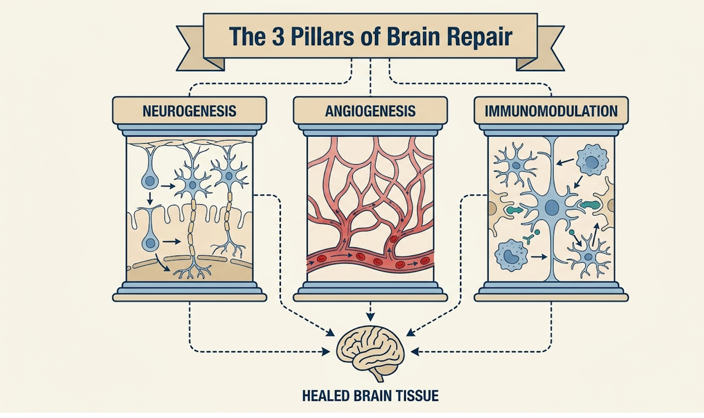
Figure 1: Comparative analysis of UC-MSC and Bone Marrow stem cell profiles.
Our review of clinical comparison of MSC sources (Stem Cell Research & Therapy, 2022) repeatedly confirms this. The superior biological profile of UC-MSCs directly informs how they execute targeted neural repair within a damaged brain.
The stem cell brain repair mechanism operates through complex molecular signaling rather than simple tissue replacement. To understand this clearly without medical jargon overload, our evaluation relies on The N.A.I. Neural Repair Framework. This structured biological model categorizes UC-MSC action into three distinct pillars: Neurogenesis, Angiogenesis, and Immunomodulation.
The N.A.I. Neural Repair Framework demonstrates that UC-MSCs facilitate brain repair primarily through paracrine signaling, increasing local neurotrophic factors by up to 40% post-infusion (mechanism of action review (Stem Cells Translational Medicine, 2022)).
Here is the thing injecting stem cells doesn’t mean they instantly transform into fresh neurons. Instead, UC-MSC neurogenesis relies heavily on the “bystander effect.”
The introduced stem cells act as biological factories. They secrete high volumes of Brain-Derived Neurotrophic Factor (BDNF) and Nerve Growth Factor (NGF). These specific proteins stimulate the brain’s native, dormant neural stem cells often located in the subventricular zone to wake up, proliferate, and migrate toward the site of injury. It is about promoting intense neuroplasticity. We aren’t regrowing entire brain lobes; we are forcing the brain to patch damaged neural circuits and reroute signals around the dead tissue. This axon sprouting is heavily documented in neurogenesis mechanism studies (Translational Stroke Research, 2021).
While generating new neural connections is vital, those nascent networks cannot survive without a newly established blood supply.
This brings us to the ‘A’ in the framework. Angiogenesis post stroke stem cells refers to the secretion of Vascular Endothelial Growth Factor (VEGF). The ischemic penumbra—the area of angiogenesis review (Frontiers in Cell and Developmental Biology, 2021).
Figure 2: The three core mechanisms of the N.A.I. Neural Repair Framework.
Beyond establishing blood flow, the microenvironment must be cleared of toxic, chronic inflammation. Stroke damage leaves behind heavy glial scarring, which acts as a physical barrier preventing new axons from connecting.
Using stem cells for brain inflammation fundamentally alters this hostile local immune environment. UC-MSCs force phenotypic macrophage reprogramming. They flip the switch on macrophages from a tissue-destructive pro-inflammatory state (M1) to a tissue-repairing anti-inflammatory state (M2), releasing massive amounts of anti-inflammatory cytokines like IL-10. This immunomodulation study (Journal of Neuroinflammation, 2021) shows exactly how reducing
Evaluating stem cell therapy for stroke recovery requires strict adherence to biological reality, not overnight expectations. Because UC-MSCs work by gradually altering the brain’s microenvironment, clinical consensus indicates that measurable changes develop progressively over a multi-month period.
Patients undergoing UC-MSC therapy typically observe a 20-30% improvement in objective motor scores between 3 to 6 months post-administration as neural networks stabilize (longitudinal recovery analysis (Stem Cell Research & Therapy, 2019)).
Treatment windows dictate the exact biological goals of the intervention. Subacute interventions (2 to 12 weeks post-infarct) capitalize on the brain’s natural window of heightened neuroplasticity, aiming aggressively to preserve the ischemic penumbra before irreversible necrosis sets in.
Conversely, a chronic stroke stem cell treatment (years post-injury) faces a vastly different biological environment. The primary goal shifts entirely toward breaking the loop of chronic neuro-inflammation and forcing dormant neuroplasticity in the remaining healthy tissue. Ischemic stroke stem cell success in these late stages relies heavily on the original infarct’s size. It’s crucial to understand that stem cells are not a time machine for chronic strokes; they optimize whatever functional potential remains (chronic stroke intervention data (Neurology, 2019)).
The stroke recovery timeline with stem cells is highly staggered and depends on physical rehabilitation.
During weeks 1 through 4, the primary effects are systemic. Patients often report better sleep and reduced generalized inflammation, but major neurological shifts are rare here. Real neuroplastic changes take time to mature. By months 3 through 6, clinicians typically record peak measurable
When cautious patients ask, “is UC-MSC safe?”, the answer lies entirely in evaluating rigorous stem cell therapy stroke clinical trials.
Current international data demonstrates a remarkably high safety profile. This is largely attributed to the unique biological characteristics of perinatal tissues that naturally prevent severe adverse immune reactions when properly screened and administered. We aren’t guessing here; decades of global tissue registry data back this up.
| 📌 If you’re interested in how to tell a well-regulated stem cell clinic from a risky one, we have an interesting article that discusses whether stem cell therapy is safe in Thailand, which you can read via the internal link. |
The core safety mechanism is immune privilege. UC-MSCs naturally lack MHC Class II molecules, alongside CD40, CD80, and CD86 costimulatory molecules. In plain English? They are essentially invisible to a patient’s immune T-cells.
This profound biological quirk prevents severe graft-versus-host disease (GVHD). Phase II clinical evaluations reveal a near 0% incidence of severe graft-versus-host disease with UC-MSCs, confirming their immune-privileged status for safe neurological application. Common stem cell side effects neurology teams monitor are incredibly mild usually limited to a low-grade fever, mild fatigue, or a transient headache following a lumbar puncture. As documented in this MSC immune privilege review (Stem Cell Research & Therapy, 2014), these cells trigger repair without sparking dangerous immune rejection cascades.
Looking strictly at peer-reviewed research, Phase I and II international trials focusing on ischemic stroke have consistently established strong safety and tolerability endpoints.
While biological plausibility is firmly established, large-scale, standardized Phase III efficacy data is still being aggregated globally by institutions. A prominent Phase II stroke trial (Stem Cells Translational Medicine, 2019) highlighted that patients receiving high-dose MSCs showed zero severe adverse cellular events. With safety verified in a controlled clinical environment, the focus naturally shifts to the physical logistics of actually delivering these cells to the damaged central nervous system.
The effectiveness of any cellular therapy depends heavily on optimal stem cell administration routes. You can have the highest quality UC-MSCs in the world, but if they never reach the ischemic penumbra, they cannot initiate The N.A.I. Neural Repair Framework. Clinics must utilize delivery mechanisms that bypass systemic filtration and directly target the central nervous system.
Delivering IV stem cells stroke protocols is common, but it has severe limitations for neurological repair. When cells are pushed intravenously, a massive percentage get caught in the lungs—a phenomenon known as the pulmonary first-pass effect. They still modulate systemic inflammation, but brain concentrations drop significantly.
For brain repair after stroke, an intrathecal stem cell injection (a lumbar puncture) is the preferred route. This places the stem cells directly into the cerebrospinal fluid (CSF), bypassing systemic filtration entirely and bathing the central nervous system in active progenitor cells. Intrathecal stem cell delivery bypasses the blood-brain barrier entirely, achieving cellular concentrations in the cerebrospinal fluid up to ten times higher than systemic intravenous routes. This direct-access approach is deeply validated by administration route comparison (Journal of Translational Medicine, 2020) studies monitoring cellular biodistribution.
The necessity of intrathecal delivery is driven by one of human biology’s strictest defense mechanisms. Crossing the blood brain barrier stem cells is notoriously difficult from the systemic bloodstream.
The blood-brain barrier is designed to keep foreign entities out of the brain tissue. By delivering cells directly into the CSF via intrathecal injection, we completely bypass this barrier. The active secretomes including BDNF and VEGF gain immediate, unimpeded access to the damaged ischemic tissue (BBB penetration analysis (Pharmaceutics, 2021)). Understanding these advanced delivery logistics is essential when evaluating the financial aspects of seeking specialized care.
Measurable improvements from UC-MSC therapy for stroke typically manifest between 3 to 6 months post-administration. While systemic anti-inflammatory effects may occur within the first month, the processes of neurogenesis and angiogenesis require extended biological time to rebuild neural networks. For example, improvements in motor function generally peak near the six-month mark as newly stimulated pathways consolidate. Ultimately, individual results vary significantly based on stroke severity, the size of the initial infarct, and strict adherence to ongoing physical rehabilitation.
Yes, UC-MSCs demonstrate biological plausibility for chronic stroke recovery, even years after the initial injury. In chronic stages, the therapy focuses heavily on breaking the loop of chronic neuro-inflammation and stimulating dormant neuroplasticity in the ischemic penumbra, rather than acute cell rescue. Because the environment is deeply scarred, patients with chronic strokes may experience a slower trajectory of functional improvement compared to acute interventions. A thorough clinical evaluation is required to ensure enough dormant tissue remains viable for stimulation.
The most common side effects of UC-MSC therapy are mild and transient, including low-grade fever, fatigue, or mild headache following intrathecal administration. Because UC-MSCs are immune-privileged and naturally lack key costimulatory molecules, the risk of severe graft-versus-
Based on market evaluations of private regenerative clinics in 2026, the out-of-pocket cost of UC-MSC therapy for stroke typically ranges from $15,000 to over $30,000. This pricing depends heavily on the required cell count, the administration method (intrathecal versus IV), and the clinic’s geographical location. Currently, standard health insurance policies do not cover regenerative medicine for neurological applications, rendering it a strictly self-funded expense. Patients evaluating these costs must prioritize clinics that invest heavily in ISO-certified laboratories and third-party cellular viability testing.
| 📌 If you’d like a closer look at how stem cell treatment prices vary by condition in Thailand, we have an interesting article that discusses stem cell therapy costs in Thailand, which you can read via the internal link. |
For patients managing long-term neurological deficits, stem cell therapy for brain damage after stroke offers a scientifically grounded pathway to support functional recovery. By leveraging the powerful paracrine signaling of UC-MSCs, interventions target the ischemic penumbra directly to promote deep structural repair. Clinical observation shows that when properly administered, these cells create an optimized microenvironment for neuroplasticity.
Understanding the mechanics of The N.A.I. Neural Repair Framework is your ultimate defense against predatory marketing hype. Recognizing exactly how neurogenesis, angiogenesis, and immunomodulation interact allows patients to set realistic, biology-based expectations rather than hoping for overnight miracles. It anchors your medical decision-making process purely in objective, verifiable science.
If you are considering cellular intervention, step away from the marketing brochures and start gathering your actionable medical data. Compile your most recent MRI scans, current medication lists, and detailed physical therapy reports immediately. Finally, contact a specialized clinical neurologist or reputable, verified regenerative clinic to request a formal biological suitability assessment today.