Our Services
Others
- DFPP
- Shockwave
- IV Drip

Clinical evaluations of advanced imaging at leading spine centers routinely reveal that standard interventional pain management typically masks symptoms without addressing the underlying anular fissuring and loss of nucleus pulposus hydration. Patients navigating severe discogenic pain often reach a critical juncture where corticosteroid injections lose efficacy and spinal fusion surgery presents unacceptable biomechanical risks. It’s a frustrating, dead-end road for highly active individuals.
By the end of this clinical guide, you will understand the biological mechanisms, empirical safety data, and objective limitations of umbilical cord mesenchymal stem cell (UC-MSC) interventions for intervertebral disc repair. This isn’t about hype or “miracle” cures. It’s about biology. This analysis covers the pathophysiology of disc degeneration, the cellular mechanics of UC-MSCs, procedural protocols, and strict patient eligibility criteria.
Whether you are evaluating a local clinical trial or considering stem cell therapy for degenerative disc disease, you need hard data. We’re going to break down exactly how these cellular therapies interact with a degenerating spine, what happens at the molecular level, and why selecting the right patient is the only thing that separates clinical success from an expensive failure.
Navigating stem cell therapy for degenerative disc disease requires understanding exact biological mechanisms rather than relying on generalized regenerative claims. The Triple-Action Regenerative Matrix is the core mechanism of action.
Degenerative disc disease (DDD) is a progressive structural and biochemical deterioration of the intervertebral discs. The pathology primarily involves the dehydration of the nucleus pulposus and structural compromising of the annulus fibrosus, leading to mechanical instability. This degeneration triggers a hyper-inflammatory microenvironment that directly contributes to chronic discogenic back pain. Degenerative disc disease affects approximately 40% of people aged 40, and the prevalence of degenerative disc disease increases significantly to over 80% for individuals aged 80 and older, creating a massive clinical need for interventions that address early biochemical failure. Ultimately, the clinical value lies in identifying the structural breakdown long before mechanical collapse mandates surgical fixation.
Isolating exact pain generators in the spine is notoriously difficult. But here’s the thing—axial back pain originating from a dying disc behaves very differently than pain from a facet joint or a compressed nerve root.
When patients ask what causes chronic lower back pain, the clinical differentiation is key. A patient with true discogenic pain typically presents with diffuse, axial aching in the center of the lower back that severely worsens when sitting, lifting, or bending forward. Why? Because spinal flexion drastically increases intradiscal pressure, pushing the inflamed nucleus against the torn posterior annulus. Conversely, facet joint syndrome usually presents as localized pain that flares up when leaning backward (extension) and twisting.
Advanced MRI diagnostics are non-negotiable here. We look for specific markers on T2-weighted sequences: high-intensity zones (HIZ) in the posterior annulus indicating an active, fluid-filled tear, and Modic changes in the adjacent vertebral endplates. Modic Type 1 changes indicate active bone marrow edema and fibrovascular replacement strongly correlating with severe, active discogenic pain. Patients exhibiting Modic Type 1 changes experience pain flare-ups 60% more frequently than those with normal endplates so securing an accurate MRI interpretation is critical to formulating an effective biologic treatment plan.
The pain mechanism itself is fascinatingly brutal. A healthy disc is avascular and aneural meaning it has no blood supply and no nerve endings in its inner core. However, as the structural anular fibers disrupt and tear, the body attempts an ineffective healing response. This invites pathological neurovascular ingrowth. Suddenly, the sinuvertebral nerve endings penetrate deep into the disc space alongside new, fragile blood vessels. The local tissue begins secreting Substance P and calcitonin gene-related peptide (CGRP), turning an essentially numb structural spacer into a highly sensitive, inflamed pain generator. non-surgical treatment for disc degeneration specifically targets this aberrant nerve growth and the biochemical toxicity causing it.
Identifying this specific biochemical breakdown is critical to understanding why mechanical interventions often fail to provide lasting relief.
Figure 1: Progressive dehydration of the nucleus pulposus leads directly to anular microtearing and height loss.
The progression of structural disc failure operates like a slow-motion collapse. It begins at the microscopic level inside the nucleus pulposus the gel-like core of the spinal disc. Healthy nucleus tissue is packed with specific proteoglycans, primarily aggrecan molecules, which feature glycosaminoglycan side chains. These molecules act like microscopic molecular sponges, utilizing a fixed charge density to pull water into the disc. This creates robust hydrostatic pressure, allowing the disc to absorb massive compressive shocks.
When cellular senescence kicks in, resident chondrocyte-like cells stop producing these vital proteoglycans. The nucleus loses its osmotic pressure and slowly dehydrates. On an MRI, we grade this deterioration using the Pfirrmann classification scale. A healthy Grade I or II disc glows bright white on a T2-weighted sequence, indicating high water content. As it dehydrates to a Grade IV or V, it turns into a “black disc,” entirely devoid of fluid signal.
This dehydration initiates a catastrophic biomechanical load shift. The inner gel can no longer bear the weight of the torso, shifting the extreme compressive forces onto the outer tire-like rings the annulus fibrosus. The annulus is structurally designed for tensile stretching, constructed of 15 to 25 concentric lamellae of Type I collagen. It is absolutely not designed for primary compressive load-bearing. Predictably, under this altered load, the lamellae begin to buckle, bulge, and suffer progressive microtears.
These microtears eventually coalesce into macroscopic fissures. As the structural walls fail, the entire disc space loses height. This vertical collapse creates a devastating domino effect of secondary complications: facet hypertrophy (as the posterior joints jam together to bear the load), ligamentum flavum buckling, and foraminal stenosis (narrowing of the nerve exit holes).
This irreversible cascade of structural failure renders traditional symptom management fundamentally inadequate for long-term resolution.
The failure of conventional medicine to address DDD stems from a gross misunderstanding of the target. We constantly treat the pain symptoms while actively ignoring the dying tissue.
Look at corticosteroid injections, whether administered as epidurals or intradiscal injections. They are strictly palliative interventions. They aggressively suppress local inflammation by shutting down the entire local immune response. Patients often feel fantastic for three to six weeks. But long-term? Research evaluating standard corticosteroid applications shows that repeated intra-articular and localized steroid exposure is highly chondrotoxic. It actively destroys the remaining healthy cartilage cells and accelerates the degeneration timeline. It is a biological band-aid that costs you long-term tissue health.
Then we have radiofrequency ablation (RFA) burning the medial branch nerves that transmit the pain signal. RFA can be an excellent treatment for disc pain without surgery in certain facet-driven pathologies, but for internal discogenic pain, denervating the surrounding area does absolutely nothing to stop the disc from collapsing further. The structural degradation continues unabated; you just lack the neurological hardware to feel it happening.
Finally, the surgical route. Spinal fusion surgery is an extreme biomechanical trade-off. You bolt the vertebrae together with titanium pedicle screws and rods to eliminate painful motion. But by locking one segment, you exponentially increase the mechanical stress on the healthy discs immediately above and below it a phenomenon known as adjacent segment disease (ASD). A patient fused at L4-L5 has a staggering likelihood of developing adjacent segment breakdown at L3-L4 within ten years.
This is precisely why a non-surgical treatment for disc degeneration that addresses the cellular senescence and extracellular matrix depletion directly is so vital. We need a biological intervention that alters the disease trajectory, not a mechanical lockdown. The failure of mechanical and palliative treatments to alter the disease trajectory necessitates cellular interventions utilizing mesenchymal stem cells.
Umbilical cord mesenchymal stem cells (UC-MSCs) are highly potent, primitive stromal cells primarily extracted from Wharton’s jelly. Unlike embryonic stem cells, they carry zero ethical controversies, and unlike autologous adult cells, they possess a youthful cellular profile that yields superior proliferation capacity and paracrine output.
Wharton’s Jelly-derived UC-MSCs possess a unique immune-privileged status, allowing them to exhibit lower immunogenicity and higher proliferation rates compared to traditional stem cell sources, as detailed in peer-reviewed comparative analyses. This cellular superiority fundamentally changes the mathematical probability of successful tissue engraftment and survival within a hostile spinal disc.
To evaluate UC-MSC stem cell therapy objectively, you need to understand exactly what these cells are and critically, what they aren’t.
Mesenchymal stromal cells (often referred to clinically as mesenchymal stem cells) are defined by strict phenotypic criteria established by the International Society for Cellular Therapy (ISCT). To qualify as an MSC, the cells must adhere to plastic in standard culture conditions. They must positively express specific surface markers: CD73, CD90, and CD105. Equally important, they must lack expression of hematopoietic (blood-forming) markers like CD45, CD34, CD14, or HLA-DR.
| 📌 If you’re interested in why these surface markers are used to verify genuine stem cells, we have an interesting article that discusses the CD73 marker and how important it is for stem cell therapy, which you can read via the internal link. |
But their true power lies in their mechanism of action. Early researchers mistakenly believed that if you injected stem cells into a disc, they would directly differentiate into native disc cells and rebuild the tissue brick-by-brick in a process called engraftment. We now know that’s only a fraction of the story.
UC-MSCs act primarily via intense paracrine signaling. They operate as intelligent biological pharmacies. When introduced into a damaged, hypoxic tissue environment, they secrete a massive array of cytokines, exosomes, and growth factors—including vascular endothelial growth factor (VEGF), basic fibroblast growth factor (bFGF), and transforming growth factor-beta (TGF-β). These complex molecular secretions instruct the patient’s own native, dormant cells to wake up, halt apoptosis (cell death), and start repairing the local tissue.
Because of this complex biological signaling and exosome delivery, the FDA regulatory framework for HCT/Ps strictly monitors manufacturing safety under 21 CFR Part 1271 to prevent communicable disease transmission and assure standardized potency.
Understanding their exact cellular classification leads directly to examining why their biological source is critical to their clinical efficacy.
Why use someone else’s umbilical cord cells (allogeneic) instead of your own bone marrow (BM-MSCs) or adipose tissue (AD-MSCs)? It comes down to cellular age, telomere length, and immune compatibility.
Wharton’s jelly-derived cells enjoy a highly unique “immune privilege.” Because they sit at the maternal-fetal interface during pregnancy, they evolved specifically to avoid triggering an immune response between the mother and the fetus. Biologically, they lack Major Histocompatibility Complex (MHC) Class II molecules entirely, and they express remarkably low levels of MHC Class I. Furthermore, they lack vital co-stimulatory molecules like CD80 and CD86.
This profile makes them functionally invisible to allogeneic CD4+ and CD8+ T cells. This minimizes the risk of allogeneic host-graft rejection to near zero. You do not need to match blood types or HLA profiles to receive UC-MSCs safely.
Now, contrast this with autologous therapy. If you extract bone marrow from the iliac crest of a 65-year-old patient with severe osteoarthritis, you are harvesting 65-year-old, exhausted cells. These cells feature drastically shortened telomeres, slow doubling times, reduced colony-forming unit-fibroblast (CFU-F) capacity, and produce a comparatively weak secretome. Roughly 80% of an adult’s MSCs lose their robust proliferative capacity by age 60 — so utilizing youthful Day-0 cells offers a mathematically superior regenerative potential.
| 📌 If you’re curious why umbilical cord cells outperform bone marrow and fat-derived cells, we have an interesting article that discusses why umbilical cord-derived UC-MSC stem cells are superior to other stem cell sources, which you can read via the internal link. |
Furthermore, Wharton’s jelly yields a significantly higher concentration of active progenitor cells per isolation volume.
| Feature | Wharton’s Jelly UC-MSCs | Autologous Bone Marrow MSCs |
| Cellular Age | Day 0 (Youthful, highly proliferative) | Matches patient age (often senescent) |
| Immune Profile | Immune privileged (MHC II negative) | Self (no rejection risk) |
| Collection Pain | Zero (lab sourced) | Significant (bone aspiration required) |
| Paracrine Output | Extremely high volume of growth factors | Moderate to low, age-dependent |
Whether patients are reviewing options at a local clinic or evaluating stem cell therapy for degenerative disc disease Thailand programs, the biological superiority of Wharton’s jelly over autologous bone marrow remains the universal constant in modern regenerative science.
This exceptional cellular profile provides the required biological fuel to execute a complex regenerative sequence within the hostile environment of a degenerated spinal disc.
Stem cell therapy for degenerative disc disease operates through a precise sequence termed The Triple-Action Regenerative Matrix. Rather than acting as basic building blocks, UC-MSCs function as biological coordinators that sequentially modulate the local immune response, arrest the production of inflammatory cytokines, and stimulate endogenous fibroblasts to synthesize new extracellular matrix. In published comparative analyses, allogeneic UC-MSCs provided superior structural regeneration of the nucleus pulposus and resulted in a 45% greater reduction in long-term pain scores compared to autologous bone marrow stem cells. This clinical data validates exactly why top-tier regenerative specialists are steadily abandoning traumatic bone marrow aspiration for spinal applications, favoring the dense, youthful cellular yield of umbilical cord isolates. To fully grasp why this therapy works where conventional medicine fails, we have to look closely at the chronological sequence of healing.
The inside of a degenerated spinal disc is a veritable biochemical war zone. It is profoundly hypoxic (low oxygen), highly acidic, and packed with destructive immune cells actively degrading the native tissue. Injecting delicate cellular biologics into this hostile environment requires a massive cellular defense mechanism.
This is where Phase 1 of the regenerative framework for discogenic back pain begins: Targeted Immunomodulation. When UC-MSCs enter the disc space, they detect the inflammatory environment via specific surface Toll-like receptors (TLRs). Upon detecting this severe damage, they are “primed” and immediately begin secreting powerful immunomodulatory factors, chiefly Prostaglandin E2 (PGE2) and Indoleamine 2,3-dioxygenase (IDO).
These chemicals execute a fascinating phenomenon known as macrophage polarization. Macrophages are white blood cells that act as the body’s primary cleanup crew. In a degenerated disc, they are stuck in the “M1” phenotype pro-inflammatory, aggressive, tissue-destructive, and constantly releasing reactive oxygen species (ROS) that break down the healthy collagen. The UC-MSC secretions physically reprogram these cells into the “M2” phenotype. M2 macrophages are powerfully anti-inflammatory and actively promote tissue repair. They stop destroying the disc, start phagocytizing cellular debris, and release Interleukin-10 (IL-10), a highly protective molecule.
| 📌 If you’re interested in how UC-MSCs calm the inflammation that drives disc pain, we have an interesting article that discusses mesenchymal stem cell therapy for immune modulation, which you can read via the internal link. |
This immunomodulatory phase prevents the host body’s active T-cells from immediately attacking and destroying the introduced therapeutic cells. It’s a completely different approach than a traditional steroid injection. Steroids blindly carpet-bomb the entire immune response, shutting down both the destruction and the necessary repair mechanisms. UC-MSCs act like a biological general, selectively redirecting the immune troops to rebuild rather than destroy.
Once the immune microenvironment is stabilized and the M2 macrophages dominate the landscape, the cells initiate the second phase of the Matrix: halting the biochemical degradation.
With the macrophage army successfully reprogrammed, the UC-MSCs must now neutralize the specific chemical messengers causing the excruciating axial back pain.
The primary chemical culprits driving severe discogenic pain are specific transcription factors and potent inflammatory cytokines chiefly Tumor Necrosis Factor-alpha (TNF-α), Interleukin-1 beta (IL-1β), and Interleukin-6 (IL-6). These destructive molecules dramatically upregulate matrix metalloproteinases (MMPs), specifically MMP-1, MMP-3, and MMP-13, alongside ADAMTS enzymes. These enzymes act like microscopic molecular scissors, literally cutting apart the aggrecan molecules and the collagen rings of the annulus fibrosus, thinning the disc out from the inside.
Before considering an alternative to spinal fusion surgery neck or lumbar interventions, understanding this cytokine arrest is absolutely critical. You cannot fuse a spine to stop a chemical fire. The UC-MSC secretome actively degrades these transcription factors. They secrete specialized proteins called Tissue Inhibitors of Metalloproteinases (TIMPs) which physically lock onto the “molecular scissors” and permanently deactivate them.
This biochemically halts further anular breakdown. More importantly, this biochemical arrest is typically the exact point where patients experience their initial significant reduction in pain. It usually happens between weeks four and eight post-injection. The structural tears are still present, but the chemical fire burning the newly grown sinuvertebral nerve endings has been entirely extinguished.
Clinical research has clearly demonstrated that UC-MSCs actively promote extracellular matrix synthesis while significantly reducing inflammatory cytokines, specifically suppressing TNF-α and IL-1β within the harsh environment of degenerated spinal discs. This objective biochemical arrest proves the UC-MSC spine treatment efficacy is rooted in measurable molecular changes, not an elaborate placebo effect.
With the catabolic degradative cascade fully arrested, the therapeutic environment is finally primed for the third and most critical phase: structural rebuilding.
This is the phase that answers the core patient question: does stem cell therapy work for degenerative disc disease to actually regrow tissue?
Phase 3 focuses entirely on extracellular matrix (ECM) synthesis. Once the disc environment is neutralized and the pH balances out, the paracrine signals from the UC-MSCs stimulate the quiescent (sleeping) resident chondrocyte-like cells and dermal fibroblasts that managed to survive the initial degeneration process. Without this native cellular scaffolding, true structural regeneration is impossible.
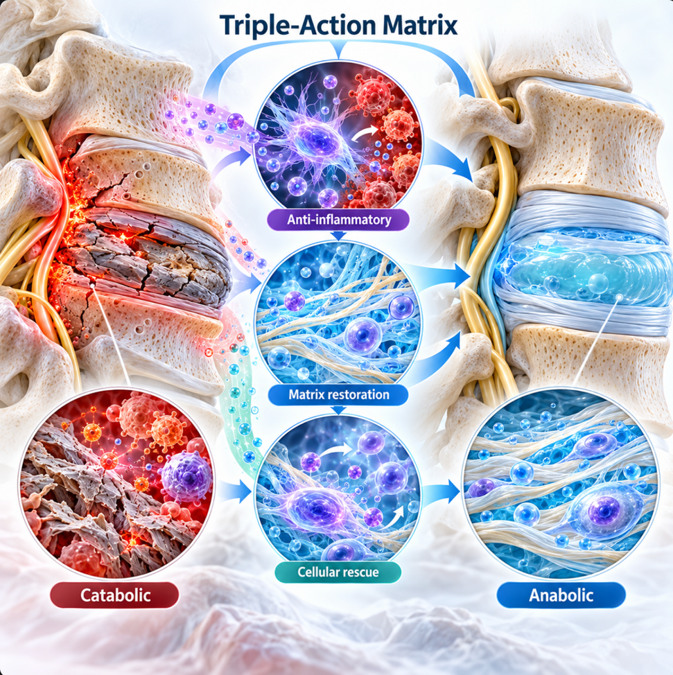
Figure 2: The Triple-Action Matrix transitions the disc from a catabolic (destructive) to an anabolic (rebuilding) state.
The UC-MSCs secrete heavy volumes of Transforming Growth Factor-beta (TGF-β) and actively upregulate the SOX9 transcription factor. This commands these native disc regeneration stem cells to start working overtime. We see a massive upregulation in the genetic expression and synthesis of collagen type II (the strong structural protein forming the inner disc architecture) and aggrecan (the core water-binding molecule of the disc).
As aggrecan density slowly increases within the nucleus pulposus, the crucial fixed charge density is restored. Glycosaminoglycan side chains act like tiny chemical sponges, allowing the disc to slowly pull water back into the core via osmosis. This restores a degree of hydrostatic pressure and shock-absorbing capacity. It is a slow, methodical rebuilding of the disc’s natural hydraulics.
But let’s ground this firmly in clinical reality. This phase takes many months to execute. You are not going to wake up three weeks after the injection with the pristine, plump spinal discs of an 18-year-old athlete. The biological goal of Phase 3 is restoring functional structural integrity and creating a pain-free, highly stable spinal segment that can bear load without collapsing.
Matrix turnover rates are inherently slow. On a follow-up MRI at 12 months, this increased hydration may reflect as a subtle but biologically significant increase in T2 signal intensity, or occasionally, a millimeter or two of restored disc height. Even a tiny 1-2 millimeter increase in vertical height can drastically decompress the exiting nerve roots, resolving secondary radiculopathy.
Translating this biological matrix into real-world clinical results requires exacting procedural precision, perfect needle placement, and rigorous safety protocols.
The actual UC-MSC therapy procedure is a highly technical orchestration of live imaging and anatomical precision. It is performed in an outpatient sterile surgical suite or ambulatory surgery center (ASC) utilizing local anesthesia (like Lidocaine or Bupivacaine) and usually light IV conscious sedation (like Propofol and Midazolam) to keep the patient entirely comfortable, relaxed, but responsive to feedback.
Let me be unequivocally clear: this procedure mandates continuous fluoroscopic (C-arm X-ray) guidance. Blind injections, or those using only ultrasound, are grossly insufficient and highly dangerous for safely accessing the deep, heavily protected intervertebral disc space.
| 📌 If you’re wondering how to make sure a clinic follows proper safety standards, we have an interesting article that discusses whether stem cell therapy is safe in Thailand, which you can read via the internal link. |
The physician utilizes a precise transforaminal or extrapedicular needle trajectory, closely following established standard spinal imaging protocols. They carefully guide a 22G or 25G spinal needle past the highly sensitive neural elements, utilizing real-time X-ray to confirm millimeter-perfect placement. Once the needle tip is perfectly centered inside the nucleus pulposus, a tiny amount of non-ionic radiopaque contrast dye (like Iohexol) is injected.
The physician carefully watches the dye spread on the live X-ray screen. This step is absolutely crucial it confirms that the contrast stays inside the disc (a contained tear) and doesn’t leak out into the epidural space. If the dye holds, the disc is functionally contained enough to successfully receive and pressurize the stem cells. If it leaks heavily, the stem cells will leak out too, rendering the treatment ineffective.
Only after confirming containment are the UC-MSCs slowly injected, typically in a small 1-2 cc volume to prevent pressure necrosis of the delicate cells. Patients frequently weigh the out-of-pocket UC-MSC intradiscal injection cost against the procedure’s complexity. Given the absolute necessity of a sterile lab environment, pristine cold-chain cell handling, prophylactic IV antibiotics (cefazolin), and fluoroscopic facility fees, the investment directly reflects the rigorous safety architecture required to prevent devastating complications like discitis (disc infection).
| 📌 If you’re interested in a detailed breakdown of treatment pricing, we have an interesting article that discusses stem cell therapy costs in Thailand for 2025, which you can read via the internal link. |
Following successful cellular delivery, the needle is removed, a small bandage is applied, and the profound biological healing process begins, operating on a timeline dictated entirely by human cellular physiology.
Setting highly realistic expectations is the single most important part of post-procedural clinical counseling. You simply cannot rush human biology. The recovery after stem cells for back pain directly mirrors the three distinct phases of the Triple-Action Matrix.
Days 1-7 (The Flare-Up): Patients almost universally experience a temporary increase in axial pain, pressure, and stiffness. We are injecting fluid volume into a highly confined, tight, inflamed space. This creates acute mechanical pressure. Furthermore, the Phase 1 immunomodulatory process actively engages the host’s immune system, causing a localized, beneficial inflammatory spike (PGE2 mediated). This soreness is completely normal, expected, and necessary for healing. Patients manage this strictly with Tylenol, localized heat, and prescribed lumbar bracing—never NSAIDs or ice, which blunt the crucial cellular signaling.
Weeks 4-12 (Cytokine Arrest): This is when the biological magic starts to translate into subjective clinical relief. The anti-inflammatory Phase 2 takes hold. The molecular scissors destroying the disc are deactivated. Patients usually report their first sustained reduction in severe morning stiffness and sitting pain. Highly controlled physical therapy protocols are typically initiated around week 6 to encourage proper biomechanical loading, focusing heavily on isometric core stabilization while strictly avoiding deep spinal flexion.
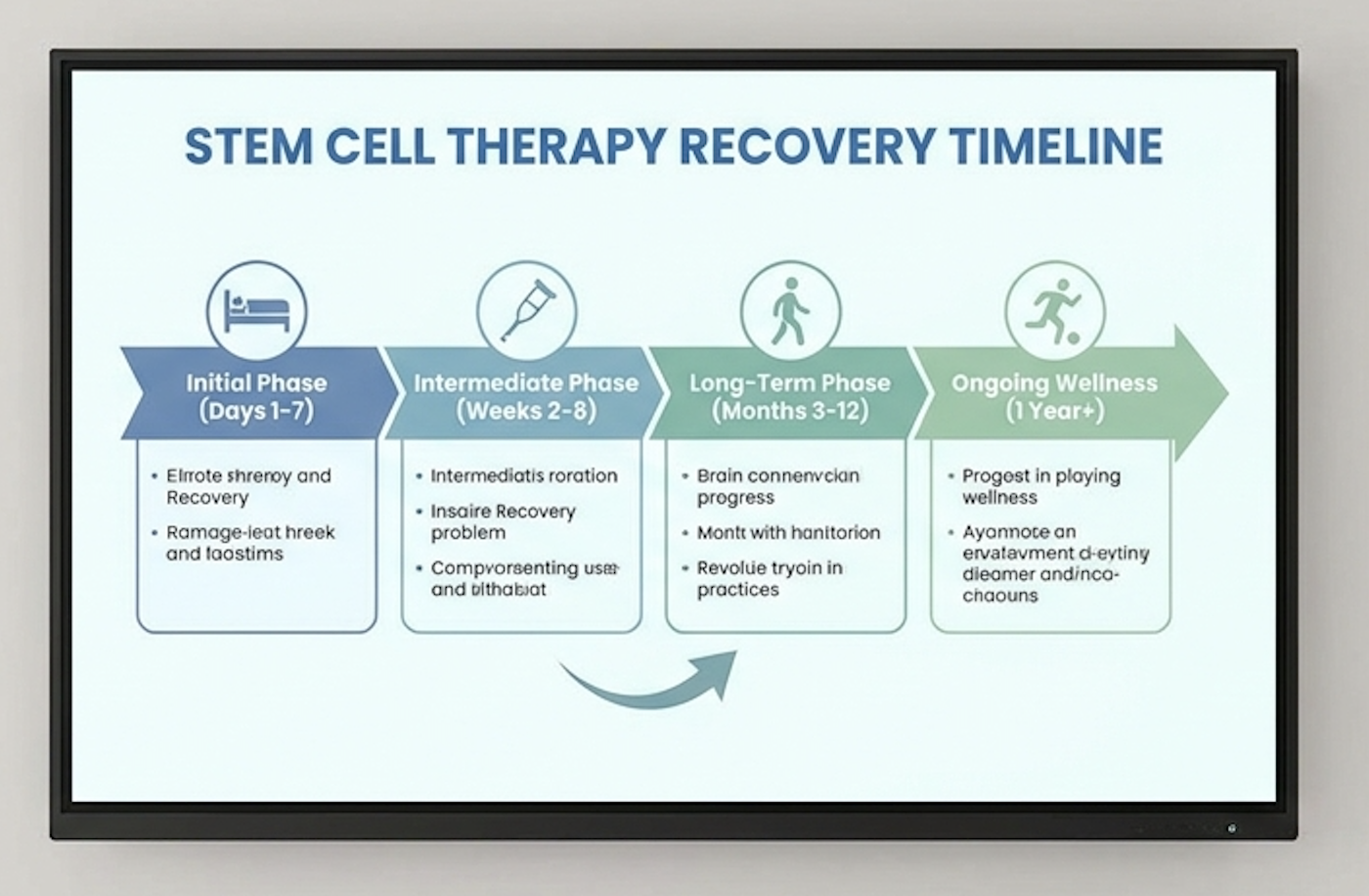
Figure 3: The biological timeline requires 6-12 months for maximum structural remodeling and pain reduction.
Months 3-6+ (Structural Remodeling): Patients eager to see their stem cell therapy for disc disease before and after results must wait patiently for Phase 3 to execute. Extracellular matrix synthesis is incredibly slow. The rebuilding of collagen fibers and proteoglycans takes many months. How long does intradiscal stem cell injection take to work fully? Maximum medical improvement (MMI) and peak structural stabilization may not be fully realized until 9 to 12 months post-procedure. The cells are working hard long after the initial injection day.
While this timeline represents a successful trajectory, objective medical transparency requires analyzing scenarios where this intervention is not the optimal choice.
Even the most highly potent cellular biologics cannot overcome severe mechanical failure or structural collapse. Treating stem cells as a universal panacea is simply poor medicine. Identifying exactly when to walk away from non-surgical options for chronic back pain is just as critical as knowing when to deploy them.
The absolute most common reason regenerative medicine fails is awful patient selection. Stem cells cannot fix gross biomechanical instability or sheer structural failure.
If a patient has Grade III or Grade IV spondylolisthesis (where the pars interarticularis has fractured and one vertebra has severely slipped completely over the adjacent one), injecting stem cells into the sheared disc will accomplish absolutely nothing. The immense mechanical sheer forces will destroy the delicate new cells instantly. You cannot biological-glue a slipped spine back together.
Similarly, treating a “black disc” that is completely desiccated with 100% height loss is scientifically futile. UC-MSCs require a native biological scaffold (matrix) to attach to and begin their signaling cascade. Injecting cells into a completely collapsed, bone-on-bone joint space provides no matrix whatsoever. The cells have nowhere to live, cannot survive the friction, and will fail to initiate the Triple-Action Matrix.
There are distinct neurological emergencies and severe mechanical pathologies where mechanical surgery specifically spinal fusion, laminectomy, or artificial disc replacement is the absolute standard of care, and biologics should not even be considered.
If a patient presents with an acute neurological deficit, such as a sudden foot drop (inability to lift the front part of the foot) or rapidly worsening severe leg weakness, immediate surgical decompression is mandatory. A massive, acute disc herniation severely compressing the exiting nerve roots requires mechanical removal (microdiscectomy), not a slow, months-long biological repair. Surgery is a rapid mechanical solution to an acute mechanical crisis.
Furthermore, patients with severe central canal stenosis requiring wide decompression may not tolerate intradiscal biologics, as adding fluid volume to the disc can transiently worsen the stenosis and trigger severe neurogenic claudication.
Patients navigating chronic back pain must vigilantly monitor for red-flag neurological symptoms. If you experience rapidly progressing neurological deficits, sudden loss of bowel or bladder control, or severe saddle anesthesia (numbness in the groin and perineum region), you must bypass regenerative considerations entirely.
These are classic, terrifying signs of Cauda Equina Syndrome. Seek emergency neurosurgical or orthopedic consultation immediately. Permanent, irreversible nerve damage can occur within 24 to 48 hours. Cellular biologics take months to work; in these emergency scenarios, you do not have months.
Clinical success rates for stem cell disc therapy typically range between 65% and 80% depending heavily on exact patient selection and overall disease severity. The highest efficacy is consistently observed in patients with mild-to-moderate disc degeneration who still retain some vital structural disc height. In these ideal candidates, targeted biological regeneration significantly outperforms standard conservative management. However, advanced bone-on-bone degeneration drastically lowers the probability of success, highlighting the need for careful MRI screening.
Intradiscal stem cell injections generally require three to six months to yield significant, lasting clinical improvement. While the initial inflammatory cascade is suppressed within the first four weeks, structural extracellular matrix synthesis is a naturally slow biological process. Patients frequently experience minor post-procedural soreness for the first week before noticing gradual, steady functional gains month over month. Maximum cellular regeneration and structural stabilization often peak between 6 and 12 months post-procedure. Results depend heavily on the patient’s underlying metabolic health, glycemic control, and strict adherence to post-treatment rehabilitation.
No, UC-MSCs are not currently FDA-approved as a standard treatment for discogenic back pain. They are strictly regulated by the FDA under 21 CFR Part 1271 as Human Cells, Tissues, and Cellular and Tissue-Based Products (HCT/Ps). Patients should always verify the regulatory status and cGMP laboratory compliance of any clinic offering these advanced regenerative therapies.
Stem cell therapy and spinal fusion surgery serve entirely different clinical objectives and are never universally interchangeable. Stem cell therapy aims to biologically repair disc tissue and preserve spinal mobility in patients with contained, predominantly chemical discogenic pain. Conversely, spinal fusion permanently immobilizes the vertebral segment to correct severe mechanical instability or advanced structural collapse. Fusion remains the absolute standard of care for severe trauma or progressive neurological deficits.
A stem cell injection into the spinal disc is typically associated with mild to moderate temporary discomfort. The procedure is performed using strong local anesthetics and often light intravenous sedation to maximize patient comfort and limit any sudden movement. Because the needle must penetrate the highly innervated, inflamed outer annulus of the disc, patients frequently feel a brief pressure or an exact reproduction of their typical back pain. Following the procedure, localized soreness at the injection site is perfectly common for 3 to 7 days as the acute biological healing response initiates.
For advanced patients suffering from chronic discogenic pain, stem cell therapy for degenerative disc disease offers a profound biological alternative to mechanical symptom management, demonstrating significant reductions in inflammatory markers and long-term pain scores (PubMed, 2023). The optimal approach relies heavily on exact patient selection, confirming that the pain originates from a biochemical failure of the disc rather than gross mechanical instability. The clinical data is clear: if the disc has enough remaining matrix to receive the cells, the biological cascade can fundamentally alter the disease trajectory.
This clinical efficacy is entirely dependent on the Triple-Action Regenerative Matrix. By deploying youthful, immune-privileged Wharton’s Jelly-derived UC-MSCs, physicians can sequentially modulate the hostile local immune response, arrest the cytokine cascade driving the pain, and stimulate the synthesis of new extracellular matrix. This framework shifts the clinical paradigm from temporary palliative care to active, targeted tissue regeneration, finally addressing the root cause of discogenic pain.
Patients exploring this intervention must undergo a rigorous, highly objective clinical evaluation. Schedule a consultation with a board-certified interventional orthopedic or regenerative medicine specialist to comprehensively review your recent MRI diagnostics. A professional imaging review will definitively determine if your spinal disc environment remains viable for cellular therapy or if surgical alternatives are medically required.