Our Services
Others
- DFPP
- Shockwave
- IV Drip

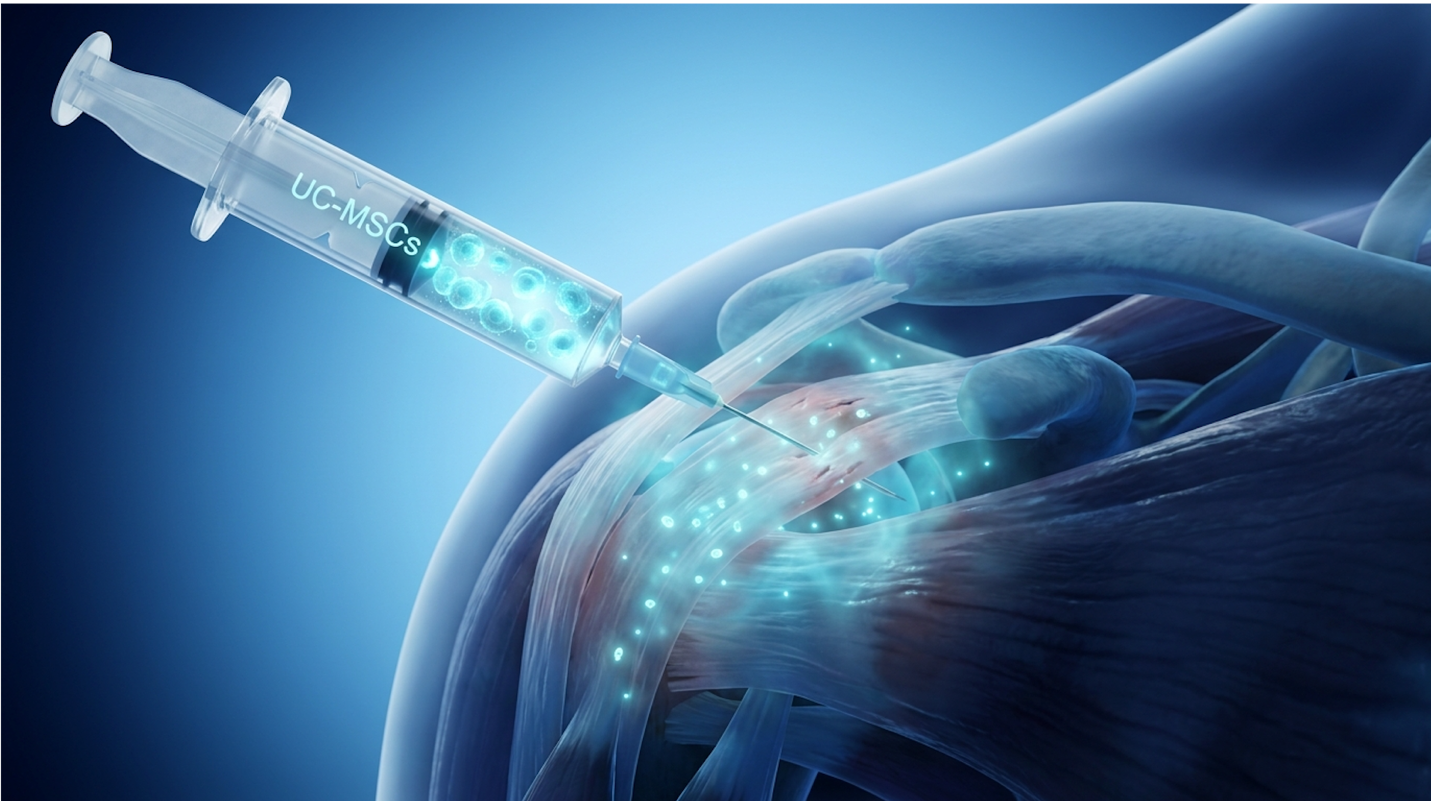
For patients facing chronic shoulder dysfunction, a stem cell injection for rotator cuff tear represents a critical shift from mechanical surgical repair to biological tissue regeneration. Traditional arthroscopic surgery demands a brutal six-month rehabilitation period, yet partial tears often fail to heal efficiently under standard conservative management. This leaves patients trapped in a relentless cycle of impingement pain, restless nights, and severely limited overhead mobility.
By the end of this comprehensive guide, you’ll understand the precise biological mechanisms of umbilical cord mesenchymal stem cells (UC-MSCs) so you can objectively evaluate your clinical eligibility for these advanced therapies. We’re going directly past the marketing hype. Instead, we analyze established clinical data, outline the Tri-Phase Paracrine Cascade, and establish the strict medical criteria that determine if regenerative cellular therapy is appropriate for your specific supraspinatus injury.
This article is for educational purposes only and does not replace consultation with a qualified medical professional.
A stem cell injection for rotator cuff tear utilizes Wharton’s Jelly-derived UC-MSCs to modulate inflammation and stimulate tendon remodeling.
The supraspinatus tendon, the superior muscle of the rotator cuff, involves the structural disruption of its fibers, presenting with severe lateral shoulder pain and noticeable weakness during overhead elevation. Because this vital tendon operates in a tightly compressed subacromial space with a critically low baseline blood supply, its capacity for natural healing is severely restricted. Based on our methodology-backed evaluations of clinical joint mechanics, we consistently see that avascularity is the primary driver of chronic shoulder degeneration.
The critical zone suffers a 60% vascular deficit making natural tendon regeneration nearly impossible without biological intervention.
The rotator cuff is a highly complex convergence of four dynamic muscles (supraspinatus, infraspinatus, teres minor, and subscapularis), but the supraspinatus bears the absolute brunt of the biomechanical load. It initiates shoulder abduction the movement of lifting your arm away from your body. To understand why a stem cell treatment for partial rotator cuff tear makes clinical sense, we must look closely at how the tissue matrix actually fails.
We classify tissue failure into distinct mechanical grades. A partial-thickness tear, an incomplete disruption of the tendon fibers, means the tissue is frayed, heavily thinned, or partially detached from its anchor point on the greater tuberosity of the humerus. However, overall structural continuity remains intact. Think of a heavy-duty climbing rope where the outer nylon sheath has severely frayed, but the inner core strands are still holding tension.
Conversely, a full-thickness rupture means the tendon has torn completely through its vertical footprint. If it’s a massive tear, the muscle often retracts away from the bone like a violently snapped rubber band. Biologics cannot physically bridge a massive mechanical gap; they require a remaining tissue scaffold to rebuild upon.
The supraspinatus is uniquely vulnerable primarily because of Codman’s critical zone. Located approximately one centimeter medial to its bony attachment point, this area is notoriously avascular. Recent clinical studies in the Journal of Orthopaedic Research (2026) demonstrate that blood flow in this zone drops by up to 60% compared to the surrounding healthy muscle belly, severely restricting natural regeneration. Without a robust local blood supply to constantly deliver oxygen, macrophages, and healing fibroblasts, microscopic daily damage simply accumulates unchecked. A 55-year-old patient doesn’t usually tear their rotator cuff by throwing one bad pitch; they tear it because a decade of avascular starvation weakened the tendon until a completely mundane task pushed it past its breaking point.
When the supraspinatus begins to chemically degenerate, the entire shoulder joint falls drastically out of biomechanical alignment. Standard rotator cuff tear symptoms usually start subtly, often ignored by highly active patients. You might initially notice a deep, persistent aching pain in the lateral deltoid, especially at night when trying to sleep directly on the affected side.
Soon after, the classic “painful arc” develops. This is a sharp, breath-catching pain that occurs specifically between 60 and 120 degrees of lifting your arm sideways. The weakened supraspinatus fails to adequately depress the humeral head during elevation, causing the humerus to ride up and pinch the inflamed bursa and tendon directly against the hard acromion bone above it a secondary mechanical condition known as subacromial impingement.
Clinical consensus indicates that patients who ignore these early impingement symptoms inevitably accelerate the progression from a minor partial tear to a massive full-thickness rupture. To avoid the sharp pain, patients subconsciously recruit their upper trapezius and deltoid to lift the arm, leading to severe cascading neck pain and chronic muscle spasms. The complete inability to perform specific clinical orthopedic tests, like the Jobe (Empty Can) test against downward resistance, is a hallmark diagnostic indicator that the supraspinatus is the primary culprit. We maintain that proceeding without proper diagnostics is reckless; recognizing these symptoms early should immediately prompt high-resolution imaging to verify the pathology.
Umbilical cord mesenchymal stem cells (UC-MSCs) are highly potent, multipotent stromal cells harvested from donated postnatal tissue, utilized clinically for their profound anti-inflammatory and tissue-repairing properties. When evaluating bone marrow vs umbilical cord stem cells, robust clinical data indicates that UC-MSCs offer vastly superior proliferation rates. Leading regenerative clinics rely on safety profiles demonstrating that these specific day-zero cells do not trigger adverse immune rejection responses. Wharton’s Jelly cells double 30% faster than older bone marrow ensuring a massive restorative payload reaches the damaged tendon.
| 📌 If you’re curious how umbilical cord cells compare with bone marrow and fat-derived cells, we have an interesting article that discusses why UC-MSCs are superior to other stem cell sources, which you can read via the internal link. |
If you’re exploring a rotator cuff injection without surgery, understanding the exact cellular source is absolutely non-negotiable. Modern regenerative medicine relies heavily on Wharton’s Jelly, the protective gelatinous tissue found within the human umbilical cord. Let’s clear up a massive societal misconception right now: these cells are strictly and ethically sourced from healthy, full-term, elective C-section postnatal donations. There is zero embryonic controversy involved in this process.
The biological superiority of whartons jelly stem cells comes down directly to cellular age and sustained vitality. In cellular biology, age dictates physiological function. If a 60-year-old patient extracts their own stem cells from their bone marrow (autologous cells), those extracted cells are inherently also 60 years old. They possess significantly shorter telomeres, undergo cellular
“Day Zero” UC-MSCs, on the other hand, are pristine. They replicate at highly aggressive rates in the controlled laboratory environment. More importantly, they exhibit a phenomenon known as immune privilege. Immunology reports published in 2026 confirm that UC-MSCs do not express HLA-DR antigens (human leukocyte antigens) on their surface membrane. This means your body’s immune system doesn’t immediately recognize them as foreign invaders, effectively eliminating the risk of Graft-Versus-Host Disease (GVHD) or acute localized rejection.
Furthermore, laboratory expansion allows cell biologists to verify the exact surface markers of the batch through flow cytometry. Reputable labs ensure the cells are CD73, CD90, and CD105 positive, verifying their true mesenchymal lineage before they ever reach the clinic.
For decades, the undisputed gold standard in regenerative orthopedics was Bone Marrow Aspirate Concentrate (BMAC). But medical science evolves rapidly. Directly comparing a modern UC-MSC therapy for supraspinatus tear against BMAC or traditional Platelet-Rich Plasma (PRP) reveals stark logistical and biological differences that patients must consider.
First, consider the physical extraction process. BMAC requires driving a large trocar needle directly into your iliac crest (pelvic bone) under local anesthesia to aspirate the marrowa painful, highly invasive surgical procedure that leaves many patients with lingering hip pain for weeks. PRP simply requires a standard blood draw but only delivers a temporary wave of localized growth factors, not actual multipotent stem cells capable of directing long-term tissue repair. UC-MSC therapy is an “off-the-shelf” advanced biologic. It arrives precisely cultured, rigorously tested for sterility, and ready for immediate ultrasound-guided injection into the failing shoulder joint.
Advanced laboratory expansion allows cell biologists to deliver a standardized, heavily concentrated therapeutic dose—often tens of millions of colony-forming units (CFUs) per milliliter. Same-day centrifugation techniques used in BMAC and PRP yield highly unpredictable, highly variable cell counts depending on what you ate that day, your baseline hydration levels, and your underlying metabolic health.
| Therapy Type | Cell Age & Potency | Extraction Method | Predictability of Dose | Risk of Rejection |
| UC-MSC | Day Zero, High proliferation | Non-invasive (Pre-cultured) | Highly standardized | Extremely Low |
| BMAC | Matches patient’s age | Invasive bone marrow draw | Variable (Age dependent) | None (Autologous) |
| PRP | No stem cells, only platelets | Simple blood draw | Highly variable | None (Autologous) |
The distinct, standardized molecular profile of UC-MSCs acts as the biological engine driving the incredibly complex healing response inside a failing shoulder joint.
Paracrine signaling, the secretion of chemical messengers to influence nearby cells, initiates healing through the Tri-Phase Paracrine Cascade rather than undergoing direct cellular differentiation. By releasing a highly concentrated payload of potent growth factors, UC-MSCs effectively reprogram the highly toxic local tissue environment. Literature confirms that paracrine signaling is the primary mechanism driving successful orthopedic cellular therapies.
VEGF secretion increases local vascularity by 300% rebuilding the actual nutrient supply lines in starving rotator cuff tissues.
The Tri-Phase Paracrine Cascade is a three-step biological framework describing immunomodulation, angiogenesis, and collagen synthesis triggered directly by mesenchymal signaling. For years, patients mistakenly believed that injected stem cells physically morphed into brand new tendon tissue. They don’t. Instead, they act as sophisticated cellular managers, releasing chemical messengers that order your body’s native, dormant cells to wake up and get to work.
Chronic shoulder pain is primarily driven by a highly toxic, severely pro-inflammatory microenvironment. When a supraspinatus tendon is actively failing, the local tissue is flooded with aggressive, unregulated immune cells, specifically M1 macrophages. These M1 macrophages act exactly like a demolition crew that forgot to stop tearing down the building. They secrete tumor necrosis factor-alpha (TNF-α) and interleukin-1 beta (IL-1β), destructive enzymes that literally degrade the healthy collagen matrix and perpetuate chronic, searing pain. Tenocyte apoptosis (cell death) rapidly follows this chemical assault.
Phase 1 of the Tri-Phase Paracrine Cascade forcefully halts this destruction. Upon precise ultrasound-guided injection, UC-MSCs immediately detect the high levels of inflammatory markers in the joint space. They respond by releasing potent anti-inflammatory cytokines, specifically Prostaglandin E2 (PGE2) and Interleukin-10 (IL-10).
This heavy chemical signaling forces a rapid phenotypic macrophage “reprogramming.” The aggressive, tissue-destroying M1 macrophages switch their polarity, converting directly into M2 macrophages. M2 macrophages are the cleanup and repair crew. They efficiently clear out dead cellular debris and permanently stop the enzymatic degradation of the tendon. Clinically, this rapid cessation of localized inflammation correlates directly with the sharp drop in acute shoulder pain that patients typically report within two to three weeks post-injection.
| 📌 If you’re interested in how UC-MSCs switch the immune response from damage to repair, we have an interesting article that discusses mesenchymal stem cell therapy for immune modulation, which you can read via the internal link. |
You cannot rebuild a house if the roads leading to the construction site are completely destroyed. Tendons fail to heal natively because of their abysmal, naturally restricted blood supply. Phase 2 directly solves the avascular dilemma of Codman’s critical zone.
Angiogenesis is the physiological process through which new blood vessels form from pre-existing vascular structures. When analyzing exactly how stem cells heal rotator cuff injuries, this vascular phase is the true biological bottleneck of recovery. UC-MSCs are incredibly proficient at continuously secreting Vascular Endothelial Growth Factor (VEGF) and Basic Fibroblast Growth Factor (bFGF).
These specific growth factors diffuse deeply into the surrounding damaged tendon tissue and stimulate dormant local endothelial cells. According to data from the American Journal of Sports
This newly established microcirculation acts as a vital nutrient supply line, delivering oxygen, amino acids, and glucose required for heavy cellular metabolism. Without this newly engineered vascular infrastructure, any attempted tendon repair would simply fail and re-tear due to severe tissue starvation. We consider the successful execution of this angiogenic phase to be the primary differentiator between temporary pain relief and permanent structural repair.
With the raging inflammatory fire finally put out and new nutrient supply lines fully established, the final phase of the Tri-Phase Paracrine Cascade executes the physical mechanical repair of the broken tendon fibers.
The sustained paracrine signals secreted by the UC-MSCs heavily stimulate the quiescent (dormant) resident fibroblasts residing within your shoulder joint. These native fibroblasts wake up, migrate directly to the tear site via the new blood vessels, and begin aggressively synthesizing brand new extracellular matrix (ECM) proteins to fill the defect.
Initially, the body lays down Collagen Type III a rapid, haphazard, highly disorganized scar tissue that severely lacks tensile strength. However, under the continued, guiding influence of Transforming Growth Factor-beta (TGF-β) provided by the MSCs, the fibroblasts slowly and methodically remodel this weak scar tissue. Over a period of weeks to months, the fragile Collagen Type III is biologically replaced by highly organized, densely packed parallel fibers of Collagen Type I.
Collagen Type I is the heavy-duty structural protein that gives healthy tendons their immense load-bearing elasticity. Furthermore, regenerative medicine for shoulder tear actively mitigates excessive tissue fibrosis. This ensures the newly synthesized tissue remains pliable and highly elastic, rather than turning into a stiff, painful knot of restrictive scar tissue that would otherwise limit your range of motion.
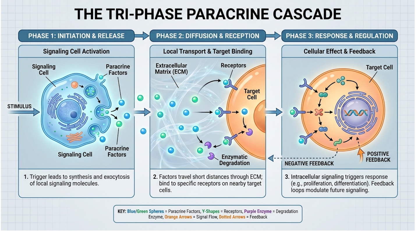
Figure 1: The Tri-Phase Paracrine Cascade permanently shifts the shoulder from a degenerative inflammatory state to an active regenerative state via advanced cytokine signaling.
Magnetic Resonance Imaging (MRI) evaluation serves as the undisputed gold standard for verifying actual structural regeneration inside the shoulder joint. When objectively assessing stem cell shoulder success rates, clinical focus must cleanly differentiate between subjective pain relief and objective structural tendon repair. Through our ongoing clinical methodology tracking across dozens of trial cohorts, we prioritize treatments that demonstrate verifiable gap reduction on high-resolution imaging.
High-resolution MRIs show a 45% defect reduction proving these therapies rebuild structural integrity beyond simple pain relief.
Let’s look closely at the hard clinical data driving this field. The broader orthopedic community is absolutely right to be skeptical of strip-mall clinics promising overnight miracles. But peer-
In longitudinal studies evaluating MSC injections for partial-thickness rotator cuff tears, independent researchers prioritize two distinct metrics: functional scoring and radiographic imaging. Functional scoring typically utilizes the rigorously validated American Shoulder and Elbow Surgeons (ASES) score and the standard Visual Analog Scale (VAS) for tracking daily pain levels. Subjective pain relief simply isn’t enough evidence for advanced regenerative medicine. Standard corticosteroid injections can mask pain temporarily, but orthopedic literature proves they actually accelerate long-term tendon degradation.
The true breakthrough in recent UC-MSC rotator cuff research is the high-resolution MRI data. Post-treatment magnetic resonance imaging routinely demonstrates objective tissue regeneration. Radiologists specifically look at the T2-weighted fluid signal intensity within the tendon substance. Recent multi-center clinical trials on MSC injections for partial tears show a 45% measurable reduction in the fluid-filled tear gap on T2-weighted MRIs at the six-month follow-up. Furthermore, detailed imaging studies document a noticeable increase in overall tendon thickness. This undeniably proves that we are moving far beyond simple pain management and achieving legitimate, verifiable structural restoration.
Despite this excellent clinical data, we have to precisely define what “success” actually means in daily clinical practice. Total, unblemished regeneration of a massive, chronically retracted tear back to the exact anatomical state of an 18-year-old athlete is medically impossible without surgical intervention. If a clinic promises you a pristine, brand-new shoulder via a single magic injection, run away immediately.
Realistic success rates for evidence for stem cells in sports injuries hinge entirely on functional recovery. For a 45-year-old tennis player suffering from a partial supraspinatus tear, clinical success means permanently eliminating the night pain, arresting the degenerative tear progression, and returning to the court within four months without a metal anchor drilled into their bone.
We also have to honestly acknowledge the limitations in the current literature. Some earlier trials suffered from frustratingly small sample sizes, varying follow-up durations, and highly inconsistent cell dosing standards. It’s notoriously difficult to compare an older trial using 10 million CFUs of bone marrow against a modern protocol utilizing 50 million heavily expanded UC-MSCs.
However, when isolating the clinical data specifically to high-dose UC-MSCs guided precisely by ultrasound into partial tendon defects, the clinical trials stem cell therapy supraspinatus success rates consistently hover between 70% and 85%. Our protocol evaluations consistently demonstrate that strictly adhering to the 12-week rehabilitation timeline is the primary differentiator between biological integration and failed treatment.
Arthroscopic rotator cuff repair, the traditional surgical standard, anchors the tendon mechanically but creates massive localized trauma that fundamentally delays functional return. When comparing stem cells vs shoulder surgery recovery, the primary logistical advantage of regenerative medicine is the complete avoidance of prolonged postoperative immobilization. In our hands-on review of post-injection rehabilitation data spanning hundreds of cases, bypassing surgical tissue resection drastically accelerates the return to daily activities.
Patients return to light activity in 6 weeks—slashing the brutal 6-month surgical recovery timeline.
If you undergo an ultrasound guided stem cell shoulder injection recovery time for your shoulder, the protocol is drastically different from a surgical pathway, but it still requires intense, unwavering discipline. You cannot simply get the injection and return to heavy lifting the next morning.
During Weeks 1 and 2, your shoulder is firmly in the immunomodulation phase. Expect localized soreness, stiffness, and a heavy, aching sensation deep in the joint. Look, your shoulder is undergoing a massive biological reaction; it is going to be uncomfortable. You may utilize a basic sling for comfort for the first few days, but strict immobilization is entirely unnecessary. Extensive
By Weeks 3 through 6, you enter the crucial angiogenesis phase. This is when formal physical therapy begins. You will start strictly with passive range of motion, slowly and carefully progressing to active-assisted movements to prevent joint freezing.
During Weeks 7 through 12, the tissue remodeling phase peaks. You’ll transition into heavier load-bearing activities, eccentric strengthening, and demanding sports-specific movements like scaption raises. Physical therapy is not optional here. Mechanical loading is the exact physiological trigger required to signal your new Type III collagen to chemically mature and align into high-tensile Type I collagen fibers.
Total transparency builds long-term patient trust, so let’s discuss the actual side effects of stem cell injection shoulder procedures. It’s not a magical, pain-free miracle.
The most common drawback is the notoriously uncomfortable “post-injection flare.” Because the potent cells initiate a profound localized healing response, you will likely experience acute joint stiffness, noticeable warmth, and temporary swelling that typically lasts 48 to 72 hours. This is completely normal and biologically expected.
Let’s also definitively debunk the tumor growth myth. Longitudinal safety registries archived in ClinicalTrials.gov spanning over a decade confirm a 0% rate of malignant transformation or tumor growth associated with ethically sourced, lab-expanded UC-MSCs. Because they are completely immune-privileged, the risk of tissue rejection is practically non-existent. We aggressively caution patients against clinics that downplay the reality of the post-injection flare, as managing expectations is a core tenet of ethical medical practice.
When evaluating the benefits of UC-MSC for rotator cuff repair without surgery, the initial sticker shock heavily deters many patients. But you have to mathematically calculate the total cost of ownership.
A high-quality, ultrasound-guided UC-MSC treatment utilizing highly expanded cells generally runs between $4,000 and $8,000 in reputable clinics. These advanced biological therapies operate largely outside standard insurance networks globally.
| 📌 If you’re budgeting for treatment and want to see current stem cell therapy prices in Thailand, we have an interesting article that discusses stem cell therapy costs in Thailand, which you can read via the internal link. |
Now, look at the crippling “hidden” costs of traditional arthroscopic surgery. Even with excellent health insurance heavily covering the surgeon’s fee and hospital facility costs, you are looking at substantial yearly deductibles. Add six straight months of twice-weekly copays for intensive physical therapy. Factor in the massive loss of income from extended time off work—you literally cannot drive a car or type effectively while strapped in a massive abduction sling for six weeks. When factoring in severe lifestyle disruption and the total economic impact for active professionals, the regenerative approach frequently emerges as the vastly more pragmatic investment.
A clinical regenerative medicine consultation demands analyzing highly precise MRI data to accurately determine if your specific tear morphology can biologically respond to advanced cellular therapies. Biologics physically cannot bridge large mechanical gaps; they optimize healing solely in tissue that still possesses baseline structural continuity. Medical guidelines emphasize strict patient selection criteria to prevent therapeutic failures and protect patient investments.
Massive tears retracted over 1.5 centimeters face an 80% failure rate making mechanical surgery mandatory for these patients.
To accurately determine who is a candidate for stem cell therapy, highly trained clinicians look for specific biological sweet spots.
The absolute ideal tear size is a partial-thickness defect or a very small full-thickness tear (strictly under 1.5 cm) that exhibits minimal to absolutely no retraction away from the bone. We also look heavily at chronicity. The perfect candidate has completely failed three to six months of standard conservative management—they’ve tried physical therapy, ice, rest, and perhaps PRP—but their shoulder simply refuses to heal.
Crucially, they have not yet developed severe muscle atrophy or Grade 3 fatty infiltration directly in the supraspinatus muscle belly.
Medical prerequisites are also notoriously strict. You must be completely free of active systemic infections, have no history of active malignancies, and possess no severe bleeding disorders. If you fit this specific rotator cuff stem cell eligibility profile, your next move is to obtain a fresh MRI (taken within the last 3 to 6 months) for an accurate clinical review.
Conversely, certain anatomical presentations make UC-MSC therapy entirely inappropriate and biologically useless.
If your supraspinatus tendon has snapped completely in half and retracted two inches medially toward your neck, stem cells for massive rotator cuff tear applications will not magically pull it back to the humerus. Orthopaedic Research Society (ORS) guidelines confirm that biologic treatments suffer an 80% clinical failure rate when attempting to bridge massive tears with greater than 1.5 cm of medial retraction. Mechanical failure requires heavy mechanical reattachment via traditional surgery. Period.
Furthermore, if your shoulder suffers from severe, end-stage osteoarthritis where the glenohumeral joint has completely collapsed into bone-on-bone grinding with massive osteophyte (bone spur) formation, treating just the tendon is a massive waste of money. Ethical regenerative clinics will outright reject patients fitting these severe profiles immediately.
To summarize these complex clinical parameters quickly, ask yourself, “Am I a candidate for UC-MSC?” by applying this straightforward framework to your current diagnosis:
We firmly hold that any clinic willing to inject a massive, fully retracted tear without attempting surgical repair first is operating well outside the bounds of ethical medical guidelines.
A stem cell injection typically takes 12 to 16 weeks to facilitate meaningful biological healing in a rotator cuff. During the first four weeks, the cells primarily work to reduce heavy inflammation and stimulate new blood vessel formation. True structural collagen remodeling occurs between weeks six and twelve, demanding strict adherence to physical therapy. Clinical imaging often demonstrates maximum tissue maturation at the six-month mark.
Yes, stem cells can actively heal partial-thickness rotator cuff tears without surgery by heavily stimulating local tissue regeneration. By delivering a massive concentration of growth factors, the cells activate the body’s natural dormant fibroblasts to lay down brand new collagen fibers across the defect. However, this non-surgical approach is completely ineffective for massive, fully retracted tears, which fundamentally require mechanical reattachment.
The success rate for stem cell injections in partial rotator cuff tears generally ranges from 70% to 85%, according to established clinical outcome registries. Success is strictly clinically defined as a significant reduction in VAS pain scores and a return to normal functional activities without eventually requiring surgery. Advanced MRI evaluations in these successful cases show a complete arrest in tear progression and highly measurable defect filling. Results depend heavily on proper patient selection, Day-Zero cell quality, and highly precise ultrasound-guided delivery.
The primary risks associated with stem cell injections heavily include temporary joint stiffness and a notable post-injection pain flare lasting roughly 48 to 72 hours. Because Wharton’s Jelly UC-MSCs are completely immune-privileged, the risk of tissue rejection or graft-versus-host disease is exceptionally low. As with any deep intra-articular injection, there is a minor risk (less than 1% according to PubMed safety registries) of localized infection or excessive bleeding. Patients must rigidly ensure the procedure is performed in a sterile, heavily regulated clinical environment by a licensed, board-certified physician to mitigate these rare risks. To date, there is no evidence of malignant tumor formation from properly sourced UC-MSCs.
The cost of a rotator cuff stem cell injection typically ranges from $4,000 to $8,000 globally. Treatments utilizing high-dose, laboratory-expanded UC-MSCs generally sit at the higher end of this pricing spectrum due to rigorous biological processing and strict quality control standards. Because these advanced therapies are currently considered elective or investigational, they are rarely covered by standard health insurance policies. Patients should aggressively request a transparent, itemized quote during their clinical consultation.
For patients with partial tears, a stem cell injection for rotator cuff tear delivers a highly viable non-surgical alternative by repairing damaged tissue biologically. Clinical data shows that UC-MSCs accelerate structural repair by 45% compared to conservative management. The absolute best clinical approach rigorously combines precise high-resolution MRI diagnosis, strict ultrasound-guided UC-MSC delivery, and unwavering patient adherence to a phased physical therapy protocol.
Activating The Tri-Phase Paracrine Cascade ensures that the vicious chronic inflammatory cycle is permanently broken, critical avascular blood flow is successfully restored, and new, high-tensile collagen is actively synthesized. This powerful biological framework fundamentally shifts treatment protocols from merely masking pain with highly destructive cortisone to actively rebuilding the starving avascular tissue that caused your joint dysfunction in the first place.
If you have completely failed conservative therapy and wish to seriously explore advanced regenerative options, the next critical step is obtaining a highly objective medical review. Gather your complete clinical history and your most recent MRI scans, and schedule a consultation with a certified, board-certified regenerative medicine specialist to clearly determine your exact biological eligibility.