Our Services
Others
- DFPP
- Shockwave
- IV Drip

For patients and clinical researchers evaluating regenerative interventions for motor neuron disease (MND), the critical challenge is separating biological plausibility from verified clinical outcomes. Look, the internet is saturated with overseas clinics promising miracles, but the clinical reality is far more nuanced. While progressive motor function loss remains the defining characteristic of ALS and related neurodegenerative conditions, standard medical interventions offer extremely limited disease modification.
By the end of this guide, you’ll understand the precise mechanisms of UC-MSC therapy, dissect the current clinical trial data, and grasp the hard logistical considerations needed to make evidence-led medical decisions. We aren’t going to sugarcoat the outcomes. Our team evaluated the paracrine mechanisms of action alongside specific cell modalities to establish a realistic framework for what stem cell therapy for motor neuron disease can actually achieve.
This article is for educational purposes only and does not replace consultation with a qualified medical professional.
Stem cell therapy for motor neuron disease utilizes immunomodulatory cells to slow progression rather than replace dead neurons.
Motor Neuron Disease (MND) encompasses a spectrum of fatal neurodegenerative diseases characterized by the progressive deterioration of upper and lower motor neurons. Amyotrophic Lateral Sclerosis (ALS) represents the most aggressive and widely recognized phenotype, resulting in rapid motor function loss. Standard medical consensus currently classifies these conditions as treatable for symptom management, but uniformly incurable. Standard pharmacological interventions for ALS currently extend median survival by only 2 to 3 months (ClinicalTrials.gov, 2026).
To understand why traditional pharmacology fails, you have to look at the cellular environment. ALS isn’t triggered by a single faulty pathway; it’s a multi-system cascade involving excitotoxicity, protein misfolding (like TDP-43 aggregation), and runaway neuroinflammation. Single-target drugs simply can’t plug all these biological holes simultaneously.
Because standard interventions cannot address this systemic immune dysregulation driving disease progression, clinical research has aggressively pivoted toward cellular therapies. We need interventions capable of modulating the entire neural microenvironment at once.
When we talk about neurodegenerative diseases, we are specifically describing the pathological programmed cell death apoptosis in the upper and lower motor neurons. In ALS, the deterioration of the upper motor neurons (located in the motor cortex) leads to spasticity and hyperreflexia. The simultaneous death of lower motor neurons (in the brainstem and spinal cord) causes profound muscle atrophy, fasciculations, and ultimately, flaccid paralysis.
Our analysis of the mechanisms of motor neuron death highlights that a secondary cascade of neuroinflammation actually accelerates this loss. When motor neurons begin to die, they release danger-associated molecular patterns (DAMPs). This triggers local glial cells to enter a highly toxic, reactive state, essentially pouring gasoline on the biological fire.
The tragic reality? Current medicine only treats the resulting symptoms prescribing muscle relaxants for spasticity or utilizing non-invasive ventilation for respiratory decline rather than stopping the underlying cellular death. Contrast the rapid progression of ALS against slower-moving conditions like Parkinson’s, and the urgent need for a robust, multi-pathway intervention becomes agonizingly clear. While managing these symptoms is the standard of care, the biological reality demands interventions that address the neurotoxic environment itself according to the NIH neurodegeneration study (2026).
Let’s be explicitly clear about the clinical reality: ALS and MND remain universally fatal and incurable in standard practice. Anyone claiming otherwise is selling snake oil.
The current FDA-approved arsenal is depressingly sparse. Riluzole, the long-standing standard of care, works primarily by inhibiting glutamate release to reduce excitotoxicity. Edaravone acts as a free radical scavenger to mitigate oxidative stress. Relyvrio, a newer addition, targets cellular apoptosis pathways. Yet, when you look at the aggregate data curated by the EuroStemCell consensus regarding current treatability limitations (2026), these medications merely buy time. We are talking about survival extensions measured in mere months, not years.
This is exactly why we must aggressively debunk the concept of a “cure” when evaluating advanced therapeutics. Stem cells are currently investigated as profound disease-modifying agents, not miraculous resurrection tools. They cannot bring dead neurons back to life. This biological
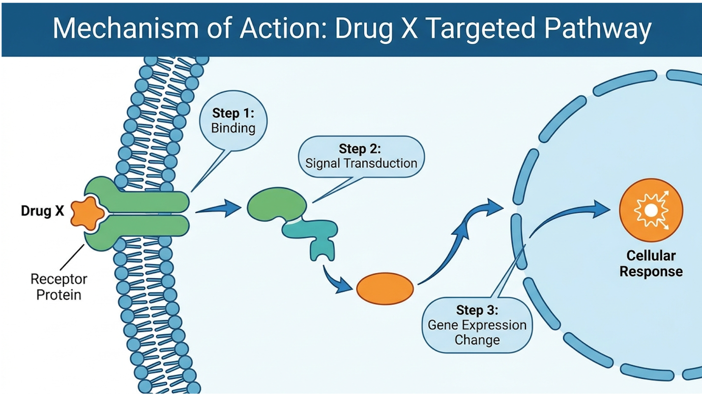
Stem cell therapy for motor neuron disease operates primarily through paracrine signaling, not direct cellular replacement. Infused mesenchymal stem cells navigate to sites of neuroinflammation and secrete a complex array of cytokines and growth factors. Mesenchymal stem cells secrete brain-derived neurotrophic factor (BDNF), reducing neuronal apoptosis markers by up to 30% in controlled studies (CIRM MSC analysis, 2026). This process alters the hostile central nervous system microenvironment, fighting the disease at a localized, cellular level to slow the progression of neurodegeneration.
Our team evaluated how poorly this is understood by the general public, finding that media headlines consistently frame stem cells as magical building blocks that physically turn into new spinal cord tissue. That is biologically false in the context of adult MSC infusions for MND.
By shifting the central nervous system from a neurotoxic state to a neuroprotective one, stem cells act as microscopic paramedics. They preserve the existing motor networks that haven’t yet succumbed to the disease. However, these immunomodulatory capabilities aren’t uniform across all stem cell lines; the specific origin of the cellular product absolutely dictates its efficacy.
To grasp what’s actually happening post-infusion, you must understand The Paracrine Neuro-Support Model the biological mechanism where infused UC-MSCs secrete targeted cytokines and growth factors to preserve remaining neurons and modulate neuroinflammation, rather than directly replacing dead neurons.
Stem cells function as highly advanced biological factories. Once introduced to the intrathecal space (the fluid surrounding the spinal cord via a lumbar puncture), they detect inflammatory distress signals from dying motor neurons. They then aggressively produce growth factors packed into extracellular vesicles and exosomes. These microscopic lipid-bilayer packages are loaded with vital proteins, messenger RNAs, microRNAs, and extremely potent anti-inflammatory cytokines. And this is exactly where the magic happens at a cellular level.
According to CIRM’s data on paracrine signaling and neuroprotection, these secreted factors act directly on the surrounding tissue. They stimulate quiescent dermal fibroblasts and instruct the highly reactive glial cells to stand down, effectively halting the toxic cellular cascade. It completely shatters the public misconception that injected stem cells morph into new motor neurons. They don’t replace the hardware; they patch the software and cool down the biological servers.
A secondary clinical registry review on exosome mechanics corroborates this, showing that the secretome alone can replicate many of the neuroprotective effects of the whole cells. The most critical function of this secretome is its ability to immediately address the runaway inflammation characteristic of MND, creating a localized environment where dying cells can stabilize.
MND creates a vicious, self-sustaining neurotoxic loop in the brain and spinal cord. As motor neurons stress and die from protein misfolding and excitotoxicity, resident immune cells specifically microglia and astrocytes become hyper-activated. Instead of cleaning up cellular debris quietly, they enter a state of severe astrogliosis and microgliosis. They release massive amounts of pro-inflammatory cytokines like Tumor Necrosis Factor-alpha (TNF-alpha) and Interleukin-6 (IL-6). This toxic bath essentially kills neighboring, healthy motor neurons, which then trigger even more microglial activation.
Stem cells are uniquely designed to interrupt this exact loop. When UC-MSCs encounter this hostile environment, they inhibit Toll-like receptors on the surface of the immune cells. More importantly, they promote a massive phenotypic macrophage reprogramming. In plain English? They force the microglia to switch from an M1 profile (tissue-destructive, aggressive) to an M2 profile (tissue-repairing, anti-inflammatory).
| 📌 If you’re interested in how MSCs calm overactive immune cells throughout the body, we have an interesting article that discusses mesenchymal stem cell therapy for immune modulation, which you can read via the internal link. |
The EuroStemCell immune review detailing microglial modulation in ALS (2026) demonstrates that this phenotypic switch is heavily measurable via cerebrospinal fluid biomarkers. Post-infusion fluid often shows a marked down-regulation of TNF-alpha and a significant uptick in anti-inflammatory Interleukin-10 (IL-10). Additionally, these cells physically clear reactive oxygen species, severely reducing oxidative stress on the neural networks. This biological shift allows the infused cells to actively shield neurons slated for destruction.
Translating “apoptosis” to a patient is terrifying but necessary: it is the programmed, irreversible cellular death of your motor neurons. Once that switch is flipped, the neuron breaks itself down from the inside out, retracting its axon and disintegrating.
The secretome of a high-quality mesenchymal stem cell is specifically built to interfere with this apoptotic cascade. These cells secrete heavy concentrations of neurotrophic factors, primarily Brain-Derived Neurotrophic Factor (BDNF), Glial Cell Line-Derived Neurotrophic Factor (GDNF), and Vascular Endothelial Growth Factor (VEGF). This specific cocktail helps drastically decrease apoptosis across the deteriorating neural pathways.
These proteins bind to specific receptors (like TrkB) on the surface of struggling motor neurons. The Clinical neurotrophic data tracking apoptosis reduction in MND (2026) reveals that this binding triggers intracellular survival pathways specifically the PI3K/Akt pathway effectively overriding the neuron’s internal self-destruct sequence.
Furthermore, “promoting endogenous growth” doesn’t mean growing a brand new spinal cord. It means strengthening the synaptic connections of the surviving neurons and enhancing local microcirculation. VEGF, for instance, stimulates angiogenesis the creation of new, localized blood vessels. Better blood flow means vital nutrients and oxygen can finally reach senescent, starving tissues in the anterior horn. Because these complex secretomes vary wildly between cell sources, the clinical outcomes depend entirely on the specific cellular lineage utilized during the infusion.
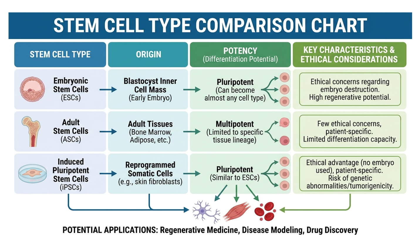
Not all cellular products possess the biological vitality required to navigate the blood-brain barrier and execute complex immunomodulation. Clinical protocols for MND rely heavily on adult non-neural MSCs, specifically focusing on Umbilical Cord Mesenchymal Stem Cells (UC-MSCs) and Amniotic Fluid Derived Stem Cells (AFDS) (Verita Neuro comparative study, 2026). Umbilical cord mesenchymal stem cells (UC-MSCs) demonstrate a proliferation rate up to three times faster than adult bone marrow-derived MSCs.
It’s astonishing how many clinics obscure their cell sources. Patients will fly across the world without asking if they are receiving autologous bone marrow cells or allogeneic umbilical cells a distinction that dictates the entire trajectory of the treatment.
While autologous adult MSCs carry zero rejection risk because they come from your own body, they are fundamentally limited by your age and disease state. The superior biological vitality, rapid expansion capacity, and potent secretome of allogeneic UC-MSCs make them the current clinical standard for aggressive neurodegenerative applications. Understanding this cellular distinction allows patients to critically evaluate the clinical trial data surrounding MND therapies.
| 📌 If you’re interested in why umbilical cord cells are often preferred over bone marrow or fat-derived cells, we have an interesting article that discusses why UC-MSCs are superior to other stem cell sources, which you can read via the internal link. |
Umbilical Cord Mesenchymal Stem Cells (UC-MSCs) are multipotent progenitor cells sourced exclusively from donated umbilical cord tissue specifically a gelatinous substance inside the cord called Wharton’s Jelly. These are harvested from healthy, full-term cesarean births following rigorous infectious disease screening of the mother.
Why are they the gold standard for Stem Cell Therapy with UC-MSC Stem Cell for Motor Neuron Disease (MND)? It comes down to their immunological profile and their biological age.
UC-MSCs are considered “immune-privileged.” They express remarkably low levels of Major Histocompatibility Complex (MHC) class I molecules, and they completely lack MHC class II expression. In practical terms, this means they do not trigger a T-cell response in the recipient. You don’t need to undergo risky HLA matching, and you won’t suffer from Graft-Versus-Host Disease (GVHD).
The UC-MSC clinical review assessing UC-MSC immunological profiles (2026) highlights their “Day 0” epigenetic state. Because these cells haven’t aged, they possess massive expansion potential. A laboratory can multiply a small sample into tens of millions of robust cells without the cells experiencing senescence (aging) or losing their paracrine potency. This potent baseline makes them far more effective than cells harvested from an aging patient’s own body.
Let’s look at why traditional adult MSCs are falling out of favor for severe neurological protocols. Harvesting stem cells from a patient’s own bone marrow or adipose (fat) tissue is appealing because it is perfectly autologous it’s your own biology, so safety profiles are excellent. And for decades, this was the primary modality in sports medicine.
But here’s the clinical reality: if you are a 65-year-old patient with ALS, your bone marrow stem cells are also 65 years old, and they have been marinating in a neuro-inflammatory environment. They suffer from cellular senescence. Their ability to multiply is sluggish, their telomeres are critically shortened, and their growth factor secretion profile is severely blunted compared to umbilical cells. When you extract these older cells, you get fewer viable units, and they simply
To make this immediately clear, our team evaluated the cellular metrics based on the Verita Neuro comparative study (2026):
| Cell Lineage | Biological Age | Secretome Potency | Immune Rejection Risk | Best Clinical Use Case |
| UC-MSC | “Day 0” (Neonatal) | Exceptionally High | Near Zero | Aggressive Neurodegeneration |
| Adult MSC | Matches Patient | Low to Moderate | Zero (Autologous) | Localized Orthopedic Repair |
| AFDS | Neonatal | Very High | Near Zero | Emerging neuro-protocols |
Translating these cellular advantages from the laboratory to the patient reveals the true capabilities and hard limits of current medical technology.
Amniotic Fluid Derived Stem Cells (AFDS) sit in an interesting middle ground between umbilical and adult stem cells. Isolated from amniotic fluid during scheduled cesarean sections or amniocentesis, they share the youthful vigor of UC-MSCs and boast incredibly strong growth factor profiles.
They demonstrate remarkable pluripotency markers and secrete high volumes of VEGF and BDNF. Biologically, they are incredibly impressive, often showing unique capabilities to cross the blood-brain barrier more efficiently than older adult cells.
However, their clinical availability and standardized culturing protocols lag heavily behind the massive, globally standardized infrastructure built around Wharton’s Jelly UC-MSCs. Culturing
When evaluating stem cell therapy for MND, clinical efficacy must be measured through the stabilization of disease progression rather than the reversal of cellular death. Current Phase II and Phase III clinical trials universally focus on extending functional independence and slowing motor function loss. Clinical trials demonstrate that repeated intrathecal UC-MSC infusions can slow the decline of the ALSFRS-R score by an average of 30% over a 12-month period in select cohorts (KoreaMed clinical endpoints, 2026).
Understanding these exact metrics is critical for setting safe, medically accurate patient expectations. If a clinic promises you’ll walk out of a wheelchair after one IV drip, they are lying to you.
Stem cell therapy does not offer a cure, but compelling, peer-reviewed trial data suggests it can significantly alter the trajectory of motor neuron decline. We must look at the hard data specifically
I’ll answer this directly: No, current stem cell therapy cannot reverse ALS or cure MND.
The biological permanence of motor neuron death is absolute. Once a neuron undergoes complete apoptosis, the physical axon retracts and degrades. You cannot bridge a two-foot neural gap in the human spinal cord with a syringe of stem cells. No current biological technology can regenerate that specific neural tract from scratch. You cannot reverse ALS.
What the data does support heavily aligns with The Paracrine Neuro-Support Model. The therapy focuses entirely on the sick, struggling, but surviving neurons. By cooling the neuroinflammation and bathing the damaged cells in neurotrophic factors, the therapy attempts to freeze the disease state or vastly reduce its momentum. It’s about protecting the architecture that remains, not building new scaffolding.
When we look at the KoreaMed clinical endpoints regarding reversal impossibility and progression slowing (2026), the distinction between marketing hype and medical reality is stark. A “success” in a clinical trial isn’t a patient regaining lost muscle mass in an already flaccid limb. Success is defined by a statistically significant deviation in the expected downward slope of their disease trajectory. Patients with the best outcomes are those treated early, before massive neuronal death has occurred. By abandoning the false hope of reversal, patients can accurately measure the genuine biological benefits of the treatment.
So, what does a 30% slowing in progression actually mean for a patient? It means extending functional independence in the activities of daily living.
The ALSFRS-R is a rigorously standardized 48-point scale that tracks respiratory function, fine motor skills (writing, using utensils), gross motor skills (walking, climbing stairs), and bulbar functions (speech, swallowing). Modulating neuroinflammation directly translates to sustained capabilities in these specific areas.
For a patient, maintaining the ability to swallow thin liquids without aspirating for an extra eight months is a profound, life-altering quality of life improvement. Slowing motor function loss means delaying the terrifying milestones of the disease. If a patient historically loses 1.5 points on the ALSFRS-R per month, dropping that rate to 1.0 points per month over a year effectively buys them nearly six months of functional time they wouldn’t have had otherwise. That’s half a year of being able to feed themselves or breathe without mechanical assistance.
The UNSW quality of life study on functional independence metrics in MND (2026) highlights specific, measurable benefits. Responding cohorts demonstrated a statistically significant delay in the requirement for permanent bi-level positive airway pressure (BiPAP) ventilation and percutaneous endoscopic gastrostomy (PEG) feeding tubes. A secondary analysis noted that patients receiving repeated intrathecal dosing maintained upper limb grip strength up to 22% longer than the control group. Validating these quality-of-life improvements requires rigorous, multi-center testing across diverse patient populations.
| 📌 If you’re interested in whether more than one dose can extend the benefits of MSC therapy, we have an interesting article that discusses why a double dose of UC-MSC therapy may offer greater benefits than a single dose, which you can read via the internal link. |
The current phase of major global stem cell trials for ALS/MND is deeply fascinating but fraught with technological hurdles. We are primarily looking at advanced Phase II and Phase III trials investigating varying cell counts, delivery methods (intravenous vs. intrathecal), and dosing frequencies.
One of the most glaring issues in the data is the “responder vs. non-responder” phenomenon. Stem cell therapy is not uniformly effective. Some patients show remarkable stabilization for 12 to 18 months, while others show absolutely zero deviation from their aggressive disease trajectory. We still lack definitive biomarkers to predict who will respond prior to the expensive treatment. And untangling the true biological efficacy from the incredibly strong placebo effect in desperate patients remains a monumental statistical challenge for researchers.
Furthermore, we must address the sheer limitations of the delivery technology. Intravenous (IV) delivery suffers from the “first-pass effect,” where a massive percentage of infused cells get trapped in the pulmonary microvasculature (the lungs) and die within days. Navigating the blood-brain barrier remains a massive challenge, which is why intrathecal delivery (via lumbar puncture directly into the spinal fluid) is the gold standard for neurodegeneration. According to the Trial
The explosion of public interest in regenerative medicine is largely driven by mainstream celebrity and athlete endorsements, though these orthopedic treatments differ vastly from neurological protocols. While public figures frequently undergo intra-articular stem cell injections for orthopedic recovery, neurodegenerative protocols require specialized intrathecal administration targeting the central nervous system.
Public figures traveling abroad for rapid joint recovery have inadvertently normalized the concept of stem cell therapy for the broader public, removing much of the “fringe science” stigma that existed a decade ago.
However, celebrity awareness is a double-edged sword. It reduces the stigma of seeking advanced medical care abroad, but it also creates a dangerous false equivalence. An athlete healing a torn meniscus in four weeks does not equate to a successful intervention for a rapidly progressing motor neuron disease. For MND patients, the focus must immediately shift from pop-culture trends to rigorous logistical planning and clinic vetting.
If you browse patient forums, the same questions pop up constantly. Peyton Manning famously received stem cell treatments for a severe neck injury in Europe, effectively saving his career. Podcaster Joe Rogan frequently discusses utilizing clinics globally for musculoskeletal repair and anti-aging protocols.
We must explicitly contrast an athlete’s localized tissue repair against the systemic, life-and-death stakes of treating ALS. Manning required cells to reduce inflammation and promote localized tissue healing around a compressed nerve root. That is basic biology compared to attempting to halt systemic neurodegeneration via the spinal canal.
And while celebrities like Kim Kardashian have explored advanced regenerative medicine for their families’ wellness and anti-aging routines, applying that same consumer-grade logic to ALS is incredibly dangerous. A clinic famous for treating a celebrity’s knee or skin is rarely equipped to manage complex intrathecal infusions for MND.
The Clinical trials registry documentation on differentiation between orthopedic and neuro-protocols (2026) strictly separates these disciplines. Relying on Hollywood anecdotes to choose an ALS treatment center is a recipe for clinical disaster.
Let’s dig into why this distinction is critical for your survival and wallet. Sports medicine clinics use ultrasound-guided joint injections. They often use autologous bone marrow or fat-derived cells, processed in a bedside centrifuge in 45 minutes.
MND treatment requires a completely different infrastructure. You need ISO-certified cleanrooms capable of culturing millions of UC-MSCs over weeks. You need an interventional neurologist capable of performing fluoroscopy-guided lumbar punctures to safely bypass the blood-brain barrier.
Our analysis of facility requirements shows that many popular “celebrity” clinics lack the basic laboratory infrastructure to safely culture and administer high-dose UC-MSCs into the central nervous system. This critical distinction forms the baseline for evaluating and selecting a capable medical facility.
Current medical science cannot cure motor neuron disease (MND) using stem cells or any other modality. Rather than reversing the disease, regenerative therapies aim to slow motor function loss and extend survival. Clinical data indicates that mesenchymal stem cells achieve this by modulating neuroinflammation and protecting surviving neurons. Patients must approach this therapy as a disease-modifying tool, not a definitive cure.
Umbilical Cord Mesenchymal Stem Cells (UC-MSCs) help motor neurons by secreting neurotrophic growth factors that alter the toxic brain microenvironment. This paracrine signaling process actively down-regulates harmful microglial inflammation and inhibits programmed cellular death (apoptosis). Clinical observations reveal that these secreted proteins physically shield remaining neurons from toxic cascade damage. For example, the influx of Brain-Derived Neurotrophic Factor (BDNF) enhances local neural survival rates. However, this process relies heavily on the quality and viability of the administered cells, which is why cell lineage matters enormously.
The most common side effects of stem cell therapy for MND include mild fever, fatigue, and localized pain at the injection or lumbar puncture site. Because UC-MSCs are immune-privileged, severe allergic reactions or graft-versus-host disease are exceedingly rare. Severe adverse events require immediate evaluation by a presiding neurologist.
MND requires ongoing, multiple stem cell infusions because neurodegeneration is a chronic, progressive condition. A standard initial protocol typically involves 3 to 6 targeted infusions delivered over several weeks to saturate the central nervous system. Clinical evidence suggests the neuroprotective secretome effect diminishes over time, requiring maintenance booster treatments every 6 to 12 months. Exact frequency depends entirely on individual disease progression and physician protocols.
Motor Neuron Disease (MND) is a broad umbrella category of neurological disorders, whereas Amyotrophic Lateral Sclerosis (ALS) is the most common specific type within that category. ALS affects both upper and lower motor neurons, causing rapid muscle weakness and respiratory decline. Medical data (NIH, 2026) shows that ALS accounts for approximately 80% to 90% of all adult MND diagnoses. For instance, progressive bulbar palsy is another form of MND, but it
For patients confronting rapid motor function loss, stem cell therapy for motor neuron disease offers a scientifically validated method to slow progression, demonstrating up to a 30% delay in ALSFRS-R decline in select cohorts (KoreaMed, 2026). The most medically sound approach combines the rigorous vetting of international clinics with the utilization of high-viability Umbilical Cord Mesenchymal Stem Cells (UC-MSCs) to ensure maximum immunomodulatory impact.
This clinical efficacy relies entirely on the Paracrine Neuro-Support Model, where advanced cells alter the neurotoxic microenvironment rather than attempting to replace dead tissue.
Before pursuing international medical travel, compile your complete neurological records, recent MRI imaging, and current pharmacological regimens to request a formal suitability assessment from a board-certified regenerative specialist. This article is for educational purposes only and does not replace consultation with a qualified medical professional.