Our Services
Others
- DFPP
- Shockwave
- IV Drip

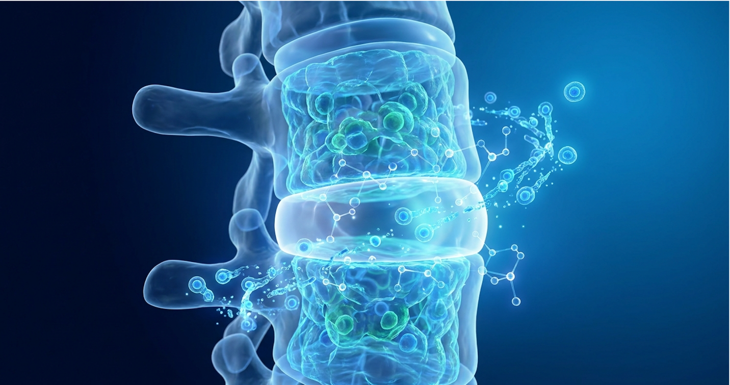
Look, for patients facing chronic lumbar or cervical pain, standard interventions often culminate in surgical fusions or a persistent dependence on analgesics. That grim reality prompts many to seek an advanced evaluation of regenerative orthopedics. But we need to separate the marketing hype from the biology. Stem cell therapy for herniated disc treatment isn’t magic. It is a highly specific, localized biological intervention.
While biological therapeutics offer promising mechanisms for tissue repair, the massive gap between commercial clinic marketing and peer-reviewed reality leaves desperate patients vulnerable to experimental procedures lacking true clinical validation. You don’t want a sales pitch; you want the hard data. By examining the latest institutional findings and biological pathways, this clinical analysis establishes precisely how cellular therapies interact with degenerated discs, enabling patients to make evidence-informed medical decisions. The following analysis dissects cellular mechanics, institutional efficacy data, exact candidacy requirements, and documented procedural risks.
Stem cell therapy for herniated disc treatment utilizes mesenchymal stromal cells to modulate localized inflammation and stimulate extracellular matrix protein synthesis.
Stem cell therapy for spine disorders requires a rigorous understanding of molecular biology, specifically regarding how localized cell therapies interact with avascular tissues. In cases of degenerative disc disease, cellular therapies aim to halt the catabolic cascade that aggressively degrades the nucleus pulposus. Rather than acting purely as structural building blocks, therapeutic cells secrete bioactive molecules that shift the disc microenvironment from pro-inflammatory to anti-inflammatory. Mesenchymal stem cells introduced into the intervertebral disc function primarily through paracrine signaling, altering the inflammatory microenvironment rather than purely replacing structural tissue (National Institutes of Health, 2024). This biological shift dictates clinical success.
Mesenchymal Stromal Cells (MSCs) are the engine of regenerative orthopedics. Unlike embryonic stem cells, adult MSCs are multipotent cells capable of profound immunomodulation without the risk of forming teratomas. Doctors primarily harvest them from two distinct autologous sources: bone marrow aspirate and adipose (fat) tissue, and the biological differences between the two are staggering.
Bone marrow aspiration typically pulls cells from the posterior iliac crest. Physicians use a Jamshidi needle to extract marrow, which is then centrifuged at high speeds in a closed system to isolate the buffy coat. Flow cytometry shows that this bone marrow aspirate concentrate (BMAC) is exceptionally rich in CD90 and CD105 markers, which are critical for osteogenesis (bone formation) and chondrogenesis (cartilage repair). Conversely, adipose-derived cells require a mini-liposuction procedure. Fat tissue contains a massive quantity of stem cells often yielding 500 times more MSCs per cubic centimeter than bone marrow but they are heavily skewed toward vascular repair rather than structural joint regeneration. When evaluating disc height restoration, placing vascular-heavy fat cells into an avascular environment often yields inferior extracellular matrix synthesis.
| 📌 If you’re curious about the surface markers labs use to confirm genuine mesenchymal stem cells, we have an interesting article that discusses the CD73 marker and how important it is for stem cell therapy, which you can read via the internal link. |
Clinical reviews of regenerative trials over the past decade demonstrate that the extraction source drastically alters the therapeutic payload. But does that make donor cells superior? Umbilical cord tissue (UC-MSCs) is heavily utilized internationally because these cells are immunologically privileged (meaning they lack HLA-DR markers) and don’t trigger a rejection response. Patients often pursue UC-MSC therapy for spine treatments abroad because international clinics have regulatory clearance to legally expand these young, highly active progenitor cells in a laboratory a process strictly limited in the US. However, whether you use autologous bone marrow or expanded UC-MSCs, success is governed by The Spinal Cell-Viability Matrix a 3-point framework (Mechanical Stability, Inflammatory Profile, Cellular Baseline) used to evaluate if a patient’s disc microenvironment can actually support a regenerative graft.
| 📌 If you’re weighing donor umbilical cord cells against bone marrow or fat-derived cells, we have an interesting article that discusses why umbilical cord-derived UC-MSC stem cells are superior to other stem cell sources, which you can read via the internal link. |
We need to clear up a massive misconception right now. Regenerating an intervertebral disc is biologically fundamentally different from repairing a severed spinal cord. They share a geographical location in the body, but that’s where the similarities end.
The intervertebral disc is the largest avascular organ in the human body. It has almost no direct blood supply, relying entirely on the adjacent vertebral endplates for nutrient diffusion. The disc’s nucleus pulposus relies heavily on a gel-like substance composed of water and proteoglycans, specifically aggrecan. When the disc degenerates, this aggrecan degrades rapidly, causing the disc
When a patient pursues stem cell therapy for herniated disc repair, the goal is rehydration and stabilization of this extracellular matrix. Stem cells injected into this harsh space don’t just act as passive filler. They actively seek out these degraded zones and work to downregulate the inflammatory cytokines that are dissolving the disc’s structural annulus. A herniated disc retreats or stabilizes primarily because macrophage reprogramming clears out necrotic tissue. It’s a localized cleanup operation. The structural repair of the disc relies heavily on the cells’ ability to survive that initial hypoxic shock and begin producing fresh structural proteins.
| 📌 If you’re interested in how stem cells calm the inflammation that damages spinal discs, we have an interesting article that discusses mesenchymal stem cell therapy for immune modulation, which you can read via the internal link. |
Conversely, spinal cord injuries involve catastrophic damage to the central nervous system’s neuronal pathways. Attempting to bridge a severed spinal cord requires complex neural regeneration and axonal remyelination. While phase 1 trials show promise for neuronal protection, expecting stem cells to rapidly “cure” paralysis remains scientifically premature. Spine Connection regenerative protocols explicitly differentiate between the biological realism of halting disc degeneration versus the highly experimental nature of central nervous system repair.
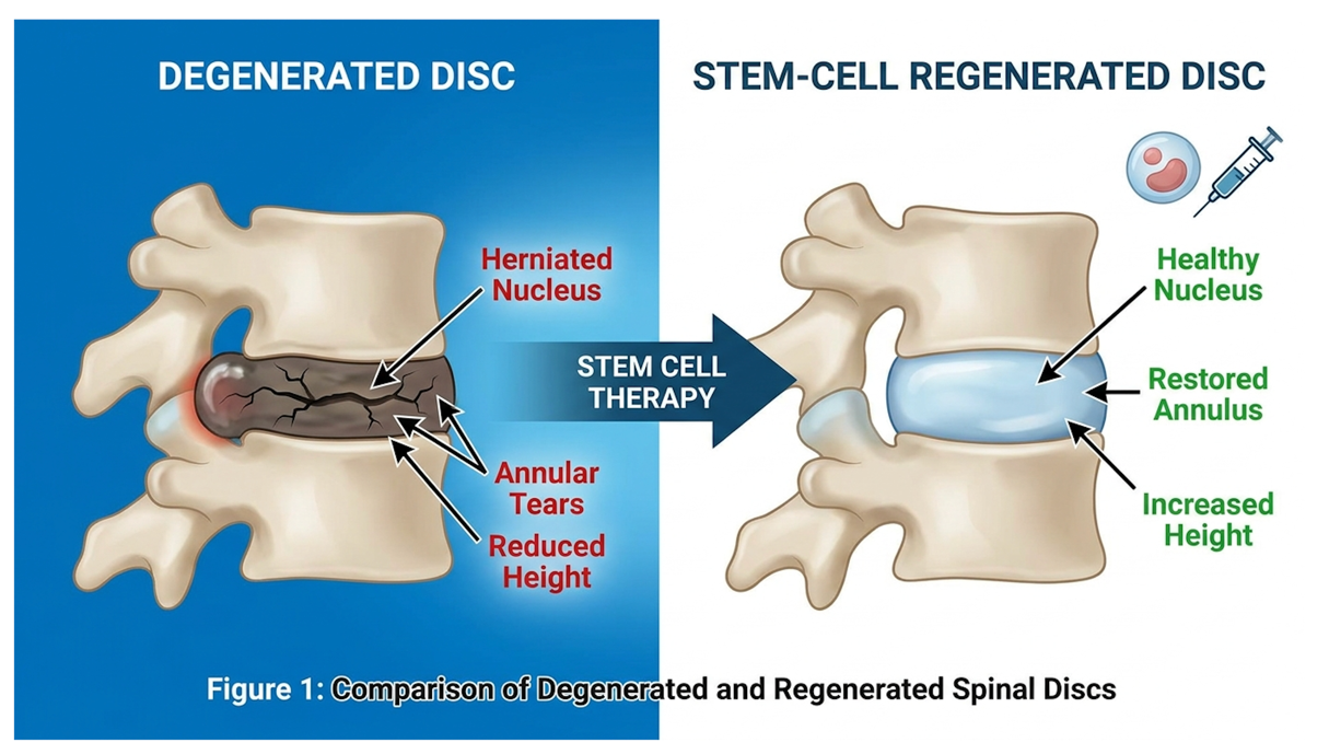
Figure 1: Notice how the stem cell-treated disc shows increased nuclear hydration and stabilized annular walls compared to the flattened, herniated baseline.
So, what are these cells actually doing once they’re sitting inside your spine? They aren’t just turning into new disc tissue. They operate as biological foremen through a process called the paracrine signaling effect.
When UC-MSCs or autologous bone marrow cells enter the hostile environment of a degenerated disc, they immediately begin secreting a potent cocktail of bioactive molecules. This “secretome” includes transforming growth factor-beta (TGF-beta), vascular endothelial growth factors (VEGF), and basic fibroblast growth factors (bFGF). These signaling molecules essentially wake up the patient’s native, quiescent dermal fibroblasts.
Once stimulated, the local cellular machinery shifts into overdrive. The fibroblasts begin synthesizing critical extracellular matrix proteins. We’re talking specifically about collagen type I and type II, fibronectin, and elastin. The ratio is vital: Collagen type II provides the tensile strength needed to resist the mechanical load of the human spine, while collagen type I forms the rigid outer boundaries. Without ample collagen type II, the annulus fibrosus remains brittle and prone to recurrent tearing. These proteins are the literal building blocks required to re-establish the structural integrity of the disc annulus, sealing micro-tears and restoring the disc’s ability to hold water.
Furthermore, this signaling cascade often triggers secondary benefits like adjacent bone regeneration and enhanced local blood microcirculation at the vertebral endplates. Open Access Government report data highlights how these cellular interventions can accelerate bone regeneration in surrounding spinal fractures. By fortifying the entire biomechanical unit and improving the endplates, the disc finally receives the nutrient diffusion it was starved of, halting the degenerative cascade at its source.
Mainstream acceptance of these therapies heavily relies on validation from tier-1 medical institutions. We aren’t just relying on private clinics publishing their own self-funded data anymore.
Consider the recent Mayo Clinic Phase 1 trial published in Nature Communications. Researchers initiated a rigorous safety profile analysis regarding intrathecal stem cell therapy for spinal cord injuries (the CELL-TOP trial). The primary objective here was not to guarantee a cure for paralysis. Phase 1 trials are strictly designed to establish safety and feasibility to ensure the injected cells don’t trigger severe adverse events, spinal fluid toxicity, or tumor growth.
The CELL-TOP trial meticulously monitored a specialized cohort of 10 patients over a multi-year period. They tracked cerebrospinal fluid (CSF) dynamics extensively. They needed absolute proof that the introduced cells did not trigger neurotoxicity, unwanted cellular proliferation, or runaway inflammatory responses. The data confirmed the exceptional safety profile of autologous bone marrow-derived cells when introduced into the central nervous system. Achieving these safety endpoints without severe adverse events is an absolute milestone for regenerative neurology. It paves the critical pathway for larger, more comprehensive Phase 2 and Phase 3 efficacy trials that will eventually test functional recovery endpoints.
And while Mayo focused specifically on cord injuries in that trial, the safety data directly bolsters the confidence of those seeking stem cell therapy for degenerative disc disease programs internationally, where the clinical applications are often years ahead of US commercial availability due to differing regulatory frameworks.
Here is one of the most confusing realities of regenerative medicine: your MRI might not look perfectly healed, even if you feel fantastic.
There is a documented disparity between imaging results and actual clinical outcomes. A patient might undergo treatment for a massive L5-S1 herniation. Twelve months later, they report a 75% reduction in pain. They are back to playing golf, sleeping through the night, and have completely discontinued opioid pain management. Yet, their follow-up MRI shows only a mild T2 signal increase meaning only slight rehydration of the disc and the bulge is only marginally smaller on the Pfirrmann grading scale.
Why does this happen? Because improvements in biomarkers specifically the downregulation of inflammatory cytokines like TNF-alpha, Interleukin-6, and interleukin-1 beta (IL-1β) do not
Ultimately, true functional benefit is the only metric that matters. If you can return to work and live without pain, the therapy worked, regardless of whether your spine looks like a 20-year-old’s on an imaging scan. This distinction between aesthetic MRI changes and meaningful functional recovery is exactly what patients need to grasp before investing heavily in these therapies.
Evaluating the stem cell therapy cost for disc regeneration requires a pragmatic assessment of out-of-pocket medical expenses and insurance exclusions. Because many cellular therapeutics exist outside of standard payer coverage, patients bear the financial responsibility entirely. Understanding exactly who is not a good candidate for stem cell therapy protects vulnerable patients from massive financial loss and ensures that advanced biological interventions are reserved only for those with the appropriate clinical baseline.
Not everyone gets better. In fact, if you inject stem cells into the wrong environment, you are quite literally throwing money into a physiological fire. This is why strict adherence to The Spinal Cell-Viability Matrix is non-negotiable for determining who should not pursue this therapy.
Let’s break down the three mandatory points of this framework:
Spine Team Texas clinical criteria outlines these exact parameters. A 45-year-old patient with an isolated, contained L4-L5 herniation is a prime candidate for stem cell therapy for a bulging disc. A 70-year-old with multi-level severe degenerative spondylolisthesis is not. Understanding where you fall on this matrix prevents predatory clinics from selling you false hope.
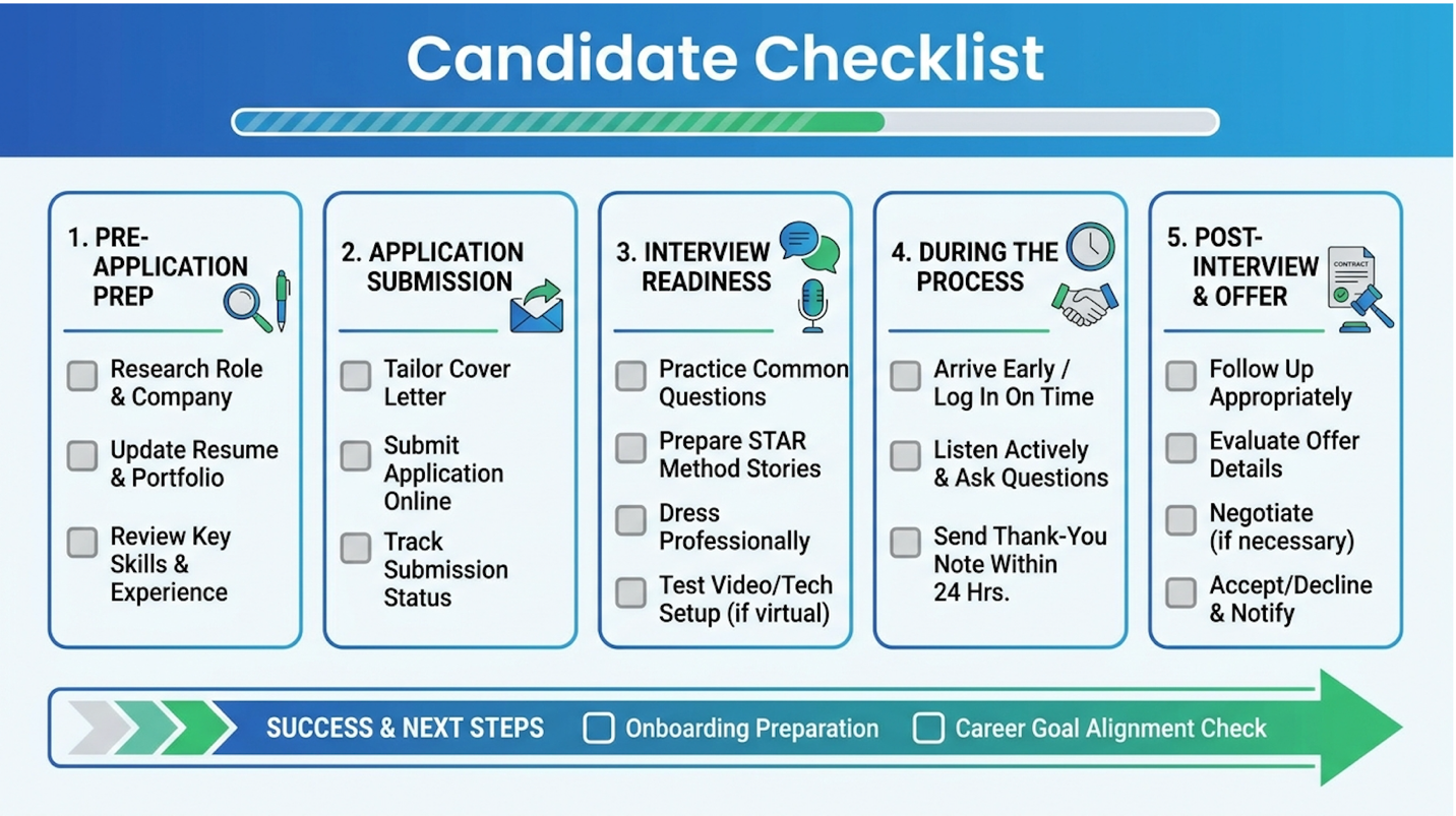
Figure 2: A contained herniation with preserved disc height offers the highest statistical probability of regenerative success.
Let’s talk about the money. Regenerative orthopedics is expensive, and you will almost certainly be paying cash.
The average cost of stem cell injections for intradiscal applications averages $5,000 to $15,000, largely because commercial insurers still classify these proven biological interventions as experimental (Atlanta Spine Clinic, 2024). Why the massive variance? It depends entirely on the cell source, the complexity of the image-guidance required, and the geographic location of the clinic.
| 📌 If you’re interested in how treatment pricing compares abroad, we have an interesting article that discusses stem cell therapy costs in Thailand for 2025, which you can read via the internal link. |
A simple bone marrow aspiration and injection into a single disc might sit around $6,000. If you are treating multiple levels, utilizing advanced fluoroscopy, and paying premium facility fees, that number skyrockets. You are paying for the physician’s expertise, the sterile lab processing, the live X-ray equipment, and the post-procedure follow-ups. While you can sometimes use HSA or FSA funds to cover the procedure, standard commercial health plans will outright deny the authorization.
But why won’t Medicare or Blue Cross cover this if the clinical trials show a 62.8% success rate? Because insurance conglomerates still classify intradiscal orthobiologics as “investigational” under current CPT billing codes. Until standard surgical fusions become cost-prohibitive for the insurers, they have little incentive to cover advanced cellular alternatives.
So, what does the actual day of a non-surgical treatment for a herniated disc look like? It’s an outpatient procedure, but it is highly meticulous.
First, the patient typically receives conscious twilight sedation. The physician extracts the cells using a Jamshidi needle inserted into the posterior iliac crest (your hip bone). The marrow is then spun in a high-speed centrifuge—often using advanced closed systems like Arthrex—to isolate the concentrated buffy coat, which is the cellular goldmine.
Next comes the critical part: the injection. A doctor cannot simply guess where your disc is. They must use live fluoroscopy (real-time X-ray) with contrast dye (like Omnipaque) to map the exact anatomy and guide a specialized needle precisely into the damaged nucleus pulposus or the epidural space. This ensures the cells are deposited directly into the site of the tear.
For the first 3 to 7 days post-procedure, you will likely experience a severe pain flare-up. This isn’t a complication; it’s the biological design. The injected cells trigger an acute inflammatory cascade
When evaluating any medical intervention, understanding the downside to stem cell therapy is paramount for patient safety. The persistent controversy in stem cell treatment often stems from predatory clinics making unsubstantiated medical claims entirely outside of regulatory oversight. However, when conducted within established clinical frameworks using properly sourced mesenchymal cells, the procedure presents a distinctly different, highly controlled safety profile that mitigates systemic risk while optimizing for localized tissue repair.
From a pure biological safety standpoint, autologous sourcing (using your own cells) is as safe as medicine gets. Because the cells are drawn from your own body, the risk of immune rejection, graft-versus-host disease, or the transmission of a communicable disease is virtually zero. Autologous bone marrow stem cell treatments maintain an excellent safety profile, with severe adverse events occurring in less than 1% of properly guided procedures (Mayo Clinic, 2023). The primary downside actually involves localized post-injection inflammation and the risk of financial loss if the therapy fails.
Allogeneic (donor) sourcing specifically UC-MSCs presents a different profile. Because these tissues are immune-privileged, they rarely trigger adverse immune reactions, making them incredibly safe for joint injections in theory. However, the safety of allogeneic cells depends entirely on the laboratory’s sterility and screening processes.
If a lab fails to properly screen the donor tissue for pathogens, the risk transfers to the patient. For instance, the FDA has historically cracked down on contaminated umbilical cord products from domestic suppliers that led to severe patient infections. This is why rigorous third-party lab testing and certification are absolute requirements before allowing anyone to inject donor cells into your spine.
The regulatory framework in the United States is fiercely complicated, heavily driven by 21 CFR 1271. The U.S. Food and Drug Administration (FDA) heavily monitors cell expansion and manipulation to protect consumers.
Under FDA Section 361, a clinic can legally harvest your bone marrow, spin it down, and inject it back into your spine on the same day. This is considered “minimal manipulation” and “homologous use.” But the moment a clinic attempts to take those cells, put them in an incubator, and grow millions of new cells (cell expansion), the FDA classifies that product as a biological drug under Section 351. Earning a Section 351 biologics license requires a multi-million-dollar, multi-year clinical trial.
This strict regulatory ceiling is exactly why patients seeking advanced stem cell therapy for sciatica from a herniated disc frequently engage in medical tourism. Jurisdictions like Thailand have entirely different national health frameworks that legally permit the clinical expansion of UC-MSCs in highly regulated, government-audited laboratories.
| 📌 If you’re considering traveling abroad for expanded UC-MSC treatment, we have an interesting article that discusses whether stem cell therapy is safe in Thailand, which you can read via the internal link. |
Despite the excellent safety profile of the cells themselves, the physical procedure carries universal physiological risks. Any time you push a needle into an intervertebral disc, you risk introducing surface bacteria into the sterile disc space. This causes discitis a severe, highly painful bacterial infection of the disc.
While exceedingly rare when strict sterile techniques, antibiotic prophylaxis, and fluoroscopic guidance are utilized, discitis remains the most significant procedural stem cell side effect. Patients must distinguish between a standard 3-day post-injection flare and the symptoms of infection, which typically present weeks later with severe localized spasms and fever.
Patients also frequently experience transient nerve irritation. If the injected fluid volume pressurizes a disc that is already bulging against a nerve root, sciatic pain can temporarily spike before subsiding. And let’s kill the tumor myth right now. Teratomas (tumors) are associated with unprogrammed embryonic stem cells. Adult mesenchymal stem cells do not form teratomas. Understanding this biological distinction is critical for your own peace of mind.
The mainstream awareness of regenerative orthopedics has been heavily driven by media coverage of elite athletes, most notably discussions surrounding Peyton Manning’s stem cell treatments for severe neck and spine injuries. Furthermore, widespread advocacy on platforms featuring Joe Rogan stem cell therapy discussions has accelerated public interest. However, patients must separate acute athletic recovery from the complex tissue remodeling required to address chronic degenerative spine disorders.
Athletes have been utilizing advanced regenerative medicine long before it hit the mainstream suburban orthopedic clinic. Years ago, Peyton Manning famously traveled to Europe to access cell expansion therapies to address severe cervical spinal issues, effectively delaying or preventing traditional cervical fusion surgery. Why travel? Because those specific expanded-cell protocols were restricted under FDA guidelines in the United States.
But we have to look at the biological rationale here. Athletes are generally dealing with massive acute inflammation from catastrophic physical trauma. Professional athletes utilize stem cell therapies to accelerate acute inflammatory recovery by up to 50%, whereas chronic degenerative patients require several months for structural remodeling (Americord, 2023). Stem cells act as incredibly powerful anti-inflammatory agents in these acute scenarios, shutting down the chemical fire and allowing the athlete’s otherwise healthy, robust cellular baseline to heal the injury.
Contrast this with a 65-year-old patient who has suffered from chronic degenerative disc disease for twenty years. The athlete has a pristine cellular baseline; the chronic patient does not.
The cultural normalization of regenerative medicine is a double-edged sword. Pop-culture icons heavily advocate for these treatments. Even mentions of Kim Kardashian stem cells on social media drive millions of Google searches. But we have to look closer at what they are actually doing. Systemic IV infusions used for general anti-aging or cosmetic recovery differ entirely from targeted, fluoroscopically-guided intradiscal injections for severe orthopedics.
These anecdotal endorsements almost never discuss the critical medical nuances: candidacy requirements, contraindications, the Spinal Cell-Viability Matrix, or the genuine risk of biological failure. Biological plausibility and celebrity endorsement do not equate to guaranteed clinical benefit for your specific spine. A podcast host telling you that MSCs cured his shoulder does not mean those same MSCs will rebuild a completely collapsed L4-L5 disc.
Patients frequently fall into highly predictable traps when navigating the regenerative medicine space. The most catastrophic pitfall is attempting to treat a completely desiccated, “black” disc on an MRI. If there is no matrix left to rebuild, cellular therapy will fail entirely—meaning the patient just burned $10,000 on a biological impossibility.
Secondly, patients are routinely misled by “stem cell soup” clinics marketing unproven amniotic or umbilical fluid products domestically as “live stem cells.” In the United States, FDA regulations require these off-the-shelf allogeneic products to be terminally sterilized, meaning they contain dead tissue and growth factors, but absolutely zero live stem cells. Finally, ignoring severe mechanical instability is a critical error. If a patient has severe spondylolisthesis (where one vertebra slips entirely over another), stem cells cannot bolt the spine back together. Structural hardware is fundamentally required.
There are distinct clinical thresholds where regenerative therapy is not just ineffective, but medically irresponsible. Surgery becomes mandatory when a patient presents with severe spinal cord compression causing progressive neurological deficits. If a herniated disc compresses the cauda equina nerve bundle, a microdiscectomy is an absolute emergency requirement. You do not have three months to wait for biological remodeling; the nerve is actively dying.
Similarly, in cases of gross spinal instability caused by severe trauma or advanced degenerative scoliosis, standard surgical options like artificial disc replacement (ADR) or spinal fusion are the
If you are currently evaluating regenerative treatments but suddenly experience progressive leg weakness (foot drop), loss of reflex function, saddle anesthesia (numbness in the groin), or any loss of bowel/bladder control, you must immediately abandon the regenerative pathway and consult a board-certified neurosurgeon. These are classic red-flag symptoms of acute neurological compromise. Regenerative therapies are designed for chronic pain management and structural rebuilding, not acute nerve rescue in the face of massive mechanical compression.
Can you recover 100% from a herniated disc? Most patients can recover significant functional capacity following a herniated disc, though perfect anatomical restoration is incredibly rare. The intervertebral disc heals by forming dense scar tissue rather than regenerating pristine native annulus fibers. Many patients achieve complete pain resolution within six months using conservative or regenerative therapies. However, individual results vary heavily based on age, tear severity, and whether central spinal stenosis is present.
Can disc bulge L4 and L5 be cured permanently? An L4-L5 disc bulge can be permanently stabilized and rendered asymptomatic, though the disc may never appear anatomically perfect on follow-up imaging. Treatments focus heavily on reducing the mechanical pressure and the local inflammation causing the debilitating sciatic nerve pain. Regenerative injections aim to strengthen the surrounding disc matrix to prevent further herniation events. Patients must maintain core strength and proper spinal biomechanics indefinitely to prevent symptom recurrence.
What is the success rate of stem cell treatment for herniated discs? Clinical data indicates a 60% to 70% success rate for patients treating contained herniated discs with targeted stem cell therapy. Success is clinically defined as a significant, sustained reduction in pain and a return to
How long does stem cell therapy for the spine take to work? Stem cell therapy for spinal disorders typically takes three to six months to yield maximal clinical benefit. Unlike steroid injections that provide immediate chemical masking of pain, cellular therapy initiates a slow, deliberate biological remodeling process. Patients often experience a mild inflammatory flare-up in the first week as the injected cells wake up the dormant fibroblasts. Following this initial shock, gradual functional improvements unfold over 12 to 24 weeks. Ultimately, the exact timeline varies heavily based on the patient’s individual metabolic baseline and adherence to post-procedure physical therapy.
Is stem cell therapy for the spine painful? The procedure involves mild to moderate discomfort during the injection and a temporary increase in localized pain for 3 to 7 days post-procedure. Physicians utilize local anesthetics and fluoroscopic guidance to minimize procedural pain. The subsequent flare-up is a normal biological response as the introduced cells trigger an acute inflammatory healing cascade. Severe, intractable pain is uncommon and warrants immediate medical consultation.
For patients evaluating advanced orthopedics, stem cell therapy for herniated discs provides a targeted biological mechanism to reduce inflammation and synthesize essential disc matrix proteins. Clinical literature documents up to a 62.8% reduction in radicular pain for qualifying candidates (Americord, 2023) meaning properly selected patients frequently avoid spinal fusion entirely. The most effective approach combines precise medical evaluation with image-guided cellular delivery, strictly avoiding unverified commercial claims.
Ensuring clinical success relies entirely on The Spinal Cell-Viability Matrix. Without adequate mechanical stability and a conducive cellular baseline, injected cells simply cannot survive the harsh avascular environment of a damaged disc. Rigorous candidate screening separates true regenerative medicine from predatory, profit-driven marketing.
Patients actively considering cellular therapy must request a comprehensive medical records review, including recent MRI imaging, from a board-certified regenerative specialist. A formal suitability assessment is the mandatory first step to determine if biological intervention actually aligns with your specific functional recovery goals.