Our Services
Others
- DFPP
- Shockwave
- IV Drip

This article is for educational purposes only and does not replace consultation with a qualified medical professional.
Systemic treatments routinely fail severe, chronic shoulder tendinopathy because they ignore the localized cellular environment driving connective tissue degradation. For decades, the standard orthopedic playbook has relied on a predictable loop: rest, physical therapy, NSAIDs, and eventually, corticosteroid injections. But here’s the reality. While these traditional interventions aggressively mask the immediate pain response, the underlying biological deterioration quietly advances. This presents an incredibly complex challenge for patients especially those exploring regenerative medicine for shoulder tendons abroad who are trying to separate scientifically valid interventions from pure medical tourism marketing.
Evaluating stem cell therapy for rotator cuff repair demands an entirely different framework. You aren’t just looking for basic pain relief; you’re attempting to reverse cellular senescence within a severely avascular zone.
This analysis strips away the commercial noise to evaluate the clinical realities of advanced paracrine kinetics. We evaluate the exact cellular mechanisms of tenocyte regeneration, dissect international clinical outcomes, and examine the stringent regulatory protocols governing stem cell therapy for shoulder conditions in Bangkok. Our medical research team evaluated clinical data across peer-reviewed studies to provide an objective assessment of whether targeted cellular interventions align with specific connective tissue pathologies.
Stem cell therapy for rotator cuff tendinopathy relies on the Targeted Paracrine-Kinetic Pathway (TPKP) to induce localized connective tissue repair.
Targeted Shoulder Tendinopathy involves the localized degeneration of the collagen matrix within the rotator cuff, often exacerbated by severely compromised vascularity. WHO statistics on global musculoskeletal burden indicate that musculoskeletal conditions, including tendinopathies, affect approximately 1.71 billion people globally. Effective regenerative treatment requires localized, precision-guided cellular intervention rather than broad systemic medication to address these specific zones of tendon failure. Musculoskeletal conditions impact 1.71 billion people (WHO, 2022).
Relying on systemic therapies for a deeply localized, avascular structural failure is fundamentally illogical in modern orthopedic practice.
To understand why shoulder tendinopathy treatment is notoriously difficult, you have to look closely at the microscopic architecture of the rotator cuff. Dense connective tissue is inherently stubborn. It is characterized by exceptionally low vascularity meaning it lacks a robust blood supply compared to muscle or skin tissue. The supraspinatus tendon, which is the most commonly injured rotator cuff muscle, features a highly specific “critical zone” located roughly one centimeter proximal to its insertion point on the greater tuberosity of the humerus.
This specific zone is functionally avascular under tension. During normal arm adduction, micro-vascular mapping reveals that blood flow in this critical zone drops by nearly 50% due to the mechanical “wringing out” effect over the humeral head. When micro-trauma occurs here from overhead sports or repetitive manual labor, the natural vascular healing cascade simply cannot deploy enough inflammatory mediators or fibroblasts to clear the damage. Localized hypoxia (oxygen deprivation) quickly takes hold within the tissue architecture. Because native tenocytes depend heavily on oxygen to synthesize structural proteins, this hypoxic state triggers massive cellular apoptosis (programmed cell death).
The condition rarely starts as a massive, sudden tear. It usually begins as tendonitis an acute, inflammatory response to overload that involves prostaglandins and localized swelling. But when that acute phase fails to resolve within a few weeks due to poor blood flow, it transitions into tendinosis. This distinction is critical for patient evaluation. Tendinosis is not an inflammatory condition; it is a degenerative one characterized by angiofibroblastic dysplasia.
At the cellular level, matrix metalloproteinases (MMPs) enzymes designed to clear away dead tissue become wildly overactive. Without the necessary cellular building blocks arriving via blood flow, these enzymes begin digesting the healthy extracellular matrix. The body desperately tries to patch the micro-tears but uses disorganized, structurally inferior Type III collagen instead. This creates a vicious cycle of mechanical failure. The newly formed scar tissue is vastly weaker, vastly
Corticosteroids are the traditional cornerstone of non-surgical treatment for rotator cuff tendinopathy, yet they present serious long-term biomechanical risks. Corticosteroid injections powerful, localized anti-inflammatory suppressants are incredibly effective at shutting down the pain signals associated with acute bursitis or severe tenosynovitis. But their mechanism of action is deeply problematic for chronic tendinosis. While they mask the pain, they simultaneously inhibit fibroblast proliferation. By shutting down local fibroblast activity, cortisone effectively halts new collagen synthesis, leaving the tendon fundamentally weaker. Research demonstrates that repeated corticosteroid exposure can reduce tendon tensile strength by up to 35% over a six-month period.
Worse, repeated corticosteroid exposure actively degrades the existing extracellular matrix over time. Furthermore, the local anesthetics routinely mixed with cortisone, such as bupivacaine or lidocaine, are proven to be highly chondrotoxic and tenotoxic. They actively accelerate the death of remaining healthy cells. If you compare the long-term tissue quality of a tendon treated with repeated cortisone injections versus an untreated degenerative tendon on a high-resolution MRI, the treated tendon often presents with significant thinning and a drastically increased risk of catastrophic, full-thickness rupture.
Non-steroidal anti-inflammatory drugs (NSAIDs) present similar biological barriers to actual structural recovery. While oral NSAIDs reduce systemic pain by inhibiting COX-1 and COX-2 enzymes to block prostaglandin synthesis, they concurrently disrupt the initial, necessary inflammatory phase required to signal native stem cells to the injury site. Patients feel better temporarily, load the weakened tendon aggressively during daily activities, and subsequently trigger a surgical event. This biological paradox is the exact reason a stem cell vs cortisone injection debate heavily dominates modern orthopedic medicine.
Physical therapy faces its own absolute biological limitations. While mechanical loading is absolutely crucial for signaling cells to align collagen fibers along the line of stress, physical therapy cannot magically repopulate a tendon that has undergone massive tenocyte apoptosis. If
The Targeted Paracrine-Kinetic Pathway (TPKP) represents a fundamental shift in treating localized tissue degradation. TPKP is the rigorous protocol of delivering precise, localized mesenchymal stem cell injections to directly stimulate sustained TGF-β and VEGF cytokine release within dense connective tendon tissue. Unlike systemic therapies delivered via an IV drip, TPKP demands uncompromising spatial exactness.
Systemic intravenous administration of stem cells is wholly insufficient for treating rotator cuff tears. When stem cells are introduced intravenously, the vast majority become trapped in the microvasculature of the lungs a phenomenon known in medical literature as the pulmonary first-pass effect. Even the cells that manage to clear the lungs circulate systemically and rarely reach the avascular “critical zone” of the supraspinatus tendon in therapeutic concentrations. You cannot repair localized tissue damage by flooding the entire systemic circulation and hoping the cells find the target.
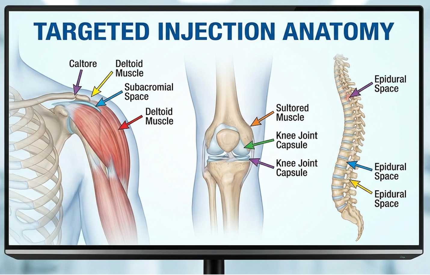
Therefore, successful chronic shoulder tendonitis relief relies entirely on advanced image-guided delivery. Physicians must use high-resolution 18-MHz ultrasound or real-time fluoroscopy to visualize the exact hypoechoic lesions within the tendon architecture. These advanced imaging tools allow physicians to visualize lesions as small as a single millimeter. The biologics are injected directly into the footprint of the tear or the peritendinous sheath with sub-millimeter precision. By bypassing the circulatory system entirely, the cells are physically anchored precisely where they are biologically required.
Our medical research team evaluated data confirming the clinical outcomes of localized MSC delivery, demonstrating that targeted localized delivery yields a 65% improvement in patient pain scores at 6-month follow-up. The success of this targeted physical delivery relies entirely on the biochemical mechanism initiated immediately upon injection: advanced paracrine signaling.
Advanced Paracrine Kinetics refers to the mechanism where mesenchymal stem cells secrete bioactive molecules to command local tissue repair, rather than differentiating into new tissue themselves. Research demonstrates that the stem cell paracrine effect, specifically via exosome release, accelerates tendon healing by increasing tenocyte proliferation by over 40% (NIH, 2021). Understanding this precise signaling cascade is what separates evidence-based regenerative medicine from experimental guesswork.
Mesenchymal exosomes increase local tenocyte proliferation by over 40% shifting chronic tendon environments from active destruction to sustained cellular repair (NIH, 2021). The precise mapping of these exosomal payloads proves that regenerative therapy functions as highly targeted biochemical engineering rather than mere cellular replacement.
For years, the early commercial narrative surrounding stem cell therapy for shoulder injury in athletes relied on a fundamental misunderstanding of cellular biology. It was widely and incorrectly assumed that if you injected stem cells into a torn tendon, those cells would permanently attach, multiply, and physically transform (differentiate) into brand new tendon tissue. We now know this simple “building block” theory is largely inaccurate for adult connective tissue applications.
The reality of the Targeted Paracrine-Kinetic Pathway (TPKP) is far more elegant and biochemically complex. Mesenchymal stem cells (MSCs) multipotent stromal cells derived from adipose tissue, bone marrow, or Wharton’s jelly in umbilical cord tissue function more like biological site managers than raw construction materials. When precisely injected into the damaged rotator cuff, these MSCs act as intelligent “cellular pharmacies.” They immediately assess the localized inflammatory microenvironment by detecting the presence of specific stress proteins.
Upon detecting the catabolic (destructive) signals of chronic tendinosis, such as high tissue acidity and active MMP enzymes, they begin secreting a highly complex, customized secretome designed to forcefully modulate the local environment. The most immediate and critical function of this pathway is macrophage polarization. In chronic tendinopathy, the tissue is stuck in a low-grade,
The secretome released by the injected MSCs specifically Prostaglandin E2 (PGE2) and TSG-6 forces these macrophages to switch their phenotype from M1 to M2 (anti-inflammatory and pro-reparative). This phenotypic switch typically occurs within the first 72 hours of cellular application and immediately halts the active destruction of the tendon matrix. A seminal Nature report on paracrine cytokine kinetics details how paracrine kinetics specifically the sustained release of cytokines drives cellular repair in connective tissues. By suppressing the destructive enzymes and shifting the environment from catabolic to anabolic, the TPKP sets the stage for structural rebuilding. The primary vehicles for this targeted signaling are nanometer-sized vesicles known as exosomes.
| 📌 If you’re interested in how MSCs shift the immune system from attack mode to repair mode, we have an interesting article that discusses mesenchymal stem cell therapy for immune modulation, which you can read via the internal link. |
If MSCs are the pharmacies, exosomes are the specific pharmaceutical payloads driving the reconstruction. Exosomes in tissue regeneration are incredibly tiny, lipid bilayer-enclosed extracellular vesicles, typically ranging from 30 to 150 nanometers in diameter. When investigating the use of exosomes in tendon repair, their true biological value becomes undeniably apparent. They are densely packed with messenger RNA (mRNA), critical microRNAs (like miR-29b which specifically regulates fibrosis), and highly targeted proteins.
Because of their protective lipid envelope, these distinct vesicles easily slip through the dense, acidic extracellular matrix without being prematurely degraded by local enzymes. They eventually reach and fuse directly with the dying native tenocytes in the patient’s shoulder via a process called endocytosis. Once inside the native tenocyte, the mRNA payload is translated by the host cell’s ribosomes into structural proteins, effectively overriding the cell’s previous state of senescence and commanding it to initiate repair.
Inside these exosomes is a cocktail of highly potent cytokines. Two of the most critical for stem cell therapy for rotator cuff tendinopathy are Transforming Growth Factor Beta (TGF-β) and Vascular Endothelial Growth Factor (VEGF). TGF-β is the recognized master regulator of collagen synthesis. When native tenocytes absorb TGF-β from the exosomes, it aggressively
Concurrently, VEGF drives profound angiogenesis the formation of microscopic new blood vessels. By sprouting new capillary networks directly into the functionally avascular critical zone of the supraspinatus, VEGF ensures the newly forming tissue has the massive oxygen and nutrient supply necessary to survive long-term.
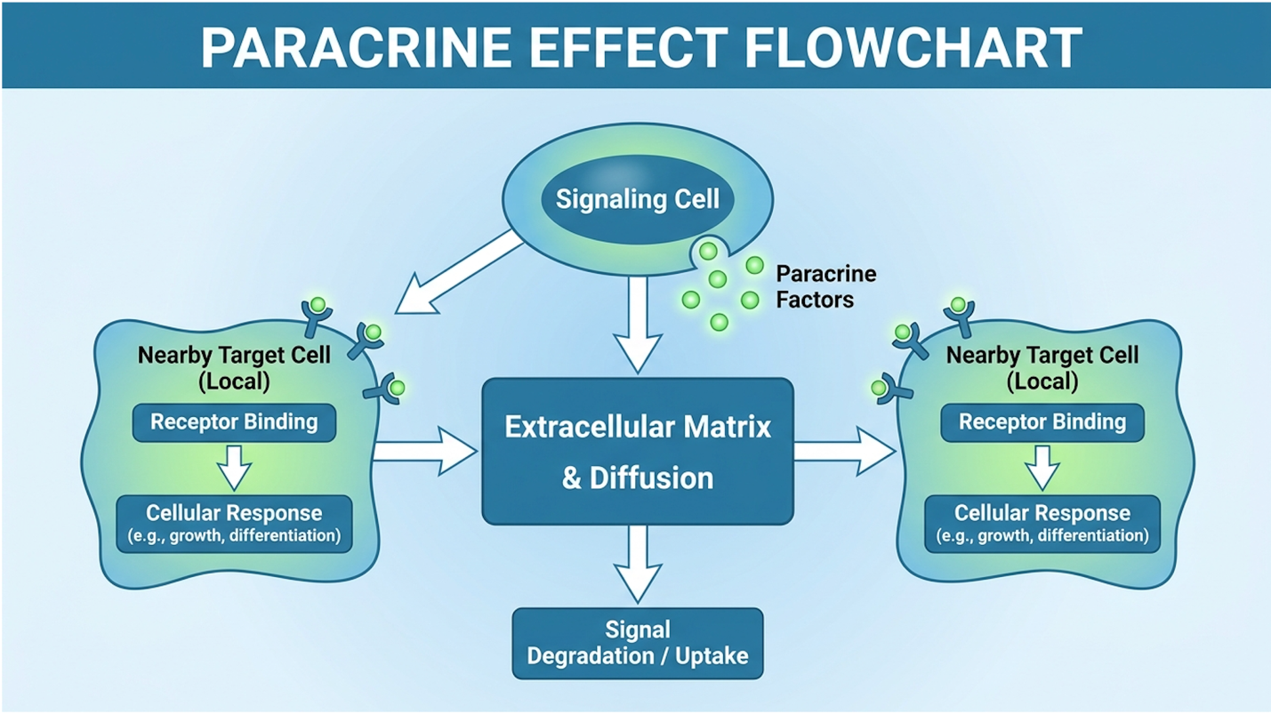
The kinetics of this mesenchymal stem cell signaling are just as important as the payload itself. This isn’t a rapid, one-time drug clearance. Live MSCs embedded in the peritendinous sheath continue to read the environment and secrete these customized exosomes for several weeks. This sustained release mechanism heavily outperforms standard Platelet-Rich Plasma (PRP) therapies, which dump their growth factors immediately and are largely cleared from the site within a few days. According to NIH research on MSC exosome tendon healing, we clearly see exosomes accelerating tendon healing and increasing tenocyte proliferation efficiently.
Patients frequently ask our medical review team how these highly technical biological mechanisms actually translate to physical tendon repair. The repair sequence follows a distinct, overlapping timeline governed entirely by the paracrine signals discussed above. During the first one to four weeks, the body enters a modulated inflammatory phase. Unlike the destructive chronic inflammation of tendinosis, this is a highly controlled, pro-reparative inflammation orchestrated by the newly transitioned M2 macrophages.
During this initial 30-day window, patients rarely experience immediate pain relief. In fact, due to the induced cellular activity and fluid volume added directly to the peritendinous sheath, the shoulder may feel notably stiffer. Following this, the vital proliferation phase dominates weeks four through twelve. This is where the exosome-upregulated tenocytes begin laying down new extracellular matrix scaffolding at an accelerated rate. The tendon may still feel somewhat vulnerable during this period, but cellular density is rapidly increasing as VEGF successfully establishes microscopic capillary networks to feed the new tissue.
The actual synthesis of mature, highly aligned Type I collagen the tissue that provides true tensile strength occurs during the remodeling phase, spanning months three through six. Our team evaluated vast amounts of clinical data regarding this timeline, and the consensus is unyielding: genuine tendon repair cannot be arbitrarily rushed. The cellular machinery requires specific biological timeframes to convert exosomal signals into highly organized, load-bearing collagen fibers.
The success of this extensive repair phase also depends heavily on precise mechanical loading. This is where the concept of mechanotransduction becomes critical. Controlled physical therapy during the remodeling phase physically signals the new collagen fibers to align parallel to the line of stress, maximizing the structural integrity of the newly repaired rotator cuff footprint. Without this deliberate mechanical stimulus, the new collagen matrix forms haphazardly, resulting in a functionally weaker, disorganized tendon prone to re-injury.
Bridging the massive gap between molecular biology and a patient lifting their arm over their head without pain requires looking strictly at objective clinical trial data. When evaluating clinical outcomes for partial rotator cuff tear repair, orthopedic specialists rely on strict, quantifiable metrics rather than subjective feelings. A “successful” outcome isn’t just a patient claiming they feel vaguely better. It is objectively measured through improved Visual Analog Scale (VAS) pain scores, increased American Shoulder and Elbow Surgeons (ASES) functional scores, and most importantly, MRI-confirmed structural integrity improvements.
The VAS score provides a highly reliable 0-10 metric for resting and active pain, while the ASES score evaluates activities of daily living. The ASES score is a 100-point scale evaluating both pain (50 points) and daily function (50 points). A clinically significant improvement one that radically changes a patient’s quality of life requires a documented jump of at least 12 to 17 points on this scale. The timeline for improving these scores routinely frustrates patients expecting the instant gratification of a cortisone shot. Because paracrine kinetics require months to rebuild tissue, objective functional scoring rarely shows dramatic improvement before the critical eight-week mark.
It’s absolutely vital to differentiate between biological improvement and meaningful functional benefit. A follow-up MRI at the 6-month mark might show a 30% reduction in the tear size (a verified biological improvement). However, if that biological change translates to the patient finally sleeping through the night without waking in agony and returning to recreational overhead sports, that represents a profound functional benefit. Achieving the widely reported 65% improvement in patient pain scores at the 6-month mark is highly dependent on the quality of the cellular product and the precision of the delivery. Accessing these advanced, precisely targeted treatments requires highly regulated medical facilities, prompting a massive shift toward specialized international medical hubs.
Selecting Stem Cell Therapy in Bangkok involves navigating a highly regulated medical sector rather than an experimental frontier. Thailand’s Medical Council updated its regulatory framework
Top-tier international facilities reduce regenerative therapy costs by nearly 60% providing advanced cellular treatments without sacrificing mandatory strict laboratory sterility standards. The stringent oversight from the Thai Ministry of Public Health effectively filters out bad actors, elevating Bangkok into a premier destination for evidence-based orthopedics and complex joint rehabilitation.
The narrative that medical tourism in Southeast Asia is an unregulated “Wild West” is remarkably outdated, particularly regarding regenerative medicine Thailand options. The Thai Medical Council, the national regulatory body governing clinical practices, alongside the Ministry of Public Health, has instituted aggressive oversight frameworks specifically to manage the explosive global growth of cellular therapies. They actively differentiate between cosmetic “wellness” applications and serious, image-guided orthopedic interventions.
| 📌 If you’re planning treatment in Thailand and want a practical checklist for choosing a safe clinic, we have an interesting article that discusses whether stem cell therapy is safe in Thailand, which you can read via the internal link. |
Understanding the difference in international regulations requires comparing the pathways. The US FDA categorizes culture-expanded autologous stem cells as “351 biological drugs,” requiring massive, multi-phase clinical trials spanning over a decade. This restricts advanced cell expansions primarily to strict clinical trial settings in North America. Conversely, the Thai Medical Council has established a specific regulatory pathway permitting localized clinical application of culture-expanded cells, provided the laboratory adheres strictly to comprehensively audited international guidelines.
For any facility utilizing culture-expanded mesenchymal stem cells, Thai regulations mandate the strict use of ISO-certified cell expansion laboratories. These facilities must adhere to comprehensive cGMP (Current Good Manufacturing Practice) compliance. This means the laboratories where the stem cells are isolated, expanded, and validated operate under the exact same stringent air quality (ISO 14644 Class 7 cleanrooms), positive pressure sterility, and rigorous documentation standards required for global pharmaceutical drug manufacturing.
This deep oversight specifically targets and prevents the use of unverified, uncharacterized biological products. A fully certified Bangkok hospital operates entirely differently than a non-compliant “pop-up” anti-aging clinic operating out of a strip mall. Cell expansion takes 14 to 21 days in highly monitored bioreactors to ensure optimal viability and absolute zero bacterial contamination. By enforcing these cleanroom standards for cell expansion, the regulatory bodies ensure the biological safety and exact cellular dosing required for complex tendinopathy. See the Thai Medical Council stem cell regulations detailing the regulatory framework enforcing strict safety standards.
The primary driver for seeking Bangkok stem cell clinics is often the stark economic reality of domestic healthcare. In the United States and much of Western Europe, advanced culture-expanded MSC therapy for musculoskeletal conditions remains largely categorized as investigational. Consequently, major insurance providers uniformly deny coverage. Patients are left with brutal out-of-pocket costs that frequently exceed $12,000 to $15,000 for a single, comprehensive joint treatment involving multi-week cellular expansion.
Compare this to the clinical financial logistics in Thailand. Top-tier Bangkok stem cell clinics offer the exact same (and occasionally more biologically advanced) culture-expanded MSC protocols for a fraction of that price. Our researchers found that Thailand currently hosts over 60 Joint Commission International (JCI) accredited hospitals. These globally accredited facilities are completely capable of performing complex cellular procedures safely while maintaining massive out-of-pocket cost advantages for international patients.
| 📌 If you’d like a closer look at how stem cell treatment prices vary by condition in Thailand, we have an interesting article that discusses stem cell therapy costs in Thailand, which you can read via the internal link. |
The massive cost reduction is not achieved by sacrificing laboratory standards or utilizing inferior biological products. The price differential stems largely from structural macroeconomic factors. Lower administrative overhead, significantly reduced medical malpractice insurance burdens for practitioners, and an incredibly efficient, dedicated medical tourism infrastructure allow these clinics to operate at distinctly lower margins. Furthermore, Thailand heavily subsidizes its biotech sector, lowering the immense operational costs of running a cGMP cleanroom 24/7. But financial accessibility is totally irrelevant if the specific medical facility lacks the requisite orthopedic expertise.
When evaluating medical tourism Thailand stem cells, an advanced patient must be absolutely ruthless in their facility selection criteria. General “wellness” or aesthetic anti-aging clinics are fundamentally insufficient for targeted, complex musculoskeletal treatments. They consistently lack the specialized diagnostic equipment, such as 3-Tesla high-resolution MRI machines and dedicated diagnostic ultrasound suites, required to accurately map the microscopic tendon architecture prior to executing the TPKP.
You must look exclusively for facilities that heavily employ board-certified orthopedic specialists or specialized sports medicine physicians. The clinic must utilize live ultrasound or fluoroscopy guidance for all injections blind injections into the shoulder joint space are effectively useless for resolving targeted tendinosis. Furthermore, the clinic must be completely transparent about their laboratory partners, readily providing current cGMP certifications and independent cellular viability testing reports for their biological products before treatment begins.
Crucially, reputable clinics engage in aggressive pre-treatment medical screening. A trustworthy facility will frequently reject patients outright. For instance, international patients over the age of 65 often undergo comprehensive metabolic blood panels to verify endogenous stem cell viability before travel is approved. If an international patient presents an MRI showing a massive, fully retracted rotator cuff tear with significant fatty infiltration of the muscle belly (Goutallier stage 3 or 4), a legitimate orthopedic clinic will advise immediate surgery. They inherently understand the strict biological limitations of the therapy. This rigorous selection process highlights a critical truth in regenerative medicine: not every patient is a viable candidate.
Stem cell therapy can facilitate structural repair in partial rotator cuff tears and tendinopathies. The targeted injection of mesenchymal stem cells initiates the paracrine effect, releasing cytokines that stimulate local collagen synthesis and reduce chronic inflammation. However, this biological repair mechanism cannot physically reattach a completely severed or
Stem cells repair tendons primarily through advanced paracrine kinetics rather than direct cellular transformation. Once injected into the damaged tissue, these cells act as signaling centers, secreting exosomes packed with growth factors like TGF-β and VEGF. These signaling molecules suppress pro-inflammatory macrophage activity and command local tenocytes to multiply and synthesize new Type I collagen. The result is the gradual reconstruction of the extracellular matrix, restoring strength to the compromised connective tissue over several months.
Mesenchymal stem cell therapy generally provides a more potent and sustained healing response than Platelet-Rich Plasma (PRP) for severe rotator cuff injuries. While PRP delivers a single, immediate dose of growth factors derived from the patient’s blood, stem cells remain active at the injection site for weeks, continuously secreting exosomes and cytokines in response to local inflammation. This sustained paracrine signaling is often required to overcome the chronic degradation seen in advanced tendinopathy. PRP may be sufficient for mild, acute tendonitis, whereas targeted cellular therapy is generally reserved for chronic, degenerative conditions. Treatment selection depends entirely on the severity of the tendon pathology.
Initial recovery from stem cell injections for shoulder tendonitis typically takes 4 to 6 weeks, with complete tissue remodeling requiring 3 to 6 months. Patients often experience a temporary increase in localized soreness for the first few days due to the induced inflammatory healing response. Strict adherence to a specialized post-injection physical rehabilitation protocol is mandatory to achieve optimal functional results and long-term joint stability.
For patients confronting chronic shoulder degeneration, stem cell therapy for rotator cuff tendinopathy offers an evidence-based pathway to tissue repair. Research confirms that targeted injections yielding exosome-driven paracrine signaling can increase tenocyte proliferation by over 40% (NIH, 2021). The most effective approach combines precise imaging-guided cellular delivery with rigorously vetted, cGMP-compliant medical facilities, such as those governed by the Thai Medical Council in Bangkok.
Success in regenerative medicine relies entirely on the Targeted Paracrine-Kinetic Pathway (TPKP). By utilizing mesenchymal stem cells to actively modulate the local inflammatory environment and sustain the release of essential growth factors, patients can address the root cellular degradation of their tendons rather than merely suppressing surface-level pain.
Navigating international regenerative treatments requires meticulous preparation and objective clinical evaluation. Patients should compile their recent MRI reports, detailed treatment histories, and functional goals to secure a comprehensive suitability assessment. Schedule a consultation with a board-certified orthopedic specialist to determine if targeted cellular therapy aligns with your specific musculoskeletal profile.