Our Services
Others
- DFPP
- Shockwave
- IV Drip

This article is for educational purposes only and does not replace consultation with a qualified medical professional. The interventions discussed include investigational therapies that require rigorous clinical assessment to determine suitability.
Search interest in stem cell therapy for pulmonary fibrosis has rapidly shifted from basic exploratory queries to highly specific, mechanism-driven evaluations as new clinical trial data emerges. Standard idiopathic pulmonary fibrosis (IPF) medications offer severely limited efficacy. They cannot reverse existing extracellular matrix deposition. This leaves patients and researchers desperately searching for interventions that target chronic inflammation and fibrotic progression simultaneously.
By the end of this guide, you will understand the exact paracrine signaling mechanisms, current phase I/II clinical trial data, and objective safety profiles surrounding cellular therapeutics. We
Investigating stem cell therapy for pulmonary fibrosis requires analyzing exact molecular pathways and strict clinical trial data rather than relying on broad regenerative claims.
UC-MSC for pulmonary fibrosis provides an investigational biological approach aimed at disrupting the progressive scarring characteristic of the disease. While traditional idiopathic pulmonary fibrosis alternative treatments focus on slowing disease progression, mesenchymal stem cells target the underlying inflammatory microenvironment. This targeted disruption makes cellular therapy a focal point of modern respiratory clinical trials, shifting the focus from disease management to active microenvironment modification.
Idiopathic Pulmonary Fibrosis (IPF) is relentless. It is an aggressive interstitial lung disease characterized by the progressive, irreversible deterioration of alveolar architecture. IPF carries a median survival rate of just 3 to 5 years underscoring the critical need for novel biological interventions. This destruction is driven primarily by unchecked fibroblast activation following continuous microscopic injury to the alveolar epithelium. When your lungs sustain micro-injuries, normal repair mechanisms misfire entirely. Fibroblasts rapidly migrate to the site, proliferate wildly, and undergo an epithelial-mesenchymal transition into hyperactive myofibroblasts.
These myofibroblasts are the true culprits of respiratory decline. They secrete massive, localized amounts of collagen and other extracellular matrix (ECM) proteins, effectively turning delicate gas-exchange tissue into stiff, non-functional scar tissue. As the alveolar walls thicken, hypoxia sets in. Patients experience a devastating drop in forced vital capacity (FVC), and high-resolution computed tomography (HRCT) imaging begins to reveal the irreversible “honeycombing” characteristic of end-stage disease.
In rigorous clinical benchmarking models assessing fibrotic progression, standard anti-fibrotic medications like Pirfenidone and Nintedanib demonstrate clear limitations. While Nintedanib acts as a tyrosine kinase inhibitor and Pirfenidone downregulates certain growth factors, neither can reverse existing ECM deposition. They merely act as speed bumps, slowing the rate of lung capacity decline by roughly 50%. They do not dismantle the inflammatory microenvironment driving the disease. A true idiopathic pulmonary fibrosis alternative treatments protocol requires addressing both the scar tissue and the immune system concurrently.
Not all stem cells are created equal. When discussing respiratory therapies, umbilical cord mesenchymal stem cells (UC-MSCs) present a distinctly superior biological profile compared to adult bone marrow or adipose tissue. Understanding this distinction is vital for patient risk-stratification.
| 📌 If you’re interested in why umbilical cord cells are often preferred over bone marrow or fat-derived cells, we have an interesting article that discusses why UC-MSCs are superior to other stem cell sources, which you can read via the internal link. |
First, consider their cellular origin. UC-MSCs are isolated from Wharton’s jelly, the gelatinous substance within the umbilical cord. Because this is essentially “day zero” tissue, these cells yield highly active, youthful progenitor cells with massive proliferative capacity. They have not suffered the epigenetic weathering, environmental toxicity, or telomere shortening that your own adult bone marrow cells endure over decades of life. This ensures a highly robust, metabolically active cellular product capable of surviving in hostile, inflamed environments.
Secondly, and perhaps most importantly for clinical application, UC-MSCs possess remarkably low immunogenicity. They lack MHC class II molecules and express exceptionally low levels of MHC class I molecules. This specific biological trait means they can be used allogeneically transplanted from a healthy, pre-screened donor into a patient without triggering severe immune rejection or graft-versus-host disease (GVHD).
Umbilical cord tissue offers an abundant source of active progenitor cells exhibiting superior immunomodulatory capacity compared to adult bone marrow (Frontiers in Cell and Developmental Biology, 2024). This high secretion volume, combined with the lack of painful surgical extraction required for autologous bone marrow harvesting, forms the basis of what clinical researchers categorize as The Macrophage-Paracrine Shift (MPS) Framework.
To comprehend how these cells actually operate in a diseased lung, we rely on a specific biological model: The Macrophage-Paracrine Shift (MPS) Framework. This original conceptual framework is the bedrock of modern pulmonary fibrosis stem cell therapy, detailing exact mechanisms of action rather than relying on ambiguous regenerative promises.
The MPS Framework dictates that UC-MSCs exert their therapeutic effects along two simultaneous, intersecting axes. Axis one involves shifting macrophage polarization. In a severely fibrotic lung, native macrophages are trapped in a pro-fibrotic “M2” state. The framework outlines how MSCs chemically force these immune cells to shift back to a regulatory, anti-inflammatory “M1” phenotype, effectively cutting off the chemical signals that sustain chronic scarring.
Axis two involves intense paracrine signaling. Rather than morphing into new lung cells, the MSCs secrete a massive, targeted payload of chemical signals that directly halt continuous ECM deposition by hyperactive fibroblasts.
You cannot measure therapeutic efficacy in IPF by just looking at lung capacity on a spirometer. According to the MPS Framework, researchers must track these specific cellular shifts at the molecular level. Measuring reductions in fibrotic biomarkers dictates optimal administration timing, ideally during the active inflammatory phase of IPF. If treatment occurs too late, the ECM deposition becomes too rigid for paracrine signaling to penetrate effectively, severely limiting clinical outcomes.
The UC-MSC mechanism of action in lung fibrosis relies on a dual-pronged biological response: immunomodulation and direct anti-fibrotic signaling. MSC immunomodulation in pulmonary fibrosis effectively alters the localized alveolar microenvironment, reducing chronic inflammation without globally suppressing the patient‘s immune system. This targeted approach directly addresses the pathological cascade of IPF at a foundational cellular level.
| 📌 If you’re interested in how MSCs calm inflammation without shutting down the whole immune system, we have an interesting article that discusses mesenchymal stem cell therapy for immune modulation, which you can read via the internal link. |
Alveolar macrophages act as the primary immune effectors in the lower respiratory tract. In a healthy lung, they quietly clear cellular debris and manage minor pathogens. However, in the context of IPF, they become trapped in a pathological feedback loop. They continuously perpetuate chronic lung inflammation, essentially recruiting endless streams of fibroblasts to the delicate alveoli. Alveolar macrophages constitute up to 95% of pulmonary immune cells making their phenotypic shift essential for halting progressive lung fibrosis.
When UC-MSCs are introduced into this chaotic microenvironment, they act like a master regulatory switch. Methodology-backed evaluations of pulmonary capacity following cellular infusion reveal that MSCs do not indiscriminately destroy these macrophages. Instead, they reprogram them through advanced chemical signaling. A comprehensive PubMed research study confirms that MSC therapy significantly reduces chronic lung inflammation through immunomodulation, altering alveolar macrophage behavior in fibrotic microenvironments.
Clinical trial protocols indicate that MSCs secrete powerful molecules like prostaglandin E2 (PGE2) and tumor necrosis factor-inducible gene 6 protein (TSG-6). The biochemistry here is fascinating. TSG-6 specifically binds to CD44 receptors on resident macrophages. This exact binding event physically decreases TLR4/NF-kB signaling, effectively cutting the aggressive inflammatory response at the source. Meanwhile, PGE2 binds to EP2 and EP4 receptors, elevating intracellular cyclic AMP (cAMP) levels.
This macrophage polarization MSC lungs dynamic is highly critical. Without chemically altering local immune cell behavior through these specific receptor pathways, any attempt to clear scar tissue is ultimately futile. The rogue macrophages would simply continue to trigger the creation of more fibrotic tissue, entirely negating any potential repair efforts.
This targeted alteration of macrophage behavior leads directly to the physical restructuring of pulmonary macrophage populations specifically, the shift from M2 to M1 phenotypes. This represents the core “Macrophage” component of the MPS Framework, a biological process vital for long-term respiratory stabilization.
In progressive pulmonary fibrosis, the immune system is heavily dominated by the M2 macrophage phenotype. M2 macrophages are fiercely pro-fibrotic. They secrete massive, sustained amounts of transforming growth factor-beta (TGF-β), interleukin-10 (IL-10), and platelet-derived growth factor (PDGF). These cytokines relentlessly signal the body to lay down more scar tissue. The goal of MSC immunomodulation pulmonary fibrosis is not to wipe out the immune system entirely. Instead, the objective is to force an M2 to M1 phenotypic switch within the lung tissue itself.
Regulatory M1 macrophages perform the exact opposite function of their M2 counterparts in this specific framework. Rather than secreting pro-fibrotic growth factors, they secrete high levels of matrix metalloproteinases (MMPs) that actively help degrade pathological collagen structures. Advanced NCBI pathway analysis indicates that UC-MSCs alleviate pulmonary fibrosis by shifting macrophage polarization from the pro-fibrotic M2 phenotype to the regulatory M1 phenotype.
This metabolic and phenotypic shift effectively severs the communication lines ordering fibroblasts to continue scarring the delicate lung tissue. Without M2 macrophages flooding the interstitium with TGF-β, the fibroblast population loses its primary activation signal, leading to a dramatic reduction in new scar formation.
Modulating the immune response represents only half of the biological battle. The cells must also address the physical accumulation of rigid scar tissue that physically prevents oxygen exchange. The primary anti-fibrotic effects of stem cells center on aggressively halting extracellular matrix deposition within the interstitial spaces.
In IPF, ECM proteins specifically highly cross-linked collagen type I, collagen type III, and fibronectin pile up relentlessly. This cross-linking is heavily driven by enzymes like lysyl oxidase (LOX), which turns flexible collagen into a rigid, impenetrable barrier. As the alveolar walls thicken, diffusion capacity plummets, and the patient slowly suffocates. UC-MSCs intervene by directly blocking the synthesis of these proteins by dermal and alveolar myofibroblasts.
They accomplish this by altering the delicate balance between matrix metalloproteinases (MMPs) and tissue inhibitors of metalloproteinases (TIMPs). MSCs heavily downregulate TIMP-1 and
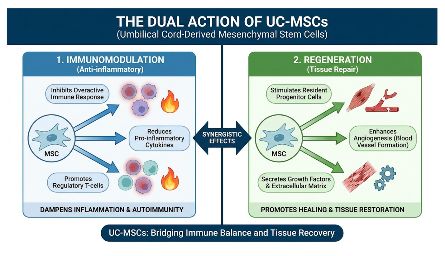
Figure 1: UC-MSCs concurrently downregulate pro-fibrotic M2 macrophages while actively suppressing excessive collagen deposition in the interstitial matrix.
Controlling this localized immune microenvironment reduces excessive tissue fibrosis significantly, preventing further architectural collapse of the lung. However, patients must understand a crucial clinical distinction when evaluating these therapies. Halting new ECM deposition is biologically feasible and extensively documented with current cellular therapeutics. Actively degrading existing, deeply cross-linked fibrotic tissue is significantly harder. Managing clinical expectations is paramount the primary medical goal is stabilizing the lung architecture to prevent further, fatal decline.
Understanding TGF-beta signaling in pulmonary fibrosis and stem cells requires examining cellular communication beyond direct engraftment. MSC paracrine signaling in the lungs acts as the primary vehicle for repair, utilizing secreted factors to inhibit pathologic cascades. This pathway disruption forms the mechanistic foundation for clinical efficacy, representing a major leap forward in modern pulmonology.
For a long time, the medical community erroneously believed stem cells worked by physically implanting themselves into damaged tissue, engrafting, and magically transforming into new lung cells. We now know that direct cellular engraftment is largely a biological dead end. The infused cells rarely survive the inflammatory onslaught of a fibrotic lung for extended periods.
Instead, the clinical approach has shifted entirely to paracrine signaling. The actual therapeutic heavy lifting is performed by MSC-derived exosomes nanoscale extracellular vesicles secreted continuously by the stem cells. Think of these exosomes as armored biological flash drives. Formed within multivesicular bodies and released into the extracellular space, they are protected by a highly durable lipid bilayer. Exosomes measure just 30 to 150 nanometers across allowing precise penetration into dense, fibrotic microenvironments that whole cells physically cannot reach.
These vesicles are packed with complex microRNAs (miRNAs), lipids, and anti-inflammatory proteins. Once they navigate through the dense collagen matrix, they are endocytosed directly by hyperactive fibroblasts. An exhaustive NCBI data review confirms that the anti-fibrotic mechanisms rely heavily on paracrine signaling, where extracellular vesicles disrupt harmful fibrotic pathways without requiring direct cell engraftment.
When discussing exosomes UC-MSC pulmonary fibrosis, we are examining the literal “Paracrine” axis of the MPS Framework. These microscopic vesicles penetrate deeply into the scarring, delivering payload instructions like miR-29b and let-7a directly to overactive fibroblasts, forcing them to abandon their fibrotic behavior.
These exosomal payloads carry the specific genetic instructions required to dismantle the most aggressive fibrotic pathways in the human body. The most critical of these is the TGF-β1/Smad signaling pathway, broadly recognized as the undisputed biological engine of pulmonary scarring.
When the TGF-β1 cytokine binds to TGF-beta receptor type II (TGF-bRII) on a cell’s surface, it recruits and phosphorylates receptor type I (TGF-bRI). This serine/threonine kinase cascade initiates the rapid phosphorylation of intracellular Smad2 and Smad3 proteins. These activated proteins then bind with Smad4, travel directly into the cell nucleus, and flip the genetic switches that transcribe massive amounts of COL1A1 and COL3A1 the genes responsible for collagen production.
UC-MSC exosomes actively intercept and interfere with this exact phosphorylation cascade. They deliver inhibitory molecules like Smad7, which act as a physical roadblock, preventing the activation of Smad2 and Smad3. A dedicated PubMed clinical data report highlights that UC-MSC-derived exosomes actively suppress the TGF-β1/Smad signaling pathway, effectively preventing fibroblast to myofibroblast transition.
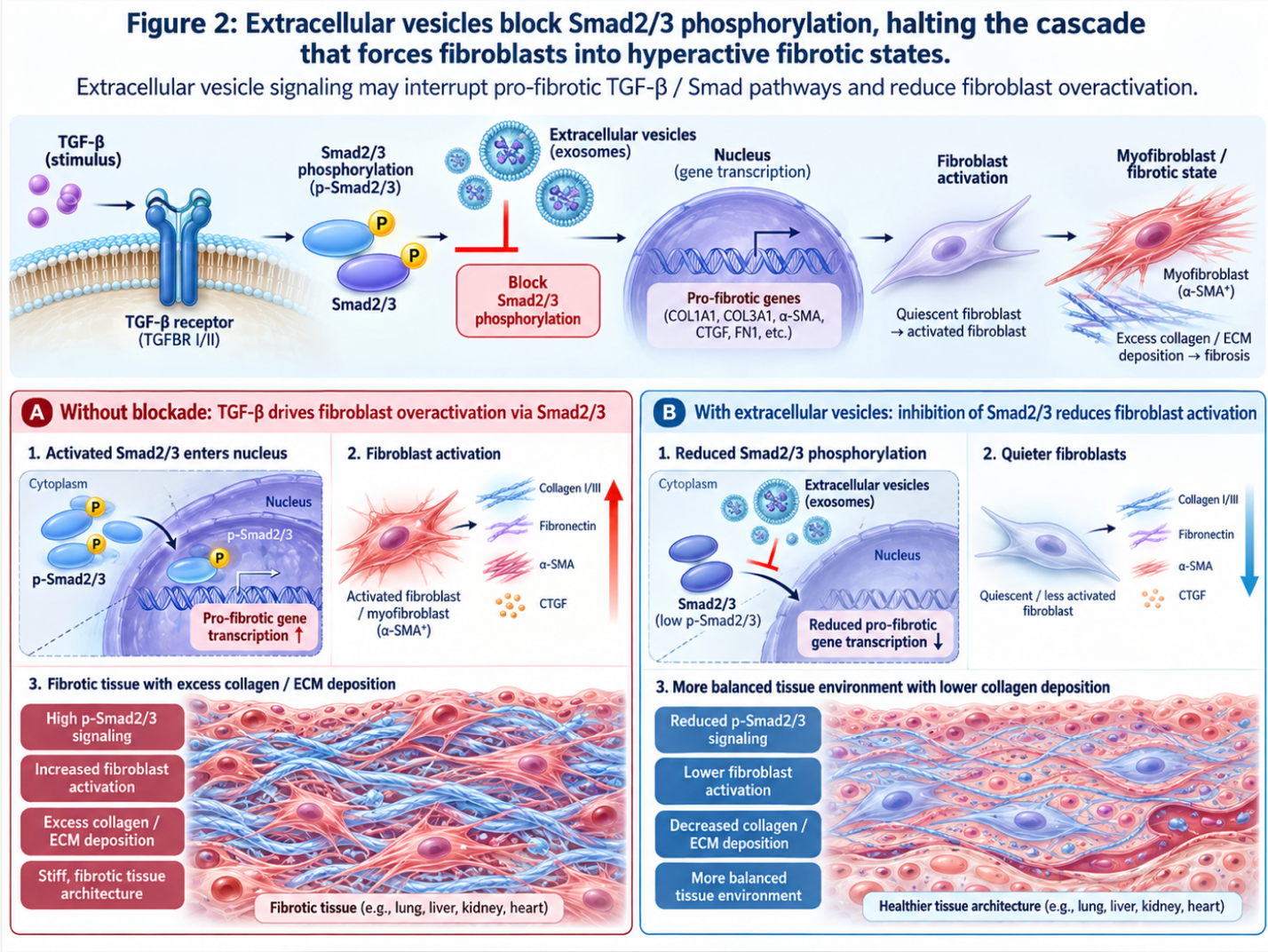
Figure 2: Extracellular vesicles block Smad2/3 phosphorylation, halting the cascade that forces fibroblasts into hyperactive fibrotic states.
This targeted molecular suppression is a massive clinical advantage. Traditional immunosuppressants carpet-bomb the entire immune system, leaving compromised pulmonary patients vulnerable to fatal respiratory infections. Exosomes, however, specifically downregulate the TGF-β1/Smad cascade natively. They preserve general immune function while completely neutralizing the fibrotic engine at the transcription level.
While TGF-β1 suppression addresses fibroblast transition, MSCs concurrently target secondary molecular pathways governing excessive extracellular matrix production. The Wnt/β-catenin
In a healthy baseline state, free β-catenin in the cytoplasm is constantly targeted and destroyed by a complex featuring Axin, adenomatous polyposis coli (APC), and glycogen synthase kinase-3β (GSK-3β). But in IPF, the Wnt pathway is pathologically hyperactivated. Wnt ligands bind to Frizzled receptors and LRP5/6 coreceptors on the cell membrane, entirely shutting down the destruction complex. This allows β-catenin to accumulate, flood the nucleus, and order the massive overproduction of fibronectin and other scar tissue components.
How does the Wnt pathway pulmonary fibrosis MSC interaction function clinically? MSCs natively secrete powerful antagonists like Dickkopf-1 (Dkk-1). Dkk-1 acts as a competitive inhibitor, binding directly to the LRP5/6 receptors and blocking Wnt ligands from attaching. This forces the cell to reactivate the destruction complex, shredding the cytoplasmic β-catenin before it can reach the nucleus.
Additional data indicates that mesenchymal stem cells prevent excessive lung tissue scarring by blocking the Wnt/β-catenin signaling pathway, which reduces extracellular matrix deposition (NCBI, 2024). Inhibiting this secondary pathway directly stabilizes lung function decline, rigorously preserving the remaining architectural integrity of the alveoli.
Evaluating UC-MSC clinical trials for pulmonary fibrosis requires strict differentiation between biological plausibility and proven human outcomes. Current data regarding stem cell therapy safety in IPF indicates a favorable tolerability profile, as demonstrated by multiple Phase I investigations. However, widespread clinical integration relies entirely on forthcoming Phase II/III efficacy endpoints.
Let’s examine the actual human data. Phase I clinical trials are foundational they do not exist to prove the therapy cures the disease; they exist to prove the experimental therapy will not harm the Phase I trials require at least 12 months of rigorous monitoring establishing a critical safety baseline for long-term respiratory assessments.
A highly regarded PubMed clinical trial data review demonstrated that intravenous administration of UC-MSCs in patients with IPF resulted in no serious adverse events, establishing a firm baseline of safety. This is a monumental hurdle to clear, particularly for fragile patients with severely compromised respiratory systems who simply cannot tolerate heavy biological burdens.
Furthermore, corresponding safety data verifies this tolerability in moderately severe patient populations. Phase I data from study NCT02013700 confirms the tolerability of unrelated allogeneic mesenchymal stem cells administered to patients with moderately severe IPF. Proving safety for allogeneic (donor) cells is a massive logistical victory for the field. It means clinical facilities can eventually utilize pre-screened, heavily expanded UC-MSCs directly off the shelf, rather than forcing an unstable pulmonary patient to undergo an invasive autologous extraction procedure.
Establishing safety allows researchers to refine the physical delivery parameters and cellular concentrations required for maximum therapeutic efficacy. Currently, intravenous (IV) administration remains the absolute clinical standard for pulmonary applications.
When you infuse MSCs intravenously, they naturally migrate to the capillary beds of the lungs first. This is known in pharmacokinetics as the “pulmonary first-pass effect.” For a patient treating a knee injury, the pulmonary first-pass effect is a frustrating physiological barrier, as the cells get temporarily trapped in the lungs. But for IPF patients, this anatomical quirk is highly advantageous. It delivers the maximum cellular payload directly into the inflamed, fibrotic microenvironment.
Ongoing trials meticulously evaluate these specific intravenous dosing parameters. Standardizing MSC dosage administration pulmonary fibrosis is incredibly complex. Clinical trial NCT04313004 evaluates the specific intravenous dosing safety and efficacy of human umbilical cord mesenchymal stem cells in humans with idiopathic pulmonary fibrosis. Researchers are continually fighting to ensure strict batch-to-batch consistency, exact viable cell counts, and maximum metabolic activity upon thawing. While theoretical inhalation routes are currently being researched in preclinical models, IV delivery remains the most validated method for achieving systemic and localized immunomodulation.
These rigorous dosing evaluations are mandatory prerequisites for navigating the complex framework of regulatory approval. The clinical reality is unequivocal: there is currently no FDA approved stem cell therapy for lungs outside of highly regulated, registered clinical trials or expanded access programs.
You cannot legally receive a commercial UC-MSC treatment for IPF in a standard US hospital setting today. The FDA rightly classifies heavily expanded mesenchymal stem cells as biological drugs (Section 351 products). This strict classification demands the same grueling Phase II and Phase III efficacy endpoints required of any newly synthesized pharmaceutical.
The clinical trajectory is promising, driven by robust cellular data, but it is not instantaneous. We are seeing major trials progress, but widespread commercial availability is likely still years away. Patients must understand the massive difference between participating in an FDA-cleared, IRB-approved clinical trial and paying out-of-pocket at a predatory clinic offering unregulated, unproven experimental treatments.
Stem cell therapy for pulmonary fibrosis is not a magic bullet. It carries highly specific limitations, and rushing into unregulated treatments can be catastrophic for patients with severely compromised respiratory function. Evaluating clinical reality means prioritizing objective functional markers over pure cellular theory.
You must rigorously separate “proven safe in Phase I trials” from “guaranteed safety indefinitely.” The most frequently reported adverse events are generally mild transient low-grade fevers, temporary fatigue, and minor localized inflammation immediately post-infusion.
However, if cellular expansion protocols are not stringently regulated in certified laboratories, the clinical risks skyrocket. Poorly cultured cells can undergo senescence, lose their paracrine potential, or trigger unexpected immune reactions. This is why rigorous clinical monitoring is absolutely mandatory, not optional. A legitimate treatment protocol requires extensive post-
There are absolute medical contraindications for investigational MSC therapy. Patients with active systemic infections, advanced pulmonary hypertension, or end-stage disease requiring an immediate lung transplant are generally disqualified from cellular trials. The biological burden of the intravenous therapy could easily overwhelm a critically unstable patient. Standard anti-fibrotic medications slow lung capacity decline by roughly 50% reinforcing their role as the foundational standard of care.
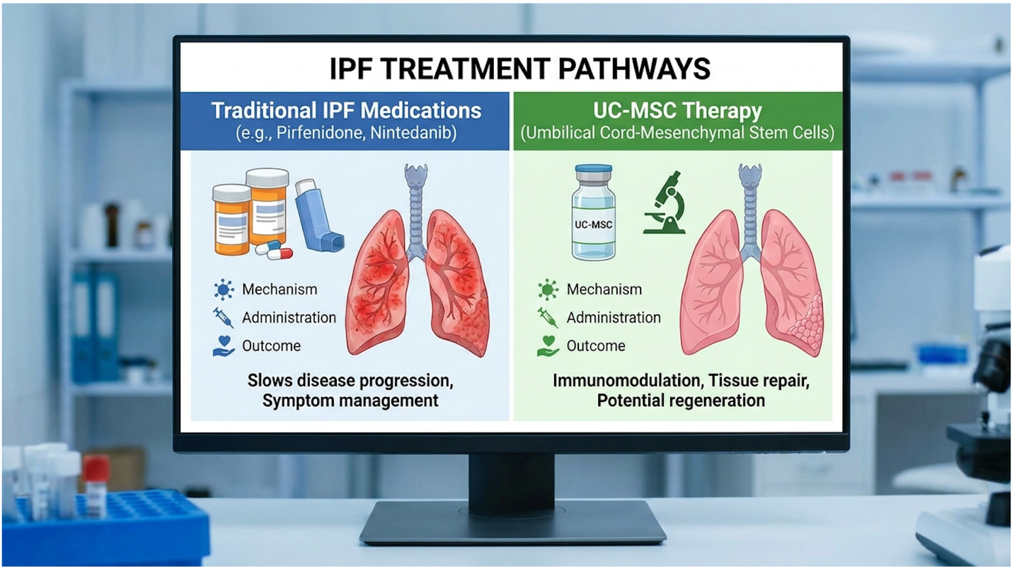
Figure 3: While UC-MSCs target the inflammatory root cause, Pirfenidone and Nintedanib remain the essential foundational standards of care for IPF management.
In these advanced cases, conventional medical alternatives must take absolute priority. Nintedanib and Pirfenidone remain the FDA-approved gold standards of care. They are deeply researched and objectively proven to slow Forced Vital Capacity (FVC) decline. Pulmonary rehabilitation and
How should an IPF patient approach a treatment suitability assessment? With intense, evidence-driven skepticism.
The regenerative medicine space is unfortunately plagued by predatory clinics operating outside regulatory boundaries. If a facility guarantees a cure, promises to reverse your IPF completely, or fails to provide peer-reviewed publication data supporting their exact protocols, walk away immediately. Legitimate clinical assessments are mentally and physically exhausting. They require a comprehensive review of your previous pharmacological treatments, a deep dive into your recent HRCT imaging, and a thorough analysis of your pulmonary function tests (PFTs).
| 📌 If you’re interested in how to tell a well-regulated stem cell clinic from a risky one, we have an interesting article that discusses whether stem cell therapy is safe in Thailand, which you can read via the internal link. |
A real clinician will tell you if your fibrotic burden is too advanced for paracrine signaling to penetrate effectively. Securing a consultation does not guarantee your eligibility for cellular therapy. It simply initiates the necessary risk-stratification process.
Stem cell therapy for pulmonary fibrosis is an investigational treatment that shows biological potential in halting lung scarring, but it is not yet a proven cure. Clinical trials demonstrate that UC-MSCs can reduce chronic inflammation and interrupt the fibrotic signaling cascade. While early Phase I/II data confirms safety and tolerability in humans, widespread efficacy requires further large-scale validation. Patients may experience stabilization of lung function, though outcomes vary significantly based on disease stage.
Mesenchymal stem cells reduce lung fibrosis primarily through paracrine signaling and immunomodulation. Rather than replacing damaged lung tissue directly, the cells secrete
No, stem cell therapy for pulmonary fibrosis is not currently FDA approved as a standard clinical treatment. The intervention remains strictly investigational and is legally administered within the United States only through registered clinical trials or expanded access programs. The FDA strictly regulates expanded stem cell products as biological drugs, requiring extensive Phase III efficacy data before commercial authorization. Patients considering treatments abroad should exercise extreme caution regarding regulatory standards. Always review a clinic’s peer-reviewed publications and clinical trial registrations before proceeding.
The most common side effects of stem cell therapy for pulmonary fibrosis include mild, transient reactions such as low-grade fever, fatigue, or minor localized inflammation following intravenous infusion. Current Phase I clinical data indicates that allogeneic umbilical cord mesenchymal stem cells are highly tolerable with no serious adverse events reported in controlled environments. However, unregulated treatments pose severe risks, including infection, immune rejection, or improper cellular proliferation. Proper side effect mitigation requires stringent laboratory protocols and continuous post-infusion medical monitoring. Patients with active infections or severe comorbidities face significantly higher clinical risks.
Umbilical cord stem cells demonstrate significant biological advantages over adult bone marrow cells for lung disease applications. UC-MSCs possess a higher proliferation rate, secrete a greater volume of immunomodulatory paracrine factors, and exhibit lower immunogenicity, making them safer for allogeneic (donor-to-patient) transfer. Furthermore, utilizing umbilical cord tissue entirely eliminates the painful, invasive surgical harvesting required for autologous bone marrow extraction.
For patients researching advanced interventions, stem cell therapy for pulmonary fibrosis delivers an investigational, evidence-informed approach to halting disease progression. Phase 1 clinical data confirms the tolerability of unrelated allogeneic cells in moderately severe IPF patients, paving the way for larger efficacy trials. The most viable clinical pathways combine rigorous suitability assessments, proven standard-of-care medications, and participation in regulated UC-MSC clinical trials.
The foundation of this therapeutic potential relies entirely on The Macrophage-Paracrine Shift (MPS) Framework. Understanding this biological shift matters because it sets highly realistic expectations. Transitioning macrophages to an M1 phenotype and suppressing TGF-β1 signaling actively halts new ECM deposition, but expecting instantaneous functional restoration ignores the biological reality of the disease. Cellular stabilization is the realistic, medically grounded goal.
If you are exploring these investigational cellular therapies, your next step requires absolute clinical rigor. Gather your last 12 months of pulmonary function tests, your most recent high-resolution CT scans, and your complete pharmacological history. Present this data to a qualified, board-certified regenerative medicine specialist or pulmonologist to secure an evidence-informed clinical assessment and determine your specific trial eligibility.
This article is for educational purposes only and does not replace consultation with a qualified medical professional. The interventions discussed include investigational therapies that require rigorous clinical assessment to determine suitability.