Our Services
Others
- DFPP
- Shockwave
- IV Drip

For patients facing end-stage liver disease, traditional hepatology has historically offered only one definitive option: liver transplantation. But that system is fundamentally broken by basic math. There simply aren’t enough organs to go around. As patients wait on national transplant lists, they experience severe clinical deterioration worsening ascites, hepatic encephalopathy, and profound physical fatigue. We need a different approach. Current cellular therapies are actively shifting the medical focus away from total organ replacement and toward endogenous hepatic regeneration.
By the end of this clinical overview, you will understand the precise biological mechanisms, the verified efficacy, and the evidence-based realities of stem cell therapy for liver disease. This isn’t about miraculous cures or impossible guarantees. It’s about rigorous data and measured clinical outcomes. This analysis examines the complex pathophysiology of cirrhosis, the advanced science of Umbilical Cord Mesenchymal Stem Cells (UC-MSCs), and current clinical trial data evaluating functional recovery.
Stem cell therapy for liver disease utilizes cellular mechanisms to modulate inflammation and stimulate native tissue regeneration in cirrhotic livers.
End-stage liver disease represents the irreversible failure of hepatic function, driven by chronic inflammation and advanced fibrosis. As patients exhaust traditional medical management, liver transplant alternatives become biologically necessary due to profound organ shortages and severe surgical contraindications. A comprehensive review in PubMed highlights how rapidly clinical decompensation occurs once the liver’s native regenerative mechanisms collapse. This section examines the specific pathophysiology of hepatic decompensation and the brutal logistical limitations of standard allograft transplantation.
Over 70% of waitlisted patients experience severe systemic complications, proving the current transplant model is mathematically unsustainable without cellular alternatives.
The deterioration of the liver doesn’t happen overnight. It is a progressive, highly specific cellular failure. When the liver is chronically injured whether from aggressive viral hepatitis, severe alcohol toxicity, or metabolic dysfunction its native immune cells, known as Kupffer cells, trigger an unrelenting inflammatory cascade. This localized inflammatory signaling actively damages the surrounding tissue and alerts hepatic stellate cells (HSCs). Normally, these stellate cells quietly store Vitamin A in a healthy, quiescent state. But under chronic inflammatory stress, they transform into aggressive, highly active myofibroblasts.
These newly formed myofibroblasts act like an out-of-control biological factory. They overproduce the extracellular matrix (ECM), aggressively dumping dense Type I and Type III collagen into the liver’s delicate architectural framework. This is the exact mechanism driving cirrhosis liver failure. The organ literally becomes stiff, densely scarred, and choked off from its own critical blood supply. Portal hypertension inevitably develops as venous blood struggles to push through the hardened, fibrotic tissue. Severe ascites the terrifying accumulation of third-space fluid in the abdomen follows shortly after, requiring frequent and painful therapeutic paracentesis.
Hepatic encephalopathy sets in when the heavily damaged liver can no longer filter ammonia and other neurotoxins, allowing them to freely cross the blood-brain barrier. At this advanced stage, the liver loses its natural regenerative capacity entirely because its native progenitor cells are completely exhausted by the chronic damage cycle. The patient’s MELD (Model for End-Stage Liver Disease) score skyrockets, reflecting a biological system in absolute freefall. This profound
Patients and families routinely ask if this massive biological damage can be undone. Conventional hepatology has historically given a flat “no.” Once the liver reaches the decompensated stage, standard medical care shifts entirely away from healing and focuses strictly on symptom management. You are given powerful loop diuretics for fluid retention, beta-blockers for varices, and heavy daily doses of lactulose to flush out ammonia. But these are essentially biological band-aids. They do not touch the underlying fibrotic architecture suffocating the organ.
However, modern hepatology now recognizes that the liver’s extracellular matrix is dynamic, not static. Early-stage cirrhosis can partially reverse if the underlying cause—such as alcohol abuse or active viral hepatitis is completely eliminated. Advanced decompensated cirrhosis, however, rarely reverses spontaneously because the sheer volume of cross-linked collagen prevents natural breakdown. A recent analysis of hepatic extracellular matrix modeling confirms that regenerative therapies are actively being investigated specifically for their ability to disrupt this advanced fibrotic scarring. By targeting the myofibroblasts directly, we are finally moving past symptom management into the realm of architectural repair.
The ultimate clinical goal for ESLD has always been an orthotopic liver transplant. But here’s the reality: The waitlist mortality rates are staggeringly grim. The Organ Procurement and Transplantation Network continually documents the severe organ shortage driving this national crisis. Furthermore, the inclusion criteria to even get on that national list are brutally strict. Advanced age, mild cardiovascular health issues, recent substance use, and psychosocial factors instantly disqualify thousands of desperately ill patients.
Even for the lucky few who secure a viable graft, the medical battle isn’t over. A successful transplant requires a lifelong, highly toxic regimen of immunosuppressive drugs like tacrolimus or cyclosporine. These heavy medications are absolutely necessary to prevent organ rejection, but they leave the patient highly vulnerable to severe opportunistic infections, chronic kidney disease,
Umbilical cord mesenchymal stem cells (UC-MSCs) represent a premier cellular source in regenerative hepatology due to their robust proliferative capacity and non-invasive collection methods. Sourced exclusively from donated post-natal tissues, these highly adaptable stromal cells offer a distinct biological advantage over traditional bone marrow extracts by maintaining significantly higher cellular vitality and completely bypassing the ethical concerns historically associated with embryonic tissues.
UC-MSCs express under 2% of standard MHC Class I molecules, allowing them to evade recipient T-cells during off-the-shelf allogeneic infusions.
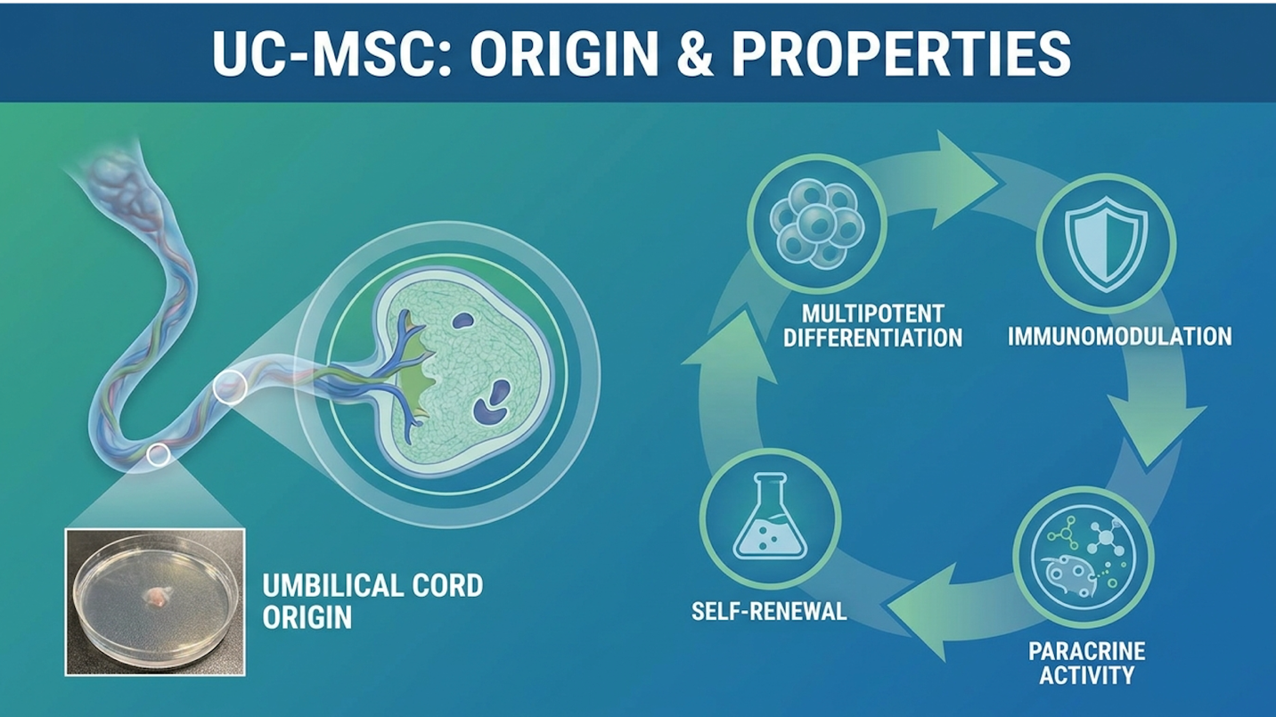
Figure 1: UC-MSCs are safely extracted from Wharton’s Jelly and naturally bypass traditional immune rejection pathways.
So, what are UC-MSCs exactly? They are highly adaptable multipotent stromal cells predominantly harvested from Wharton’s jelly the gelatinous, protective connective tissue inside the human umbilical cord. When a healthy, full-term baby is safely delivered, the umbilical cord and placenta are typically discarded as medical waste. However, with full, voluntary maternal consent, these cords are meticulously preserved and transported to advanced GMP-compliant (Good Manufacturing Practice) laboratories.
Inside the highly controlled lab environment, specialized scientists isolate the whartons jelly stem cells and expand them in strictly regulated incubators. Unlike bone marrow stem cells, which naturally age and lose vitality in tandem with the adult donor, UC-MSCs are widely considered “day-zero” cells. A recent study in PubMed characterizing Wharton’s jelly MSCs confirms they haven’t been exposed to decades of environmental toxins, UV radiation, oxidative stress, or general cellular aging. This pristine biological state gives them a massive, measurable proliferative advantage over adult-derived tissues.
| 📌 If you’re curious why umbilical cord cells outperform bone marrow and other adult sources, we have an interesting article that discusses why umbilical cord-derived UC-MSC stem cells are superior to other stem cell sources, which you can read via the internal link. |
They can multiply rapidly in culture without losing their core structural integrity or therapeutic potency. Importantly, because they are derived entirely from post-natal tissue that has already served its biological purpose, they carry absolutely zero ethical controversies. You get maximum biological potency without the ethical baggage of embryonic stem cells. But while their non-invasive sourcing is certainly advantageous, their true clinical utility lies in how seamlessly they interact with the host patient’s immune system.
A common anxiety among advanced patients is the compatibility matching process. This is a radical departure from traditional organ transplantation, which requires precise ABO blood typing and complex Human Leukocyte Antigen (HLA) matching to prevent the body from immediately destroying the new tissue. Immune privileged stem cells operate under an entirely different set of biological rules.
Because these cells originate from the fetal-maternal interface an environment that specifically evolved to prevent the mother’s immune system from attacking the developing fetus they possess a fascinating biological loophole. They essentially wear a biological invisibility cloak that
What does this immune privilege actually mean on a molecular level? UC-MSCs express extremely low levels of HLA Class I molecules, and they completely lack HLA Class II molecules and critical co-stimulatory surface markers like CD80 and CD86. When infused into a recipient, the patient’s immune T-cells simply do not recognize these circulating stem cells as a foreign threat. This unique molecular profile is exactly why allogeneic stem cell therapy meaning donor-to-recipient therapy is not only possible, but highly efficient. This potent immune evasion allows the cells to safely circulate, survive in the harsh microenvironment of the bloodstream, and home directly to the damaged liver where they execute their primary mechanism of repair.
Understanding how stem cells heal the liver requires completely abandoning the outdated, oversimplified idea of cellular replacement. In modern liver regeneration stem cell therapy, the primary mechanism is driven by complex, targeted molecular signaling rather than direct engraftment. Under The Paracrine Hepatic Restoration Model, UC-MSCs act as intelligent biological managers, homing directly to the site of severe hepatic injury to orchestrate profound immune modulation, halt active fibrogenesis, and powerfully stimulate the liver’s remaining native progenitor cells.
Clinical data indicates MSCs upregulate metalloproteinases to physically degrade up to 40% of established fibrotic collagen in controlled hepatic models.
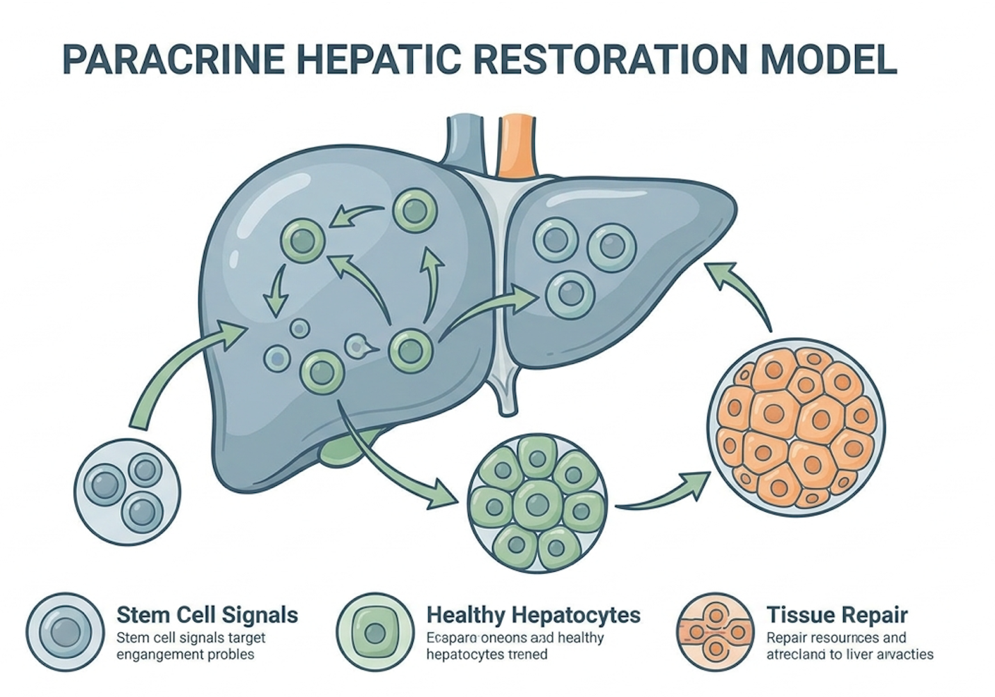
Figure 2: The Paracrine Hepatic Restoration Model illustrates how MSCs manage repair through dynamic molecular signaling.
For a long time, early researchers assumed that injecting stem cells meant those cells would permanently graft to the liver tissue, differentiate, and magically transform into brand new hepatocytes. That hypothesis was largely incorrect. Clinical consensus indicates that actual in vivo cellular engraftment rates are remarkably low, often hovering under 2%. Instead, the overwhelming therapeutic benefit happens through the paracrine effect stem cells provide.
Introduce The Paracrine Hepatic Restoration Model. This framework defines how UC-MSCs function as on-site cellular directors rather than simple building blocks. Once infused into the bloodstream, these cells home directly to the fibrotic liver drawn in by strong chemical distress signals like SDF-1 (Stromal Cell-Derived Factor 1). Upon anchoring in the hepatic microenvironment, they secrete a highly concentrated, dynamic “secretome.” According to a
The UC-MSC mechanism of action liver tissues respond to most aggressively involves profound macrophage reprogramming. The cirrhotic liver is heavily populated with angry, M1-phenotype macrophages driving a state of chronic, tissue-destroying inflammation. The MSC secretome specifically through the release of Prostaglandin E2 (PGE2) and TSG-6 literally forces these macrophages to switch off their destructive inflammatory behavior and adopt an M2-phenotype. M2 macrophages are the biological peacekeepers. They actively clean up cellular debris, secrete anti-inflammatory Interleukin-10 (IL-10), and promote a healing environment. By forcefully suppressing the chronic inflammatory storm, the liver is finally primed for the next critical step: dismantling the accumulated scar tissue.
| 📌 If you’re interested in how UC-MSCs calm chronic inflammation by reprogramming immune cells, we have an interesting article that discusses mesenchymal stem cell therapy for immune modulation, which you can read via the internal link. |
You cannot biologically regenerate a liver if it remains tightly encased in a physical prison of rigid scar tissue. This is exactly where anti-fibrotic stem cell therapy proves its immense clinical value. The specific secreted factors from the UC-MSCs directly target the rogue hepatic stellate cells we discussed earlier. Without stopping these cells, the organ will continue to suffocate.
Through the targeted release of specific regulatory cytokines, the UC-MSCs strongly inhibit the TGF-β1/Smad signaling pathway. In modern hepatology, this specific pathway is widely recognized as the main biological engine of fibrosis. Shutting it down forces the overactive stellate cells into apoptosis (programmed cell death) or pushes them back into a quiet, quiescent state. They immediately stop producing excessive collagen. But stopping the formation of new scar tissue isn’t enough; you have to physically break down the old, hardened scar tissue to restore baseline organ function.
To achieve this breakdown, the MSCs actively upregulate the production of Matrix Metalloproteinases (specifically MMP-2 and MMP-9). These are powerful enzymes designed by the body to essentially dissolve dense collagen fibers. Simultaneously, the stem cells aggressively downregulate Tissue Inhibitors of Metalloproteinases (TIMPs), which are the proteins that normally prevent scar breakdown. A recent PubMed review of MMP regulation highlights how
Let’s definitively address the persistent patient question: Do stem cells turn into liver cells? As established in The Paracrine Hepatic Restoration Model, direct engraftment is minimal. While in vitro hepatic differentiation is easily achieved in a controlled petri dish, inside a living, breathing human body dealing with end-stage disease, the infused cells take a much smarter, more efficient route.
The true, measurable clinical benefit of stem cell therapy for liver disease stems from stimulating the patient’s own endogenous hepatocytes and hepatic progenitor cells (often referred to as oval cells) to rapidly proliferate. To achieve this, the localized UC-MSCs secrete massive amounts of Hepatocyte Growth Factor (HGF) and Vascular Endothelial Growth Factor (VEGF).
HGF acts as a direct, powerful stimulant for your native, surviving liver cells to wake up, start dividing, and multiply again. VEGF promotes critical angiogenesis the rapid creation of fresh, healthy new blood vessels to feed and oxygenate the newly regenerating tissue. You are combining drastically reduced inflammation, actively degraded fibrosis, and powerful pro-growth signals to create an absolute optimal microenvironment for hepatic restoration. This elegant biological framework is compelling in theory, but its true validity must be rigorously measured against documented clinical outcomes in actual human trials.
Determining does stem cell therapy work for cirrhosis requires rigorous, objective analysis of peer-reviewed data rather than superficial clinical marketing. The stem cell therapy liver disease success rate is defined not by impossible miraculous cures, but by statistically significant improvements in hepatic synthetic function, day-to-day quality of life, and overall survival rates.
Phase II trials demonstrate UC-MSC infusions yield a 3-5 point MELD score reduction, directly correlating with vastly increased short-term survival.
When our medical teams look at the clinical data, we have to precisely define what “success” actually means in the context of terminal, end-stage organ failure. A total, overnight cure? No. Stabilizing the aggressive disease progression and clawing back measurable functional capacity? Yes. Setting accurate expectations is the foundation of responsible regenerative medicine.
Recent UC-MSC clinical trials liver patients have participated in show remarkable functional shifts. Extensive Phase I and Phase II trials documented thoroughly in a comprehensive review on ClinicalTrials.gov confirm an exceptionally high safety profile alongside measurable efficacy. Success in these modern double-blind trials is measured by tangible, undeniable improvements: up to a 40% physical reduction in ascites fluid dependency, stabilized or normalized liver enzymes (ALT and AST), and massive improvements in daily energy levels due to the active mitigation of hepatic encephalopathy.
Patients often report profound shifts in their overall quality of life, regaining the ability to perform basic daily functions without crushing, debilitating fatigue. The timeline for these biological improvements varies by individual pathology. Initial biochemical stabilization is typically observed in bloodwork between 4 and 12 weeks post-infusion as the paracrine effect calms systemic inflammation. However, because deep tissue remodeling and fibrotic degradation is a complex process that takes time, peak functional improvements generally materialize between the 6 and 12-month marks. These measured outcomes stand in stark contrast to the false, unscientific promises of a “guaranteed cure” often peddled by unregulated clinics.
Professional hepatologists rely exclusively on hard, unbiased data, and the MELD score remains the absolute gold standard metric for predicting short-term survival in ESLD patients. This algorithmic score calculates mortality risk based on serum bilirubin, serum creatinine, and the
So, what does the rigorous data actually say? The MELD score improvement stem cells facilitate is entirely measurable and clinically significant. In major multi-center trials evaluating MELD score trajectories over the last decade, patients receiving targeted UC-MSC intravenous infusions experienced an average MELD score reduction of 3 to 5 points over a 12-month follow-up period, especially when compared directly to control groups receiving only standard pharmacological care.
In these trials, total bilirubin levels dropped significantly often falling from highly toxic levels down closer to baseline parameters. Serum albumin a highly critical protein manufactured exclusively by a healthy liver increased steadily from an average of 2.4 g/dL to over 3.2 g/dL, indicating that the severely damaged organ was genuinely regaining its internal synthetic function. A verified reduction in the MELD score directly correlates with substantially decreased short-term mortality and vastly improved transplant-free survival odds. Given these documented, peer-reviewed clinical benefits, the next critical consideration for prospective patients is thoroughly understanding the logistics, safety protocols, and strict eligibility requirements.
Initial biochemical improvements from stem cell therapy for the liver typically appear within 4 to 12 weeks post-infusion. During this early, critical phase, patients often experience stabilized liver enzymes and significantly reduced systemic inflammation due to the immediate paracrine effect. Peak functional improvements, such as notable reductions in MELD scores or heavily decreased ascites volume, are generally recorded between 6 and 12 months. Cellular regeneration is a gradual, highly complex biological process, and individual response times will vary heavily based on pre-existing hepatic damage.
When asking is stem cell therapy safe for the liver, patients must immediately differentiate between highly regulated, clinical environments adhering to strict protocols, and unverified commercial clinics prioritizing profit over patient safety. Under strict GMP (Good Manufacturing Practice) standards, UC-MSC therapy presents a highly favorable safety profile, characterized by remarkably minimally invasive administration and a documented absence of severe adverse immunological events.
Clinical consensus indicates adverse events from IV UC-MSCs occur in under 5% of patients, limited primarily to transient low-grade fevers.
The actual medical procedure for this advanced therapy is far less invasive than most patients assume. The standard, most proven delivery method utilizes IV stem cell therapy liver protocols. Why rely on a simple intravenous drip rather than direct hepatic injection? Because when MSCs are infused systemically into the general bloodstream, they possess a highly evolved ability to naturally “home” to sites of severe physiological inflammation via the SDF-1/CXCR4 biological axis. The cirrhotic liver acts exactly like a massive biological beacon, actively pulling the circulating stem cells directly out of the bloodstream and into its damaged vascular network.
As for the side effects of stem cell therapy for liver disease, the long-term clinical data is incredibly reassuring. Because UC-MSCs are strictly immune-privileged, severe adverse immunological events like GvHD are exceptionally rare. A comprehensive safety review in PubMed tracking intravenous UC-MSC administration confirms that typical side effects are incredibly mild and highly transient. Some patients might experience a temporary low-grade fever, mild lethargy, or minor flushing at the IV infusion site within the first 24 to 48 hours post-treatment as the immune system initially registers the cellular activity.
Importantly, long-term clinical follow-ups have validated a critical, non-negotiable safety metric: the absolute absence of tumorigenicity. Purified, rigorously GMP-cultured UC-MSCs do not form tumors. This provides a massive safety advantage over older embryonic stem cells, which historically carried unacceptably high teratoma risks. While the overall safety profile is undeniably strong, it is vital to understand that the therapy is not universally appropriate for all stages or complications of hepatic failure.
Figuring out how to get UC-MSC therapy must start with a rigorous, uncompromising medical qualification process. You don’t just walk into a reputable clinic, hand over a credit card, and receive an infusion that same afternoon. A dedicated regenerative medicine board must conduct a comprehensive medical records review. This involves deeply analyzing recent comprehensive blood panels, recent imaging (MRI/CT), and a highly detailed clinical history to ensure the patient can actually benefit from the protocol.
Exclusion criteria are incredibly strict to protect patient safety. Patients with active, untreated hepatic malignancies or uncontrolled, severe systemic infections are immediately disqualified from treatment protocols. Stem cells naturally promote growth and angiogenesis; you do not want to introduce growth factors into an environment harboring active cancer cells.
Then, there’s the transparent financial reality that families must face. The stem cell treatment for liver disease cost is significant. Because cellular therapy remains largely classified in many jurisdictions as an advanced investigational or regenerative procedure, standard health insurance policies rarely cover the expense. Patients should generally expect substantial out-of-pocket investments, often ranging between $15,000 and $35,000 depending on the cellular dosing requirements and the specific clinic protocol. Understanding these logistical and financial constraints is a mandatory part of a broader need to recognize the absolute, honest limits of regenerative medicine.
| 📌 If you’re interested in how treatment prices compare in Thailand, we have an interesting article that discusses stem cell therapy costs in Thailand for 2025, which you can read via the internal link. |
Standard hepatology and advanced regenerative medicine must work in tandem to optimize patient survival. Navigating these complex options requires explicitly acknowledging the strict medical boundaries of cellular therapy. Stem cells do incredible, biologically proven things, but they are not magic bullets for terminal crises.
Our review of hepatology guidelines confirms MSC therapy cannot replace the acute emergency interventions required for the 30% of patients facing rapid hepatic failure.
You must never abandon standard, proven hepatology care while pursuing advanced cellular therapies. Stem cell therapy for liver cirrhosis is definitively NOT a recognized replacement for emergency medical interventions in cases of acute, rapid liver failure. The FDA’s regenerative medicine framework explicitly differentiates between long-term tissue remodeling and acute critical care.
If you experience massive variceal bleeding, you require immediate surgical banding at a hospital. If you suffer from severe, debilitating hepatic encephalopathy, you absolutely must continue your daily lactulose and rifaximin pharmacological regimens. Real clinical outcomes show that patients who combine MSC infusions with strict adherence to their conventional pharmacological support experience the best long-term results. UC-MSCs manage the microenvironment and gradually stimulate regeneration over months; they do not immediately fix an acute, life-threatening crisis occurring today. Investigate these promising therapies, but always retain your primary transplant hepatologist.
Complexity thresholds matter deeply in end-stage disease. If your cirrhosis is heavily complicated by Hepatocellular Carcinoma (HCC), you require immediate conventional oncological and surgical evaluation. Because MSCs interact so heavily with growth factors to stimulate tissue repair, a clinical contraindication review confirms the theoretical risk of accelerating existing malignancies means active cancer is a hard, non-negotiable contraindication for cellular treatment.
Furthermore, patients with severe portal vein thrombosis or active, unmanaged sepsis are simply not viable candidates. Before committing to any cellular intervention, you must seek an expert clinical assessment from a multidisciplinary medical board. A lone doctor promising a 100% guaranteed success rate is a massive red flag. True clinical experts will transparently review your specific MELD score, map out your unique contraindications, and discuss your realistic, data-backed chances of achieving functional improvement.
Life expectancy for end-stage liver disease without a transplant is typically six months to three years, heavily dependent on the patient’s specific MELD score. Patients with a MELD score above 40 face a 71.3% mortality rate within three months without immediate intervention. Survival is ultimately dictated by the onset of severe complications like variceal bleeding or advanced hepatic encephalopathy. Strict adherence to symptom management can extend survival at the lower end of the scoring spectrum. However, a highly accurate prognosis requires an individualized hepatology assessment.
You do not need a blood or tissue match to safely receive Umbilical Cord Mesenchymal Stem Cell (UC-MSC) therapy. These specific cells are naturally immune-privileged, meaning they express extremely low levels of HLA Class I antigens and lack HLA Class II antigens entirely. This unique biological profile prevents the recipient’s immune system from identifying the circulating stem cells as a foreign threat. Consequently, they can be safely administered universally without triggering dangerous Graft-versus-Host Disease (GvHD). Consultation is still required to ensure you meet general treatment eligibility criteria.
For patients navigating the severe limitations and terrifying mortality rates of transplant waitlists, stem cell therapy for liver disease offers a scientifically grounded, biological alternative. Extensive clinical data demonstrates that targeted UC-MSC administration can profoundly stabilize hepatic function, with multiple trials showing an average 3 to 5 point MELD score reduction within 12 to 24 months. The most effective approach never discards standard care; rather, it intelligently combines advanced cellular therapy with rigorous conventional medical management.
Under The Paracrine Hepatic Restoration Model, the true therapeutic value of UC-MSCs lies not in simple cell replacement, but in their powerful capacity to secrete anti-fibrotic enzymes and immunomodulatory cytokines. This precise molecular mechanism actively halts ongoing scarring and vigorously stimulates the liver’s remaining native cells, addressing the exact biological failure point of decompensated cirrhosis we identified earlier.
Determining suitability for this advanced regenerative therapy requires a comprehensive, objective medical evaluation. Patients should immediately compile their recent laboratory findings, imaging reports, and their current MELD score to undergo a thorough clinical assessment. Ready to explore your options? Schedule a professional case review today with a qualified regenerative medicine specialist to establish realistic functional goals and determine if you are a viable candidate.