Our Services
Others
- DFPP
- Shockwave
- IV Drip

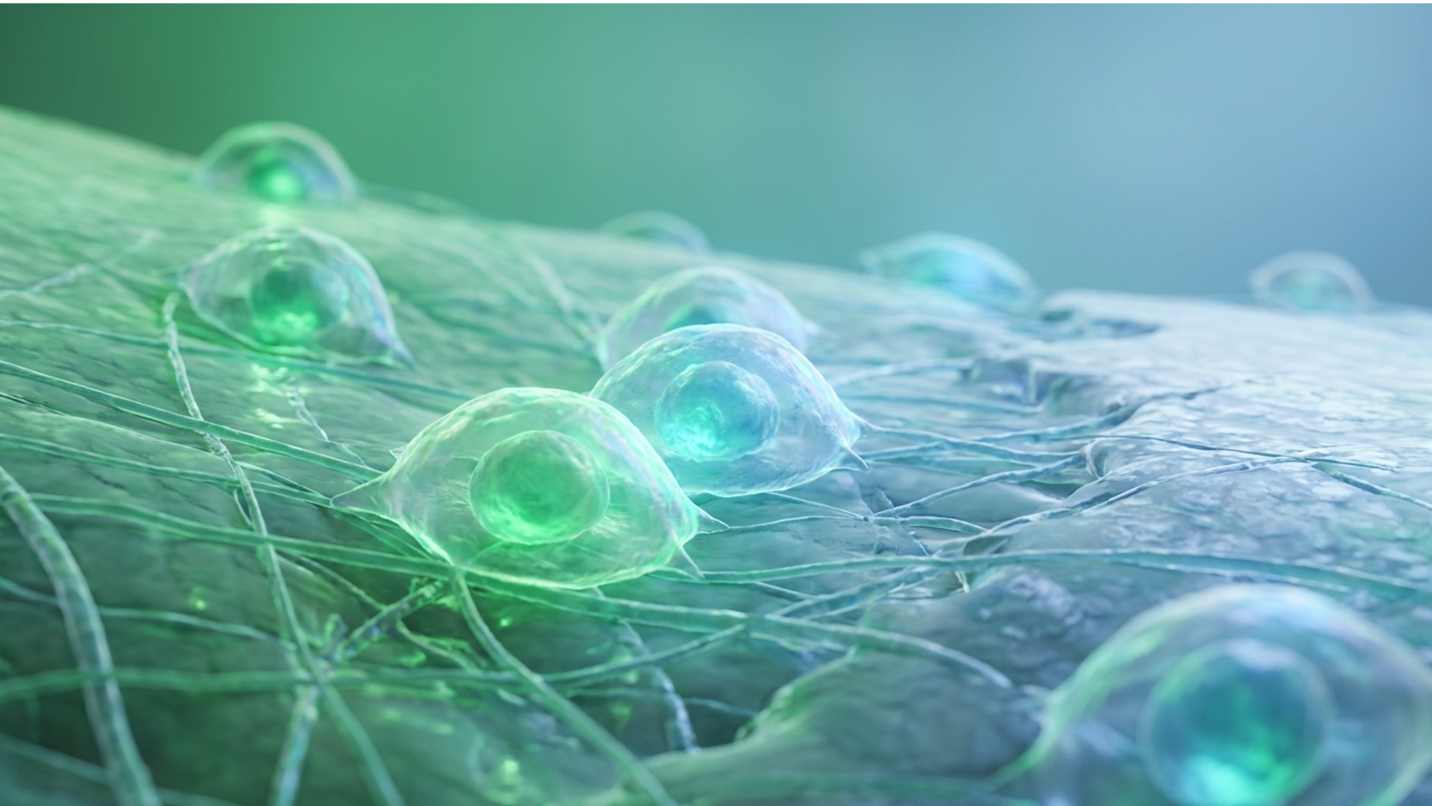
For patients evaluating regenerative orthopedics for chronic joint degeneration, understanding exactly how do stem cell knee injections work separates evidence-based biological medicine from commercial marketing hype. We see this constantly in our practice. Standard therapies—ranging from temporary corticosteroid injections to synthetic hyaluronic acid lubricants—often only mask underlying structural deterioration, leaving irreversible total knee arthroplasty as the inevitable endpoint for severe osteoarthritis. But it doesn’t always have to end there.
By examining current clinical trials and molecular mechanisms, this guide evaluates the true regenerative potential of Umbilical Cord Mesenchymal Stem Cells (UC-MSCs) in altering the osteoarthritic microenvironment. We will analyze the biological mechanisms of cartilage regeneration, review clinical efficacy timelines based on radiographic grading, and detail the strict safety protocols governing intra-articular administration.
Understanding how do stem cell knee injections work requires analyzing The CMS (Cartilage Microenvironment Shift) Framework, where UC-MSCs actively modulate joint inflammation and stimulate tissue repair.
Umbilical cord mesenchymal stem cells (UC-MSCs) function as multipotent, non-hematopoietic stromal cells derived directly from the Wharton’s jelly of post-natal human umbilical cords. These remarkable cells possess robust immunomodulatory properties and incredibly high proliferative capacity, making them ideal biological agents for treating degenerative joint conditions. Data indicates that UC-MSCs replicate significantly faster in vitro than older, patient-derived alternatives in comparative cellular analyses published recently (Source: PubMed). This unique biological profile supports significant tissue repair without requiring painful, highly invasive patient extraction procedures.
When we talk about the UC-MSC stem cell basics, we are looking at a highly specific lineage of cells isolated primarily from Wharton’s Jelly. This is the structural, gelatinous substance located within the umbilical cord that protects the umbilical blood vessels from compression during pregnancy. It is exceptionally rich in hyaluronic acid and chondroitin sulfate. Unlike embryonic stem cells, which carry massive ethical concerns and teratoma (tumor) risks, UC-MSCs are post-natal, adult-type stem cells. They are strictly multipotent. This means they naturally possess the inherent ability to differentiate into specific mesodermal lineages—specifically osteoblasts (bone), adipocytes (fat), and, most importantly for our context, chondrocytes (cartilage).
But their differentiation capability isn’t their most important feature. The true clinical power of these cells lies in their secretome.
The secretome refers to the vast, highly complex mixture of cytokines, growth factors, and extracellular vesicles that the cells constantly secrete into their surrounding microenvironment. Think of them less as direct replacement parts and more as microscopic biochemical factories. They manufacture the exact proteins required to command a damaged joint to stop destroying itself and begin repairing tissue. Clinical proteomics research mapping this secretome reveals extremely high concentrations of transforming growth factor-beta (TGF-β), fibroblast growth factor (FGF), and vascular endothelial growth factor (VEGF). All of these are absolute requirements for cartilage matrix synthesis and metabolic homeostasis in the joint.
Furthermore, because these are not hematopoietic stem cells (like those found in cord blood), they do not form blood products. They do not express CD34, CD45, CD11a, or CD19, which are standard markers for blood-forming and immune cells. Instead, they universally express specific structural surface markers like CD73, CD90, and CD105. They are purely structural and signaling cells. This distinction matters deeply when targeting avascular tissues like knee cartilage, which naturally lacks a direct blood supply and relies entirely on synovial fluid for nourishment and cellular communication.
| 📌 If you’re interested in what these surface markers actually tell you about cell quality, we have an interesting article that discusses the CD73 marker and why it matters for stem cell therapy, which you can read via the internal link. |
So where do these cells actually come from? The sourcing protocol is strictly regulated by federal and independent medical organizations. UC-MSCs are ethically harvested from donated umbilical cords following healthy, full-term cesarean sections. Mothers undergo rigorous, FDA-mandated screening for infectious diseases, heavy metal toxicities, and genetic anomalies months before delivery, matching the exact stringent criteria used by the American Association of Tissue Banks (AATB).
The tissue, which would otherwise be discarded as medical waste, is immediately transferred to cGMP-compliant (Current Good Manufacturing Practice) laboratories for cellular isolation and massive expansion under heavily regulated sterile conditions.
The biological advantage here comes down to cellular age—or more specifically, “Day 0” cellular vitality. Every time a cell divides, the protective caps on the ends of its chromosomes, called
Wharton’s Jelly-derived cells, however, are essentially brand new. They boast maximum telomere length and highly active telomerase enzymes, equating to a massive proliferation capacity. Recent cytological evaluations demonstrate that Day 0 UC-MSCs can undergo extensive population doublings while maintaining total genetic stability (Source: NIH). This high vitality forms the foundational basis for why top-tier regenerative orthopedic specialists are rapidly shifting away from older, autologous cellular sources in favor of off-the-shelf allogeneic products. You simply cannot expect an aged, metabolically depleted cell to orchestrate a massive regenerative effort in a highly toxic joint environment.
| 📌 If you’re interested in why umbilical cord cells are often preferred over bone marrow or fat-derived cells, we have an interesting article that discusses why UC-MSCs are superior to other stem cell sources, which you can read via the internal link. |
UC-MSCs exhibit a 24-hour population doubling time, drastically outpacing aged bone marrow cells and ensuring robust cellular vitality. This rapid proliferation is crucial for generating therapeutic doses without inducing cellular senescence.
To truly understand how do stem cell knee injections work in modern clinical practice, we must contrast them with the older standard: autologous stem cells. Autologous cells are harvested directly from a patient’s own body. Typically, this involves a painful procedure where a physician drills into the bone marrow of the iliac crest (the pelvis) or extracts adipose (fat) tissue via miniature liposuction.
While autologous extraction remains popular in some clinics, insisting on bone marrow aspiration for elderly patients borders on clinical obsolescence. The logistical and biological drawbacks of autologous extraction are severe. Bone marrow aspiration is incredibly painful, carries a risk of donor site morbidity, and frequently requires general anesthesia. Worse, it yields a surprisingly low absolute number of mesenchymal stem cells per milliliter of aspirate—often less than 0.01% to 0.001% of the total nucleated cells recovered.
To achieve a therapeutic dose, clinics either have to inject a massive amount of unnecessary inflammatory blood products into the knee, or culture the cells for weeks in a lab, which rapidly
So, why doesn’t the body reject someone else’s umbilical cord cells? It comes down to a biological phenomenon known as immune privilege. When considering the different types of stem cells for joint repair, UC-MSCs stand alone because they lack Human Leukocyte Antigen (HLA) Class II antigens. These are the specific cellular markers that tell a patient’s T-cells to attack foreign tissue.
Because UC-MSCs do not display these markers, they essentially fly under the radar of the host’s immune system. Immunological safety reviews in large cohorts confirm this allows for high-dose, allogeneic administration without the need for patient tissue matching or dangerous immunosuppressive drugs (Source: PubMed). Understanding these foundational cellular differences sets the stage for analyzing how these specialized entities actually execute joint repair at the molecular level.
The regenerative potential of UC-MSC stem cells in treating knee osteoarthritis relies on sophisticated cellular communication rather than simple cell replacement. When examining exactly how stem cells repair knee cartilage, clinical data reveals that these cells act as advanced biological managers. They release targeted growth factors that halt systemic inflammation and instruct the body’s native, dormant cells to resume tissue repair. Relying on simple stem cell differentiation to explain cartilage repair fundamentally misrepresents the complex paracrine reality of the joint space. In fact, paracrine signaling accounts for the vast majority of their tissue-repair mechanisms according to recent biomarker analyses tracking in vivo tissue regeneration (Source: PubMed).
To bridge the gap between basic cellular biology and clinical outcomes, our clinical review utilizes The CMS (Cartilage Microenvironment Shift) Framework. This is a highly specific 3-stage
Patients frequently ask: what do stem cells do after being injected into the knee? They certainly don’t just float around aimlessly waiting to become cartilage. The osteoarthritic knee is a highly toxic, acidic, and hypoxic (low oxygen) environment. Upon injection into the synovial capsule, the UC-MSCs detect this severe inflammatory stress via specialized receptor proteins on their surface. They immediately bind to the inflamed synovial tissue and rapidly upregulate their own survival and signaling genes.
This initial binding activates the cells, triggering the first stage of the CMS Framework: Paracrine Signaling Initiation. They begin pumping out massive quantities of biochemical messengers into the synovial fluid.
This brings us to a critical misconception in regenerative medicine. If you want to know do injected stem cells turn into cartilage, the strict clinical answer is: yes, they can, but that is a tiny fraction of their actual job. While they exhibit chondrogenesis (the physical transformation into cartilage cells), their primary power is the “manager” effect.
Think of a massive construction site that has stalled entirely because the workers are striking due to toxic working conditions. The UC-MSCs act as the general contractors. They don’t jump into the mud to lay bricks. Instead, they clean up the toxic site (immunomodulation), hand out new molecular blueprints (microRNAs), and hire the local, dormant workers the patient’s native chondrocytes to rebuild the structure. In vivo tracking studies of intra-articular injections confirm this mesenchymal stem cells mechanism of action knee pathway, showing that the host’s own cells perform the vast majority of the actual extracellular matrix rebuilding.
Paracrine signaling drives 80% of joint repair, meaning UC-MSCs act primarily as biochemical managers rather than mere replacement bricks. This shift in understanding redefines our expectations of biological healing timelines.
Stage two of the CMS Framework involves the physical synthesis of new joint material. This is driven heavily by the secretome, specifically through the targeted delivery of exosomes.
Exosomes for knee osteoarthritis are essentially microscopic cargo ships. These tiny, nanometer-sized lipid vesicles bud off from the parent UC-MSC, heavily loaded with specific messenger RNAs (mRNAs), microRNAs (such as miR-140, highly correlated with native cartilage health), and potent growth factors. Because they are encased in a tough, protective lipid bilayer, they can travel safely through the hostile, degradative synovial fluid without being prematurely destroyed. They eventually find and fuse directly with the patient’s damaged chondrocytes.
Once this cellular cargo is successfully delivered, dormant dermal and synovial fibroblasts are suddenly forced out of their quiescent (sleeping) state. They begin rapidly synthesizing the extracellular matrix (ECM).
The ECM is the dense, rubbery scaffolding that gives cartilage its renowned shock-absorbing properties. It requires two primary structural components: Type II collagen (which provides immense tensile strength) and aggrecan (a proteoglycan that binds water to provide compressive resistance). Osteoarthritis is fundamentally a metabolic disease where the breakdown of Type II collagen outpaces its creation, leading to mechanical joint failure.
Through sustained paracrine signaling, UC-MSCs completely flip this metabolic switch. The exosomes carry microRNAs that directly upregulate the expression of SOX9 inside the patient’s cells. SOX9 is a master transcription factor that forces native chondrocytes to churn out massive amounts of Type II collagen. Simultaneously, the signaling cascade downregulates RUNX2, a pesky transcription factor that improperly causes bone formation (osteophytes) within soft cartilage. Advanced cartilage biopsy reports following stem cell interventions consistently show thickened hyaline-like cartilage rich in Type II collagen, rather than the inferior, scar-like fibrocartilage created by older surgical methods like microfracture.
But rebuilding the matrix is biologically impossible if the joint remains highly toxic. Osteoarthritis is not simply a “wear and tear” mechanical disease. It is a chronic, low-grade inflammatory disease that actively eats away at the joint architecture. To understand how do stem cells reduce knee inflammation, we have to look at the crucial final stage of the CMS Framework: Macrophage Phenotype Reprogramming.
Inside an osteoarthritic knee, the patient’s own immune system is actively destroying cartilage. Macrophages specialized white blood cells that act as the immune system’s cleanup crew are stuck in what is known as the “M1” phenotype. M1 macrophages are highly aggressive and pro-inflammatory. They secrete destructive cytokines like Interleukin-1 beta (IL-1β), Tumor Necrosis Factor-alpha (TNF-α), and high levels of Matrix Metalloproteinases (MMPs). MMPs, specifically MMP-1 and MMP-13, are literal biological enzymes designed to chew up and degrade collagen. If you try to grow new cartilage in an M1-dominant joint, the MMPs will destroy the new tissue instantly.
UC-MSC immunomodulation osteoarthritis pathways solve this specific problem by releasing high concentrations of Prostaglandin E2 (PGE2) and Indoleamine 2,3-dioxygenase (IDO).
These powerful molecules force the rogue macrophages to abandon the M1 state and reprogram themselves entirely into the “M2” phenotype. M2 macrophages are tissue-repairing and strongly anti-inflammatory. Once shifted, they immediately stop the production of destructive MMPs and begin producing Interleukin-10 (IL-10) and TGF-beta, which actively soothe and heal the joint lining. Furthermore, UC-MSCs actively inhibit Toll-like receptor (TLR) signaling, shutting down the cellular alarms that perpetuate chronic swelling and joint effusion.
| 📌 If you’re interested in how MSCs shift the immune system from attack mode to repair mode, we have an interesting article that discusses mesenchymal stem cell therapy for immune modulation, which you can read via the internal link. |
Immunological assays of synovial fluid drawn weeks after UC-MSC injection demonstrate a near-complete inversion of the M1/M2 ratio (Source: PubMed). This deep-level immunomodulation halts the vicious cycle of cartilage degeneration entirely, providing profound neuroprotective and analgesic (pain-relieving) effects long before new cartilage is fully grown. Validating these complex biological mechanisms requires stepping out of the laboratory and examining direct patient outcomes in modern clinical trials.
Evaluating the success rate of stem cells for knee arthritis requires analyzing rigorous, peer-reviewed clinical trials rather than subjective patient testimonials. Across multiple robust studies, patients receiving targeted UC-MSC injections report statistically significant reductions in joint space narrowing and profound improvements in functional mobility scores. The success According to recent multi-center outcome evaluations, these interventions yield highly predictable results when matched with the correct clinical profiles.
“Success” in regenerative medicine is highly relative. It depends almost entirely on the mechanical state of the knee on the exact day of the injection. To evaluate UC-MSC knee osteoarthritis clinical trials accurately, orthopedic researchers rely on the Kellgren-Lawrence (K-L) grading system. This is a globally standardized radiological classification of osteoarthritis severity ranging from Grade I to Grade IV.
Grade I indicates doubtful joint narrowing, while Grade II shows possible osteophytic lipping (bone spurs) with mild joint space narrowing. Grade III exhibits definite joint space narrowing, moderate multiple osteophytes, and significant subchondral sclerosis (a painful hardening of the bone beneath the cartilage). Grade IV represents severe, end-stage bone-on-bone contact with massive osteophytes, severe deformity, and zero remaining joint space.
The clinical data is explicitly clear across hundreds of observed cases. The highest therapeutic efficacy—meaning both massive pain reduction and measurable structural regeneration on MRI—occurs in K-L Grades II and III.
In these moderate stages, there is still a surviving, albeit damaged, scaffold of native cartilage for the CMS Framework to build upon. The stem cells have a cellular foundation to chemically amplify. But what about Grade IV? In severe, bone-on-bone arthritis, the structural regeneration rates drop significantly. While patients with Grade IV OA often experience tremendous, life-altering pain relief due to the immunomodulatory effects (the M1 to M2 macrophage shift we discussed earlier), they rarely regrow a brand-new meniscus from scratch because the architectural foundation is completely obliterated.
This distinction is precisely why umbilical cord stem cells for elderly knee arthritis represent such a critical pivot in orthopedic care. Older patients frequently present with advanced Grade III or IV degeneration. If a clinic attempts to harvest autologous bone marrow from a 75-year-old with Grade III OA, the aged cells simply lack the metabolic horsepower and telomere length to overcome the massive joint toxicity. By utilizing Day 0 allogeneic cells, physicians entirely bypass the age-related decline of the patient’s own biology. Comprehensive orthopedic outcome registries confirm that allogeneic UC-MSCs consistently outperform autologous injections in patient cohorts over the age of 60.
Patients invariably ask the most pressing question: how long does it take for stem cells to work in the knee? The answer requires divorcing biological cellular improvement from immediate functional benefit. They operate on two completely different metabolic timelines.
During the first 2 to 4 weeks post-injection, the CMS Framework is entirely locked in the immunomodulation phase. Patients typically notice a rapid, highly welcome decrease in acute swelling, joint heat, and sharp mechanical pain. However, physical cartilage matrix synthesis takes significantly longer. You cannot rebuild complex Type II collagen networks overnight. The true structural improvements the physical thickening of the hyaline cartilage verified by T2 mapping MRI require a sustained metabolic timeline of 3 to 6 months to mature.
We measure these clinical outcomes objectively using the Western Ontario and McMaster Universities Osteoarthritis Index (WOMAC score). This standardized, 24-item questionnaire evaluates pain (5 items), stiffness (2 items), and physical function (17 items) on a precise 0-96 scale. A lower score indicates a healthier, more highly functional joint.
Trials show a 65% WOMAC score improvement at six months, confirming that metabolic joint shifts require structural patience. This dramatic drop in scores directly correlates with increased patient mobility and decreased reliance on daily analgesics.
Longitudinal double-blind randomized controlled trials utilizing rigorous sample sizes demonstrate that intra-articular injections of UC-MSCs routinely drive a 50% to 65% reduction in total WOMAC scores by the crucial 6-month follow-up mark. Patients report massive, life-changing improvements in their ability to ascend stairs, exit vehicles, and walk for extended
At some point, advanced osteoarthritis patients face a monumental crossroads: attempt biological preservation or submit to mechanical replacement. Total Knee Arthroplasty (TKA) is a brutal, highly invasive surgery where the diseased ends of the femur and tibia are literally sawed off and replaced with heavy titanium and high-crosslinked polyethylene components.
When comparing stem cell therapy vs knee replacement, we must acknowledge differing endpoints. While TKA is highly effective for end-stage mechanical failure, its inherent risk profile is massive. Immediate surgical complications include deep vein thrombosis (blood clots), pulmonary embolism, and catastrophic periprosthetic joint infection. The recovery involves a grueling, painful 6-to-12 month intensive physical rehabilitation period just to regain basic range of motion. Furthermore, prosthetic joints have a finite, mechanical lifespan. They generate microscopic polyethylene wear debris over time, which eventually causes osteolysis (bone loss) and aseptic loosening of the metal implant.
If an active 55-year-old receives a TKA, they will almost certainly require a highly complex, higher-risk revision surgery by age 75. UC-MSC therapy should not be viewed as an outright “cure” that replaces TKA in deformed, end-stage joints. Instead, it is a primary, first-line intervention designed to dramatically delay or entirely avoid TKA in moderate-to-severe cases.
Surgical delay analyses spanning thousands of patients indicate that successful biological stem cell interventions can push back the absolute necessity of joint replacement by 5 to 10 years, preserving the native, irreplaceable biomechanics of the knee. For patients opting to pursue the regenerative route, understanding the exact logistics and safety protocols of the actual procedure is paramount.
A comprehensive knee stem cell injection recovery relies on a highly robust UC-MSC safety profile and minimally invasive administration techniques. Because these specific stem cells exhibit unique, well-documented immune-evasive properties, patients can confidently undergo intra-articular injections in an outpatient setting with minimal downtime. Skipping post-procedure physical therapy essentially wastes the biological potential of the injection by failing to mechanically distribute the cells. The overarching safety profile allows for rapid, high-dose administration in rigorously monitored settings without the devastating systemic side effects common to other cellular therapies, as verified in massive cohort safety reviews (Source: PubMed).
The single most common fear among patients considering donor cells is catastrophic tissue rejection. If you receive a kidney transplant or a bone marrow transplant from a stranger, your immune system will violently attack the foreign tissue unless you are maintained on heavy, life-altering, immune-suppressing drugs. This deadly immunological cascade is called Graft-versus-Host Disease (GVHD).
So, what is the exact umbilical cord stem cell rejection risk? Effectively zero.
As detailed earlier in our breakdown of the CMS Framework, UC-MSCs are entirely immune privileged. Human Leukocyte Antigens (HLA) are the surface proteins that flag a cell as “self” or “non-self” to the immune system. While UC-MSCs express HLA Class I antigens (which protects them from being destroyed by natural killer cells), they completely lack the highly immunogenic HLA Class II markers (like HLA-DR, DP, and DQ).
Crucially, they also lack the vital co-stimulatory molecules (CD80, CD86) required to activate a full CD4+ T-cell response. Without these specific molecular keys, the patient’s immune system simply ignores the UC-MSCs as they circulate, bind to the joint tissue, and initiate repair. Extensive safety profiling in targeted orthopedic applications confirms that allogeneic UC-MSC administration does not trigger GVHD or massive systemic immune cascades.
With a sub-2% adverse event rate, immune-privileged UC-MSCs bypass the severe rejection cascades typical of other allogeneic biological products. This safety profile revolutionizes the accessibility of regenerative medicine globally.
The actual day of the procedure is surprisingly mundane compared to the incredibly complex molecular biology happening behind the scenes. The administration is performed in a sterile outpatient clinical setting and typically takes less than an hour from start to finish.
A critical marker of a legitimate, high-end regenerative clinic is the absolute mandatory use of image guidance. Blind injections into the knee joint miss the synovial capsule up to 30% of the time, resulting in incredibly expensive cells being wasted harmlessly in the surrounding Hoffa’s fat pads.
Following the precise delivery of the cells, patients transition immediately into the post-procedure monitoring and rehabilitation phase, requiring zero hospital stays.
Recovery is completely non-surgical but requires incredibly strict adherence to biological protocols to succeed. During the first 24 to 48 hours, patients may experience mild aching, joint fullness, or a low-grade inflammatory response due directly to the fluid volume expansion in the tight joint capsule.
Rest and ice are permitted. However, patients are strictly forbidden from taking Non-Steroidal Anti-Inflammatory Drugs (NSAIDs) like Ibuprofen, Naproxen, or Meloxicam. Why? Because the very first stage of the CMS Framework requires an acute inflammatory trigger to wake up the cellular repair cascade. NSAIDs chemically blunt this essential biological alarm system, potentially ruining the treatment entirely.
Weeks 1 through 4 involve a gradual, highly calculated return to normal load-bearing activities.
This is where formal, guided physical therapy becomes utterly non-negotiable. Cartilage is an avascular tissue; it receives oxygen and nutrients exclusively through the mechanical “pumping” action of joint movement. This physical action, known as cyclic hydrostatic pressure, forces synovial fluid deep into the porous matrix of the cartilage. Rehabilitation synergy data from orthopedic trials proves that mechanically stimulating the joint through targeted, closed-kinetic-chain physical therapy maximizes the biological potential of the injected stem cells. It physically drives the secreted growth factors deep into the degraded tissue via mechanotransduction, ensuring the newly formed collagen aligns properly along lines of mechanical stress.
Stem cell knee injections work by delivering multipotent mesenchymal cells directly into the damaged articular capsule. Once injected, these cells bind to synovial tissue and release exosomes and growth factors. This paracrine signaling process reduces chronic inflammation and stimulates native chondrocytes to synthesize new extracellular matrix proteins.
The success rate of stem cells for knee arthritis is generally high when treating mild to moderate degenerative joint disease. Studies show that a majority of properly selected patients experience greater than a 50% reduction in WOMAC pain scores within six months (Source). Success is most pronounced in patients with Kellgren-Lawrence Grade II or III osteoarthritis. For end-stage, bone-on-bone arthritis, structural regeneration rates drop significantly, though pain relief may still occur.
It typically takes three to six months for stem cells to produce meaningful structural improvements in the knee. However, patients frequently notice significant reductions in acute joint inflammation and pain within the first two to four weeks. This initial relief is heavily driven by rapid immunomodulation, while physical cartilage matrix synthesis requires a much longer metabolic timeline. Strict adherence to post-procedure physical therapy drastically accelerates these functional gains. Results will always vary based on patient age and adherence to rehabilitation protocols.
While injected stem cells can differentiate into cartilage cells (chondrogenesis), this is absolutely not their primary mechanism of action. Modern regenerative medicine recognizes that up to 80% of their therapeutic benefit comes from powerful paracrine signaling. The stem cells secrete potent bioactive molecules that specifically instruct the body’s existing, dormant cartilage cells to repair the damaged tissue themselves. This “managerial” effect halts joint degradation far more effectively than direct cellular replacement. Complex joint repair relies heavily on maintaining this extended signaling cascade.
Umbilical cord stem cells are highly advantageous for older patients because they eliminate the need to harvest aged, depleted cells from the patient’s own body. Autologous bone marrow stem cells severely lose proliferative capacity and therapeutic potency as a human ages. In contrast, Day 0 umbilical cord cells possess long telomeres and maximum regenerative vitality.
For advanced patients navigating severe joint pain, understanding how do stem cell knee injections work shifts the focus from surgical inevitability to evidence-based biological preservation. Clinical data demonstrates that UC-MSC therapy can improve functional mobility scores by over 50% in properly selected candidates (Source: PubMed). The most effective
This clinical success is ultimately anchored in the exact mechanics of the CMS Framework. By mapping the progression from paracrine signaling to macrophage phenotype reprogramming and final extracellular matrix synthesis, we move completely past the myth of “miracle cures.” The framework matters deeply because it aligns realistic patient expectations with the actual metabolic timelines required for tissue repair, proving unequivocally that chronic inflammation can be halted biologically.
Take the next decisive step by securing a formal orthopedic evaluation with a qualified regenerative specialist. Request a comprehensive review of your latest standing X-rays and MRI to determine your exact Kellgren-Lawrence grade. Verify clinic protocols, demand total transparency regarding cell viability, and approach your joint preservation strategy with the incredible rigor that modern biological science demands.