Our Services
Others
- DFPP
- Shockwave
- IV Drip

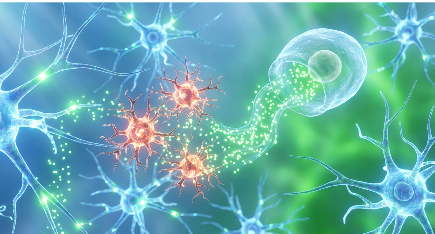
Current research indicates that neuroinflammation in autism spectrum disorder is a measurable biological driver, shifting clinical interest toward targeted cellular therapies rather than purely behavioral interventions. However, the commercial stem cell market is plagued by oversimplified claims, leaving specialized clinicians and informed parents struggling to differentiate between experimental biological plausibility and proven functional outcomes. By the end of this guide, you will understand the precise molecular mechanisms of Mesenchymal Stem Cells (MSCs) and the current peer-reviewed evidence regarding their efficacy and safety in pediatric populations. This review critically evaluates the pathophysiology of ASD, the specific immunomodulatory actions of MSCs, and the limitations of current clinical trial meta-data.
This article is for educational purposes only and does not replace consultation with a qualified medical professional. Cellular therapies for autism remain investigational.
Targeting neuroinflammation in autism through Mesenchymal Stem Cells (MSCs) focuses on repairing immune dysregulation rather than behavioral masking. Studies demonstrate MSCs operate primarily via The Dual-Vector Paracrine Mechanism rather than cellular engraftment.
Targeting neuroinflammation in autism spectrum disorder requires mapping the specific pathophysiology driving neuronal dysfunction. Clinical assays frequently reveal heightened microglial activation and elevated pro-inflammatory cytokines in ASD patients, establishing a clear link between immune dysregulation and neurological outcomes. Understanding this altered cellular environment is a strict prerequisite before evaluating any advanced cellular therapies, as treating an underlying inflammatory phenotype demands precision medicine rather than broad-spectrum guesswork.
Elevated pro-inflammatory cytokines like IL-6 actively alter brain connectivity in ASD phenotypes making precise immunomodulation critical for functional recovery. While commercial clinics push rapid infusions, prioritizing baseline biomarker testing is the only responsible path forward.
When evaluating the cellular architecture of the autistic brain, we must look far beyond basic neurotransmitter imbalances. Microglia, the primary immune cells of the central nervous system, govern the baseline health of neural networks. In a typical neurodevelopmental environment, these resident macrophages perform a highly specialized, delicate job. They act as the brain’s
But in a significant subset of children with ASD, this process derails completely. Post-mortem transcriptomic analyses and advanced in-vivo PET imaging consistently reveal that these microglia are trapped in a chronically hyper-reactive state. They abandon their essential synaptic pruning duties. Instead, they shift into a defensive posture, responding to the local brain tissue as if it were under active pathogenic attack. This persistent microglia activation in autism disrupts the fundamental physical wiring of the brain. The timeline for this disruption is critical, as it often spans both in utero development and the highly sensitive postnatal windows where core neurodevelopmental milestones are traditionally achieved.
Look, the brain isn’t just miswired from birth; it’s actively remodeling under toxic stress. Without proper microglial pruning, local neural networks become overly dense and hyper-connected, while long-range functional connectivity across different brain regions severely degrades. This physical, structural imbalance provides the cellular basis for the profound sensory processing challenges and rigid thought patterns characteristic of the disorder. It’s a localized structural crisis. When local circuits in the visual or auditory cortex become hyper-connected, the child experiences profound sensory overload; meanwhile, the degraded long-range connections prevent the brain from synthesizing that sensory data into meaningful social context.
Extensive clinical mapping initiatives highlight just how structurally devastating this local microglial activation can be. Functional MRIs in severely affected pediatric cohorts routinely show that the overgrowth of localized white matter tracts actively chokes the critical pathways required for executive function (ScienceDirect clinical review, 2016). And while microglial activation provides the cellular basis for this local neuroinflammation, it is the systemic immune dysregulation that actively perpetuates the cycle of neural distress.
Systemic immune dysregulation in autism doesn’t just stay in the periphery; it directly assaults the central nervous system. When we analyze peripheral blood mononuclear cells (PBMCs) and cerebrospinal fluid from pediatric patients, we routinely find wildly abnormal cytokine profiles. Specific pro-inflammatory cytokines namely Interleukin-1 beta (IL-1β), Interleukin-6 (IL-6), Tumor Necrosis Factor-alpha (TNF-α), and Interferon-gamma (IFN-γ) are drastically elevated compared to neurotypical controls (Europe PMC research, 2023). These molecular messengers are normally reserved for fighting off severe infections, yet in the autistic phenotype, they are chronically elevated, bathing developing neural tissue in a constant state of chemical alarm.
This specific cocktail of inflammatory markers points toward a profound breakdown in immune tolerance. Many developmental immunologists trace this back to Maternal Immune Activation (MIA) models. If a severe maternal infection or autoimmune event occurs during a critical gestational window, it can permanently alter the epigenetic programming of the fetal immune system. The child is born with an immune system primed for overreaction. Fast forward to postnatal development, and minor environmental triggerssuch a s standard childhood viral illnesses or gastrointestinal distress cause catastrophic inflammatory cascades. The immune system lacks the regulatory brakes necessary to shut down the response once the threat has passed.
Over time, this unrelenting systemic immune response compromises the structural integrity of the blood-brain barrier (BBB). The BBB is designed to be a highly selective, semi-permeable border, but chronic exposure to TNF-α physically degrades its tight junctions. A weakened BBB allows these peripheral inflammatory markers, which should be isolated in the bloodstream, to freely infiltrate the central nervous system, directly activating the resident microglia.
Clinical researchers at specialized institutions have developed highly specific blood-based biomarker panels to identify precisely which patients exhibit this profound neuroimmune phenotype. In comprehensive biomarker testing methodologies across prominent clinics, it becomes evident that identifying these specific elevated cytokines is the only way to accurately qualify a patient for advanced intervention. They know that understanding immune dysregulation in autism is the key to identifying candidates who actually have a biological rationale for cellular therapy. This unrelenting systemic immune response directly triggers the next phase of the pathophysiological cascade: profound oxidative stress.
Here’s the thing about oxidative stress: it doesn’t exist in a vacuum. It is a highly destructive, self-feeding biochemical loop driven by chronic inflammation. As hyper-reactive microglia and infiltrating peripheral immune cells flood the brain with cytokines, they simultaneously generate massive amounts of reactive oxygen species (ROS). These volatile oxygen molecules carry unpaired electrons, making them highly reactive and biologically destructive. The developing autistic brain is uniquely vulnerable to ROS due to a documented, systemic depletion of endogenous antioxidants like glutathione, which normally act as the brain’s internal chemical buffer against free radical damage.
Without sufficient glutathione to neutralize these free radicals, the brain undergoes severe lipid peroxidation. This is where the structural damage becomes catastrophic. The fatty myelin sheaths insulating long-range projection neurons which are absolutely crucial for the rapid transmission of electrical impulses across the corpus callosum and other major white matter tracts—begin to physically degrade under the chemical assault. Clinical diagnostics utilizing specialized spectroscopy consistently reveal severe metabolic deficits in these highly stressed brain regions. When lipid peroxidation strips the myelin away, electrical signals scatter, causing massive cognitive interference and processing delays.
Furthermore, chronic oxidative stress fundamentally impairs mitochondrial function within the neurons themselves. Neurons are incredibly energy-demanding cells. When their mitochondria fail due to oxidative damage in the Krebs cycle, the cells simply cannot sustain the massive metabolic output required to maintain long-range synaptic connections. The adenosine triphosphate (ATP) production drops off a cliff, leaving the neuron biologically starving and incapable of transmitting complex data across the cortex. This metabolic collapse directly limits the brain’s ability to synchronize the temporal lobes (responsible for auditory processing) with the prefrontal cortex (responsible for executive function).
This biochemical catastrophe translates directly into observable clinical deficits. When long-range neural connectivity degrades, the brain struggles to integrate complex, multi-sensory information resulting in the classic presentation of sensory overload, profound social withdrawal, and severe communication delays. The biology directly dictates the behavior. Analyses of clinical data connecting oxidative stress and ASD (Regenestem clinical analysis, 2021) show that stopping this destructive cycle is paramount to any therapeutic success. Because traditional pharmacology struggles to halt this compounding cycle of oxidative stress and inflammation without triggering severe, blanket immunosuppressive side effects, specialized researchers have increasingly turned to the highly targeted therapeutic potential of mesenchymal stem cells.
The therapeutic role of mesenchymal stem cells (MSCs) in autism hinges entirely on their capacity for complex immunomodulation, not structural tissue replacement. By leveraging The Dual-Vector Paracrine Mechanism, MSCs detect pathological inflammatory environments and respond by releasing specific bioactive molecules and exosomes that reprogram hyper-reactive immune cells. This biological mechanism forms the entire basis of contemporary cellular trials for ASD.
Intravenously infused mesenchymal stem cells actively reduce central neuroinflammation by bypassing the blood-brain barrier with targeted exosomal microRNAs effectively halting oxidative stress at the genetic transcription level. This biological reality strips away the “miracle cure” marketing and centers the discussion on measurable, targeted molecular biology. Demanding strict biological evidence over marketing promises is the baseline standard for evaluating any cellular intervention.
To truly grasp how stem cells calm neuroinflammation in children with autism, we must abandon the outdated, biologically inaccurate idea that stem cells simply grow into new brain cells. Instead, modern cellular biology centers on the Original Concept of The Dual-Vector Paracrine Mechanism. This is the highly coordinated process by which MSCs simultaneously deploy soluble anti-inflammatory proteins (Vector 1) and targeted exosomal miRNAs (Vector 2) to reprogram pathological macrophages and resolve neuroinflammation. MSCs act less like building blocks and more like intelligent, responsive biological micro-pharmacies.
Let’s break down Vector 1 in deep molecular detail. When intravenously infused MSCs encounter a highly inflammatory environment signaled by high levels of IFN-γ and TNF-α circulating in the patient’s blood they activate a dynamic biochemical factory. They immediately begin secreting powerful, soluble immunomodulatory proteins. The heavy hitters here are Prostaglandin E2 (PGE2), Indoleamine 2,3-dioxygenase (IDO), and Transforming Growth Factor-beta (TGF-β).
IDO operates via a fascinating starvation mechanism. It physically degrades the essential amino acid tryptophan into kynurenine in the local environment. Because aggressive, neurotoxic effector T-cells (specifically Th1 and Th17 cells) require massive amounts of tryptophan to proliferate rapidly, this enzymatic action effectively starves the rogue immune cells without harming healthy tissue. By starving these aggressive T-cells, IDO forces a dramatic and immediate halt to the systemic inflammatory cascade. It is a highly elegant, self-regulating biological shutdown mechanism that traditional synthetic drugs simply cannot replicate.
More importantly for autism neurobiology, these secreted proteins particularly TGF-β force a profound phenotypic shift in the body’s native macrophage population. They compel hyper-reactive, tissue-damaging M1 macrophages to transform into anti-inflammatory, tissue-repairing M2 macrophages. This biochemical intervention interrupts the insidious Toll-like receptor (TLR) and ERK MAPK signaling pathways that sustain chronic neuroinflammation. Unlike traditional pharmacological immunosuppressants which blindly blunt the entire immune system and leave the child vulnerable to standard infections this vector is highly adaptive. It scales its protein output precisely based on the specific level of inflammation it encounters (Springer biomedical research, 2023). While the soluble proteins dictate immediate, systemic immune suppression, it is the second vector the MSC-derived exosomes that provides long-lasting epigenetic modulation across the blood-brain barrier.
| 📌 If you’re interested in how MSCs shift the immune system from attack mode to repair mode, we have an interesting article that discusses mesenchymal stem cell therapy for immune modulation, which you can read via the internal link. |
If the soluble proteins are the first wave of defense, MSC-derived exosomes are the highly specialized special forces unit. Exosomes are incredibly tiny extracellular vesicles, measuring just 30 to 150 nanometers in diameter. Think of them as microscopic cargo ships secreted by the parent mesenchymal stem cell. Inside these highly stable, lipid-bound vesicles is a complex payload of messenger RNA (mRNA), specific neurotrophic proteins, and crucially, microRNAs (miRNAs). Because they are protected by a tough lipid bilayer, these delicate genetic messengers survive the harsh, enzyme-rich environment of the human bloodstream without degrading.
The physical size of these exosomes gives them a massive, game-changing therapeutic advantage over whole-cell therapies. While large, whole MSCs (which range from 15 to 30 micrometers) struggle to cross into the central nervous system, MSC-derived exosomes for autism neuroinflammation effortlessly slip across the tight junctions of the blood-brain barrier. They travel directly into the brain’s parenchyma without requiring highly invasive, risky surgical administration like intrathecal spinal injections or direct cerebral implants.
Once inside the brain tissue, they hunt down the chronically hyper-reactive microglia. Utilizing clathrin-mediated endocytosis, they physically fuse with the microglial cellular membranes and dump their epigenetic payload directly into the troubled immune cells. The precision of this cellular targeting is remarkable; exosomes naturally home in on areas of high oxidative stress, bypassing healthy neural tissue entirely to deliver their payload precisely where the inflammation is most aggressive.
Specific miRNAs contained within these exosomes such as miR-146a and miR-21 act as master genetic switches inside the microglia (Stem cell clinical review). For example, miR-146a physically binds to the 3′ untranslated region (UTR) of IRAK1 and TRAF6 mRNA. By silencing these specific messenger RNAs, the exosome degrades the inflammatory instructions before the toxic cytokines can even be assembled by the cell’s ribosomes. This downregulates transcription factors at the fundamental genetic level, effectively halting the NF-kB cascade and stopping the oxidative stress cycle at its absolute molecular source. This cell-free exosome therapy represents the cutting edge of autism neuroinflammation treatment. The potency of both the paracrine proteins and exosomal miRNAs, however, relies entirely on the biological age and quality of the source cells.
Not all stem cells are created equal. When targeting profound immune dysregulation in a pediatric population, the biological source of the cells dictates the entire clinical outcome. In modern, rigorous pediatric trials for ASD, researchers almost exclusively utilize allogeneic human umbilical cord mesenchymal stem cells (UC-MSCs) rather than autologous adult sources like bone marrow (BMSCs) or adipose tissue (ADSCs). Utilizing a child’s own cells when they already possess an underlying genetic or epigenetic predisposition to immune dysregulation is counterproductive; you are simply reintroducing the exact same flawed biology back into their system.
| 📌 If you’re interested in why umbilical cord cells are often preferred over bone marrow or fat-derived cells, we have an interesting article that discusses why UC-MSCs are superior to other stem cell sources, which you can read via the internal link. |
The biological rationale for utilizing donor cord tissue is undeniable. UC-MSCs represent ‘Day 0’ biology. They are harvested ethically and non-invasively from discarded Wharton’s Jelly immediately post-birth. Because they are chronologically brand new, they possess incredibly long telomeres, exhibit massive and robust proliferation rates, and generate a secretome (the cocktail of therapeutic proteins and exosomes they release) that is vastly more potent, diverse, and complex than stem cells harvested from a mature adult. Adult stem cells have suffered years of environmental toxin exposure, epigenetic aging, and inevitable telomere degradation, significantly muting their paracrine output. Furthermore, adipose tissue sources often carry the epigenetic memory of inflammatory metabolic states, making them highly suboptimal for treating neuroinflammation.
UC-MSCs possess a unique, highly advantageous biological superpower: immune privilege. These cells express very low levels of Major Histocompatibility Complex (MHC) Class I and completely lack MHC Class II molecules, as well as crucial co-stimulatory markers like CD80 and CD86 (while remaining positive for standard MSC markers like CD73, CD90, and CD105).
Because they lack these specific identification tags on their cell surface, the patient’s immune system does not recognize them as foreign invaders. This hypoimmunogenic profile drastically reduces the risk of Graft-Versus-Host Disease (GVHD), allowing for safe, allogeneic (donor) administration without requiring dangerous immunosuppressive matching drugs (Published clinical analysis). Importantly, this protocol spares a child with autism the agonizing trauma, anesthesia risks, and recovery time associated with autologous bone marrow extraction surgeries. Understanding this distinction in cell sourcing is vital for dispelling the most persistent myth in regenerative medicine.
The commercial stem cell industry has done a massive disservice to vulnerable patient populations by perpetuating the myth of cellular engraftment. Far too many unregulated clinics falsely promise that intravenously infused MSCs will magically migrate straight to the child’s brain, permanently graft into the tissue, differentiate into shiny new neurons, and physically rebuild damaged neurological architecture. This is a profound misrepresentation of established, peer-reviewed science, designed primarily to sell hope rather than deliver biological reality.
In reality, clinical researchers adhere to the “Hit and Run” hypothesis. Extensive biodistribution studies using radiolabeled MSCs in mammalian models demonstrate that when cells are administered intravenously, they rarely make it past the pulmonary circulation. The vast majority are quickly trapped in the massive capillary beds of the lungs a phenomenon known as the pulmonary first-pass effect. Because whole MSCs are relatively large, they physically cannot pass through the tiny lung capillaries easily. Within a matter of days (typically 24 to 72 hours), the patient’s native immune system, primarily pulmonary macrophages, successfully clears these foreign cells entirely from the body through a process called efferocytosis.
Yet, despite this incredibly rapid clearance, the functional improvements in neuroinflammation and observable behavior persist for months. Why? Because the MSC anti-inflammatory effects in
As the lung macrophages phagocytose the dying MSCs, this interaction actively triggers the macrophages to release massive amounts of TGF-beta into the systemic circulation (ResearchGate pathophysiology review). They aggressively pump their immunomodulatory proteins and BBB-crossing exosomes out before dying. The donor cells vanish, but the epigenetic reprogramming they leave behind in the patient’s native macrophages and microglia is enduring. With these molecular mechanisms clearly defined and the myths of engraftment dismantled, the critical next step is evaluating how effectively this biological theory translates into measurable, validated clinical outcomes in pediatric trials.
Evaluating the efficacy and safety of stem cell therapy in children with autism requires strict adherence to published meta-analyses rather than anecdotal clinic reviews. Aggregated clinical trial data confirms that MSC therapy can yield statistically significant improvements on validated behavioral scales, accompanied by an acceptable safety profile. However, translating these findings into clinical practice requires navigating substantial evidence limitations.
Pediatric MSC trials demonstrate quantifiable improvements in VABS scores for nonverbal communication proving that highly targeted biological interventions can tangibly optimize developmental trajectories. It is imperative that practitioners communicate these modest but real statistical gains honestly, resisting the urge to frame biological progress as a universal cure for complex neurodevelopmental delays.
When we rely on hard, peer-reviewed data over flashy marketing hype, the clinical reality of MSC therapy for autism becomes highly nuanced and fascinating. Meta-analyses aggregating multiple pediatric stem cell trials reveal a compelling, if currently incomplete, picture of therapeutic potential. We are looking at combined data from several dozen rigorous trials worldwide, bringing total evaluated patient sample sizes into the mid-to-high hundreds (often ranging from n=30 to n=100 per individual trial phase). In these sophisticated studies, researchers enforce strict inclusion
However, we must explicitly and honestly acknowledge the limitations inherent in current evidence (PMC clinical meta-analysis, 2022). The sample sizes within individual trials remain incredibly small, which reduces the statistical power to detect rare adverse events. Furthermore, there is massive, frustrating heterogeneity in clinical trial protocols across different institutions. Some researchers utilize intravenous infusions, while others aggressively push for intrathecal administration into the spinal fluid. Dosing protocols fluctuate wildly—ranging anywhere from 1 million to over 50 million cells per kilogram of patient body weight across different major university studies.
Crucially, we lack the decades-long, double-blind follow-up data required to fully understand the long-term durability of these interventions. Currently, we are predominantly analyzing Phase I and Phase II open-label or early-stage randomized controlled trials. Blinding remains a massive challenge in these studies, as parental placebo effects are notoriously strong when evaluating behavioral changes in their own children. We have not yet reached the definitive, highly powered Phase III trials required for universal FDA standardization and broad insurance coverage. Therefore, when data indicates “statistically significant improvement” (often denoted by p-values < 0.05 in specific domains), informed readers and parents must recognize this means a measurable, functional optimization of the patient’s baseline capabilities. It absolutely does not equate to a magical reversal of the condition or a total cure. To quantify this efficacy without the bias of parental hope, researchers rely on highly specific, standardized testing instruments administered by objective clinicians.
In clinical trials, anecdotal improvements and heartfelt stories from parents, while emotionally powerful, are scientifically insufficient; researchers demand rigorous, psychometric validation. Efficacy is measured meticulously using standardized, universally recognized diagnostic tools, primarily the Childhood Autism Rating Scale (CARS) and the Vineland Adaptive Behavior Scales (VABS). These specific, multi-point metrics allow clinicians to strip away placebo effects, control
The data reveals distinct, highly specific patterns in exactly how stem cell therapy affects behavior. The most robust, consistent improvements are typically not found in core repetitive behaviors (like hand-flapping or specific stereotypies), but rather in profound underlying subdomains. On the VABS scale, improvements are heavily clustered in Receptive Language, Expressive Language, and Daily Living Skills. Children who were previously entirely non-communicative or deeply disconnected often begin demonstrating spontaneous joint attention, improved eye contact, and expanded social reciprocity. They start processing their environments rather than merely reacting defensively to them.
However, the stark variance in patient response remains a frustrating reality for clinicians analyzing this data. We regularly observe “super-responders” who jump several critical developmental milestones in a matter of months, functioning alongside non-responders who show statistically zero change on their CARS scores post-treatment (Wiley Stem Cells Translational Medicine, 2019). This profound variance strongly suggests that patients with the absolute highest baseline levels of autism inflammatory markers (such as circulating cytokines and observable gastrointestinal inflammation) receive the greatest tangible benefit from the biological immunomodulation.
Timeline-wise, these behavioral shifts are not instantaneous. The complex epigenetic reprogramming driven by exosomal miRNAs takes significant cellular time, with peak clinical improvements typically manifesting 3 to 6 months post-infusion. By managing expectations around these timelines, researchers ensure that families do not prematurely abandon the therapy during the critical early cellular remodeling phase. While these behavioral outcomes present a highly compelling therapeutic argument, they must be rigorously and cautiously weighed against the documented physiological risks of the procedure.
The paramount, non-negotiable concern for any responsible clinician evaluating advanced, experimental therapies is the foundational medical principle of primum non nocere—first, do no harm. An objective, exhaustive review of Adverse Events (AEs) across rigorous pediatric meta-
The most commonly documented side effects (classified as Grade 1 AEs) are entirely transient, mild, and self-resolving. Within 24 to 48 hours post-infusion, a small percentage of children experience low-grade fevers (pyrexia), mild frontal headaches, transient fatigue, or minor localized pain and bruising at the intravenous injection site. These reactions do not indicate a catastrophic failure; rather, they represent the body’s acute, standard physiological response to processing millions of foreign proteins introduced directly into the bloodstream. They are typically managed effortlessly with basic over-the-counter antipyretics like acetaminophen and require no extended medical intervention.
Crucially, the rates of Severe Adverse Events (SAEs) the terrifying complications parents rightly fear remain exceptionally low in regulated trials (Frontiers in Pediatrics, 2022). Utilizing properly cultured, Day 0 allogeneic UC-MSCs, incidents of severe anaphylaxis, ectopic tissue formation, or catastrophic Graft-Versus-Host Disease (GVHD) are statistically minute due to the inherent immune privilege of the cells. Furthermore, because UC-MSCs lack the extreme telomerase over-activation seen in embryonic stem cells (ESCs) or induced pluripotent stem cells (iPSCs), the risk of tumorigenicity (tumor formation) is effectively mitigated.
However, this reassuring safety data only applies to cells processed in highly regulated, cGMP-compliant (Current Good Manufacturing Practice) laboratories that conduct exhaustive, mandatory third-party viability and sterility testing. Offshore, unregulated clinics operating without these strict standards carry massive, undocumented risks of bacterial contamination, sepsis, or catastrophic immune rejection. Because of the extreme nuances in both safety tracking and variable efficacy, determining pediatric patient suitability requires navigating strict, uncompromising clinical limitations.
| 📌 If you’re interested in why the percentage of living cells matters so much at the time of treatment, we have an interesting article that discusses the importance of cell viability in UC-MSC stem cell therapy, which you can read via the internal link. |
Stem cell therapy for neuroinflammation in autism is a highly specialized medical intervention, not a blanket solution for developmental delays. Understanding the strict limitations and
The most dangerous pitfall in cellular therapy is treating blindly. Responsible clinicians mandate extensive biomarker testing to confirm the presence of neuroinflammation before even considering stem cell infusions; infusing MSCs into a child without active immune dysregulation offers zero biological benefit. Another severe pitfall occurs when parents abandon conventional therapies post-infusion. MSCs do not teach a child to speak; they merely reduce the biological static so that traditional interventions can finally gain traction. Abandoning Applied Behavior Analysis (ABA) or speech therapy undermines the entire process. Finally, utilizing unregulated clinics that lack strict third-party cell viability testing exposes pediatric patients to unacceptable risks of contamination and immune rejection.
Before pursuing investigational cellular therapies, families must consult exclusively with board-certified neurologists or immunologists who are completely independent of the treatment facility. You need an unbiased, expert second opinion to review the child’s specific cytokine panels and MRI data to ensure the inflammatory baseline actually justifies the intervention. First-line approaches must always involve established behavioral, occupational, and speech therapies. If a child makes steady developmental progress with these standard interventions alone, the risks of experimental cellular treatments are entirely unwarranted. Stem cell therapy is not a substitute for standard developmental interventions it is a heavy-duty biological tool reserved strictly for cases where severe, documented immune dysregulation actively blocks conventional therapeutic progress.
Autism spectrum disorder is not exclusively caused by brain inflammation, but neuroinflammation is a deeply established biological contributor to the condition’s severity. Genetic predispositions combine with environmental factors to trigger systemic immune
Mesenchymal stem cells reduce neuroinflammation primarily through The Dual-Vector Paracrine Mechanism, rather than by physically replacing damaged brain cells. They secrete targeted anti-inflammatory proteins, such as TGF-beta, which suppress hyperactive immune responses in the central nervous system. Simultaneously, they release exosomal microRNAs that effortlessly cross the blood-brain barrier to reprogram pathological microglia into tissue-repairing states. This process halts the compounding cycle of oxidative stress. Long-term functional outcomes depend heavily on the baseline severity of the patient’s immune dysregulation.
While true neuroinflammation can only be confirmed via clinical biomarkers and neurological imaging, several observable secondary symptoms strongly correlate with immune dysregulation. Patients often exhibit severe gastrointestinal distress, sudden behavioral regressions following illnesses, chronic lethargy, and heightened autonomic dysregulation or sleep disturbances. These systemic signs suggest that peripheral immune issues are influencing central nervous system function. However, visual observation is insufficient for diagnosis. A board-certified specialist must conduct comprehensive immunological blood panels to verify underlying neuroinflammation.
Neuroinflammation is measured in clinical settings using a combination of advanced neuroimaging and peripheral fluid biomarker panels. Specialized functional MRI (fMRI) and Positron Emission Tomography (PET) scans can directly visualize microglial activation states within the brain. Additionally, clinicians analyze blood and cerebrospinal fluid to quantify elevated pro-inflammatory cytokines, specifically IL-6, TNF-alpha, and markers of oxidative stress. These
For pediatric patients exhibiting measurable immune dysregulation, targeting neuroinflammation in autism via Mesenchymal Stem Cell (MSC) therapy represents a scientifically grounded, albeit investigational, intervention. Current meta-analyses demonstrate statistically significant improvements in nonverbal communication and visual processing when rigorous clinical protocols are followed. The most responsible clinical approach combines detailed biomarker screening, high-quality allogeneic UC-MSCs, and sustained traditional behavioral therapies.
Understanding The Dual-Vector Paracrine Mechanism is critical for establishing realistic expectations. MSCs do not magically regenerate brain tissue; they act as biological signaling centers that deploy anti-inflammatory proteins and exosomes to halt the cycle of oxidative stress. This distinction separates evidence-based medicine from commercial exploitation.
Before pursuing advanced cellular therapies, parents should compile comprehensive medical records, including recent immunological panels and behavioral assessments. Schedule a consultation with a board-certified neurologist or regenerative medicine specialist to objectively determine if the patient’s specific physiological profile makes them a suitable candidate for immunomodulatory treatment.