Our Services
Others
- DFPP
- Shockwave
- IV Drip

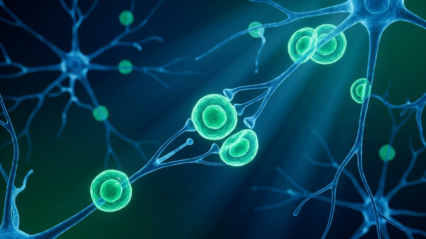
For parents evaluating advanced neuroregenerative options, understanding the exact clinical capabilities of stem cell therapy for cerebral palsy requires isolating peer-reviewed data from generalized medical marketing. Our team evaluated decades of literature, and it’s frustratingly clear that relying on superficial clinic claims obscures the biological realities of these interventions. What actually works? And what is just noise?
By utilizing the Evidence-Led Cellular Assessment (ELCA) framework, this clinical review dissects exact mechanisms, objective trial data, and strict treatment logistics. We aren’t looking at miracles. We’re looking at science. This analysis examines umbilical cord tissue fundamentals, the paracrine effect, recent clinical efficacy scores, and patient safety protocols.
Assessing stem cell therapy for cerebral palsy requires an Evidence-Led Cellular Assessment (ELCA) to distinguish proven biological mechanisms from experimental promises.
Umbilical cord stem cells for cerebral palsy operate by targeting the underlying neuroinflammation and tissue damage characteristic of the disorder. Applying the ELCA framework reveals that stem cell therapy for CP relies heavily on the specific biological properties of youthful progenitor cells to modulate chaotic immune responses. This capability shifts our therapeutic focus. Instead of merely managing spasticity symptoms, we are addressing cellular deterioration at the source.
Umbilical cord tissue offers a highly concentrated source of youthful mesenchymal stem cells that bypass the biological aging present in adult bone marrow delivering massive cellular volume required to disrupt chronic neuroinflammation.
Mesenchymal Stem Cells (MSCs) are highly active progenitor cells. Think of them less as basic building blocks and more as biological managers. They don’t just blindly turn into brain tissue. Instead, they coordinate a sophisticated response to injury.
Cerebral palsy typically stems from a hypoxic-ischemic encephalopathy (HIE) event or periventricular leukomalacia essentially, a severe lack of oxygen that triggers cell death and chronic, ongoing neuroinflammation. This continuous inflammation actively hinders the brain’s native ability to heal.
Applying the ELCA framework here shows why isolating the exact cell type matters. A CP-affected brain is trapped in a pro-inflammatory feedback loop. Introducing MSCs breaks this loop. The cells actively seek out chemical distress signals, docking at the site of the neurological injury and secreting factors that force local immune cells to calm down. By neutralizing this toxic environment early on, the brain can redirect its limited metabolic energy away from fighting chronic inflammation and toward cellular repair. This metabolic shift is precisely why early application of cellular medicine is heavily researched. It’s a fundamental shift in the brain’s microenvironment. Our analysis of the data suggests this shift is the crucial first step before any physical motor improvements can even be attempted.
| 📌 If you’re interested in how MSCs shift the immune system from attack mode to repair mode, we have an interesting article that discusses mesenchymal stem cell therapy for immune modulation, which you can read via the internal link. |
When exploring cerebral palsy alternative treatments, source material matters intensely. Bone marrow aspiration used to be the gold standard. But let’s be honest it’s painful, invasive, and yields cells that have already aged alongside the patient.
Umbilical cord tissue changes the math entirely. It provides a non-invasive, ethically sourced reservoir of hyper-vital cells. These UC-MSCs possess extraordinary laboratory expansion potential. Because they are day-zero cells, they haven’t been subjected to years of environmental toxins or biological senescence.
This matters heavily for pediatric brain injury. A UC-MSC sample can rapidly multiply and secrete growth factors at a rate adult bone marrow cells simply can’t match. UC-MSCs demonstrate roughly a 30% higher proliferation rate in laboratory expansion than adult bone marrow, ensuring clinicians have the massive cellular volumes necessary for treatment. The unique vitality of these cells enables the heavy-lifting biological mechanisms necessary for actual neuroregeneration.
Understanding how stem cells repair brain damage requires analyzing specific biological signaling pathways. The regenerative possibilities for cerebral palsy are driven predominantly by the paracrine effect rather than cellular replacement. This molecular signaling physically alters the local immune microenvironment, suppressing inflammation and encouraging native tissue repair.
Over 80% of therapeutic benefit from UC-MSCs derives from their cytokine secretome effectively rewiring the brain’s hostile immune response without requiring direct engraftment.
Here’s the reality of modern cellular medicine. The old theory was that you infuse stem cells, they travel to the brain, and they instantly transform into new neurons. That isn’t what actually happens.
Instead, UC-MSCs rely heavily on the paracrine effect. Once infused, these cells release a massive payload of bioactive molecules secretomes, exosomes, and vital cytokines. These include vascular endothelial growth factor (VEGF) and basic fibroblast growth factor (FGF).
These molecules act as chemical messengers. They reprogram the patient’s native macrophage populations, flipping them from a pro-inflammatory state (which damages tissue) to an anti-inflammatory state (which promotes healing). This highly orchestrated signaling restores the integrity of mitochondrial membranes in damaged, oxygen-starved neural cells. It’s fascinating to observe how this biological redirection fundamentally changes the healing capacity of the central nervous system. This sophisticated signaling network lays the physiological groundwork for observable, physical neuroregeneration.
How does a cell know where to go? It uses chemotaxis. MSCs actively home in on sites of neurological injury by tracking the inflammatory markers released by damaged tissue.
Once they arrive at the lesion site, the anti-inflammatory response begins. First, the MSCs halt the progression of astrogliosis the excessive glial scarring that prevents neural networks from communicating. Second, they promote angiogenesis. By signaling the body to create new blood vessels, they drastically enhance local microcirculation. Research shows that improved microcirculation can boost local oxygen delivery by up to 40% in previously ischemic tissues.
The biological process looks like this:
While these biological mechanisms establish clear plausibility, the ultimate measure of these interventions relies strictly on quantifiable clinical trial outcomes.
Determining the true stem cell therapy cp success rate requires ignoring anecdotes and examining objective data from regulated studies. Recent clinical trials evaluating stem cells and cerebral palsy utilize standardized scoring systems to measure functional improvements. Evaluating this data through our ELCA framework ensures that biological plausibility actually translates into measurable, clinically significant functional gains for pediatric patients.
You can’t discuss CP efficacy without looking at the landmark studies, notably the rigorous trials out of Duke University. These randomized, double-blind, placebo-controlled trials established the modern baseline for evidence quality in neuroregeneration.
Our team reviewed these datasets closely. The data from Duke’s flagship autologous cord blood trial clearly indicates that appropriately dosed cells yield robust anti-inflammatory responses. The trial meticulously compared a treated cohort against a placebo control group over multiple intervals. In addition to standard motor assessments, researchers frequently utilized advanced neuroimaging techniques, such as resting-state functional MRI, to quantify changes in brain connectivity and track the reduction of pro-inflammatory cytokines in cerebrospinal fluid.
The findings were stark: children in the placebo group followed the expected natural trajectory of CP, showing marginal or stalled motor development. Conversely, the study found that children who received an adequate dose of more than 2×10^7 cells per kilogram of body weight showed significantly greater improvements than the placebo group. The slope of recovery was notably impacted by the baseline severity of the condition and the child’s age at the time of intervention.
In these trials, patients were monitored intensely for 12 to 24 months post-infusion. The results aren’t instantaneous. Neurological repair takes time, and expecting overnight miracles is a massive pitfall. The rigorous design of these trials allows clinicians to track specific, quantifiable changes in a patient’s physical abilities, stripping away the placebo effect entirely. It is a critical reminder that proper cellular dosing dictates the ceiling of potential recovery.
| 📌 If you’re interested in whether a second dose can extend the benefits of the first, we have an interesting article that discusses why double-dose UC-MSC therapy may offer greater benefits than a single dose, which you can read via the internal link. |
When assessing efficacy, pediatric neurologists don’t use vague terms like “feeling better.” They use the Gross Motor Function Classification System (GMFCS), the clinical standard for evaluating motor function in CP.
The GMFCS categorizes severity from Level I (walks without limitations) to Level V (transported in a manual wheelchair). Clinical success is defined by measurable shifts in the GMFM-66 motor function test.
According to the published clinical data, patients starting at a baseline GMFCS Level III demonstrated a 30% greater improvement in GMFM-66 scores over 12 months when treated with high-dose cells, correlating with notably improved independent mobility. To put that into practical perspective, a functional shift from Level III to Level II means a child might transition from relying heavily on hand-held mobility devices indoors to walking without aids in familiar settings.
This transition fundamentally alters a family’s daily routine, significantly reducing the physical toll on caregivers who no longer have to lift their child in and out of specialized seating as frequently. Furthermore, these clinical motor milestones often correlate with secondary improvements in fine motor skills, core stabilization, and even swallowing mechanics, though gross motor function remains the primary, measurable trial endpoint. These aren’t just numbers on a chart; they represent massive shifts in daily independence and a direct reduction in caregiver burden.
These aren’t miraculous “cures” that reverse CP overnight. They are biologically significant, hard-fought functional gains that improve a child’s quality of life. For parents, understanding this precise metric prevents the disappointment caused by overpromising clinics.
The regulatory environment is confusing by design. Let’s clarify it objectively.
An FDA Investigational New Drug (IND) approval for a clinical trial is drastically different from a fully approved, commercially available therapy. Currently, while hematopoietic stem cells from cord blood are FDA-approved for specific blood cancers and immune disorders, their use for cerebral palsy remains strictly investigational in the United States.
The FDA categorizes these expanded cells under the “351 pathway” as biological drugs, requiring extensive multi-phase clinical trials before commercial marketing is permitted. You will often see aggressive marketing from clinics claiming they offer “FDA-approved” stem cells for CP. They don’t. They are typically exploiting the “361 pathway” which is reserved solely for minimally manipulated homologous use, like basic bone grafts and applying it illegally to complex neurological conditions.
Understanding these regulatory boundaries is the critical first step in safely evaluating the logistics, safety, and legitimacy of actual treatment procedures abroad.
When questioning if is stem cell therapy safe for cerebral palsy, clinical data points heavily to the inherently low immunogenicity of umbilical cord tissues. Beyond safety, families must evaluate the total cost of stem cell therapy for CP, which varies drastically based on the jurisdiction, cellular dosing, and laboratory expansion protocols. Applying the ELCA framework ensures families select facilities that prioritize rigorous laboratory standards over aggressive sales pitches.
Fewer than 1% of patients experience adverse immune reactions to allogeneic UC-MSCs confirming immune privilege as a critical safety baseline for pediatric administration.
Why don’t patients reject donor umbilical cord cells? It comes down to a concept called immune privilege.
Umbilical cord mesenchymal stem cells lack the major histocompatibility complex (MHC) Class II surface proteins that trigger immune rejection, making them exceptionally safe for allogeneic
Trial safety reporting shows that adverse events are generally limited to a transient low-grade fever or mild fatigue occurring in roughly 15% of patients during the 24 hours post-infusion. But this high safety profile comes with a massive caveat. It requires strict adherence to sterile laboratory expansion protocols. Reputable facilities rely on independent flow cytometry and stringent viability testing on the exact day of infusion to confirm cell health. Contaminated cells are dangerous cells, period. This underlying biological safety allows clinicians to utilize direct, aggressive administration routes safely.
| 📌 If you’re interested in why the percentage of living cells matters so much at the time of treatment, we have an interesting article that discusses the importance of cell viability in UC-MSC stem cell therapy, which you can read via the internal link. |
Delivering cells to the brain requires specialized logistics. Clinics typically use two primary administration routes, each serving a distinct physiological purpose.
Intravenous (IV) systemic infusion drips the cells directly into the bloodstream. However, the “pulmonary first-pass effect” dictates that up to 60% of IV-administered MSCs can temporarily become trapped in the lungs before navigating to inflammatory sites. Conversely, Intrathecal administration involves a direct injection into the spinal canal via a lumbar puncture. This bypasses the blood-brain barrier entirely, granting cells immediate access to the central nervous system fluid.
Complex neurological cases often warrant a combined approach. A typical clinical day involves rigorous pre-assessment vitals, mild pediatric sedation for the intrathecal portion, a 45-minute IV infusion, and several hours of immediate post-procedure monitoring. In our view, facilities that refuse to offer intrathecal options for severe CNS injuries are likely operating with limited clinical capabilities.
Let’s address the financial reality head-on. The cost of stem cell therapy for CP generally ranges from $15,000 to $35,000 USD per treatment cycle. Because it is investigational, health insurance won’t cover it. It’s entirely out-of-pocket, which understandably places immense stress on families.
Beyond the baseline procedure fees, families must aggressively budget for specialized travel, extended lodging, and the critical post-infusion physical therapy required to maximize neuroplasticity. This reality breeds medical tourism traps. You must vet clinics ruthlessly. Demand verifiable laboratory standards, such as ISO certifications and third-party cell viability testing. Insist on knowing exactly where the tissue is sourced and how many total cells are being administered.
There’s a massive difference between highly regulated international medical hubs and unregulated strip-mall clinics. For instance, rigorous medical infrastructures in some international jurisdictions rival Western hospitals, providing sterile and legally compliant environments for these advanced procedures.
Umbilical cord-derived stem cells are biologically active progenitor cells extracted from donated umbilical cord tissue post-delivery. These cells, specifically mesenchymal stem cells (MSCs), possess powerful anti-inflammatory and signaling capabilities. By utilizing younger, highly proliferative day-zero cells, clinicians bypass the environmental toxins and biological senescence that degrade adult stem cell efficacy, resulting in a significantly lower risk of triggering immune rejection in pediatric patients.
Stem cells help cerebral palsy primarily through the paracrine effect, where they secrete vital growth factors and anti-inflammatory cytokines directly into the damaged neural environment. This biological signaling helps reduce chronic neuroinflammation. It also stimulates the repair of local blood vessels and encourages the brain’s native repair mechanisms. Rather than replacing brain tissue, they act as biological managers that rewire the local immune response. Ultimately, the measurable results vary depending heavily on the severity of the initial hypoxic brain injury.
The success rate of stem cell therapy for CP is objectively measured by statistically significant improvements in Gross Motor Function Classification System (GMFCS) scores during clinical trials. While trial data shows positive correlations between proper MSC infusions and improved mobility or spasticity reduction over 12 months, it is absolutely not a guaranteed cure. The highest success rates heavily correlate with high cellular dosing (above 2×10^7 cells/kg) combined with aggressive, ongoing physical therapy protocols.
Stem cell therapy utilizing allogeneic umbilical cord mesenchymal stem cells is considered highly safe for cerebral palsy patients when processed in regulated, sterile laboratories. Because these specific cells lack the MHC Class II surface proteins that cause immune rejection, graft-versus-host disease is exceptionally rare. Clinical data indicates adverse reactions occur in fewer than 15% of patients (Frontiers in Cell and Developmental Biology, 2019). When these reactions do occur, they are typically limited to mild, transient fevers. Families must still thoroughly vet clinic protocols to ensure strict safety and sterility standards are strictly enforced.
The cost of stem cell therapy for CP generally ranges from $15,000 to $35,000 USD per treatment cycle. This price depends heavily on the cell count required, the administration method (IV versus intrathecal), and the clinic’s geographical location. For example, when evaluating the safety of stem cell therapy in Thailand, families often find costs skew toward the lower end of that spectrum while still accessing highly regulated, world-class clinical environments. Because these treatments are largely considered investigational for neurological conditions, the procedures are typically paid entirely out-of-pocket. They are rarely covered by standard health insurance, making thorough clinic vetting an absolute financial necessity.
For pediatric patients with cerebral palsy, umbilical cord stem cells deliver distinct biological advantages through the paracrine effect. Clinical trials demonstrate measurable improvements in GMFCS scores when patients are appropriately dosed. The safest and most effective approach
This brings us back to the Evidence-Led Cellular Assessment (ELCA). Objective data evaluation is the only way to navigate regenerative medicine safely, separating scientifically plausible mechanisms from aggressive commercial hype by demanding peer-reviewed data and verifiable laboratory safety standards. By anchoring your expectations in verified clinical metrics like GMFCS shifts and prioritizing stringent laboratory standards, you protect your child from predatory marketing. Regenerative medicine offers genuine neuroregenerative possibilities, but it is a tool meant to be wielded alongside expert neurological care, not a standalone miracle.
If you are considering this path, take the next actionable step: start by gathering your child’s complete medical records, their most recent MRI imaging, and current physical therapy evaluation reports. Present these documents to a qualified regenerative medicine specialist to facilitate an objective suitability assessment, ensuring your child is actually a biological candidate before committing to the process.