Our Services
Others
- DFPP
- Shockwave
- IV Drip


“This literature review provides a mechanistic insight into how the concentric type I collagen lamellae of the annulus fibrosus resist multidirectional torsional stresses and hoop tension from the nucleus pulposus, and how microarchitectural disruption leads to mechanical failure and discogenic low back pain in degenerative disc disease.”
Intervertebral disc degeneration is a prominent structural and biomechanical source of chronic low back pain worldwide. However, beyond nuclear dehydration and cellular phenotypic loss, which have been the focus of previous research efforts, it is also important to appreciate that the structural and functional integrity of the surrounding annulus fibrosus is critical for the mechanical stability of the spine. Microscopically, the annulus fibrosus has a complex architecture consisting of 15-25 densely packed concentric sheets of type I collagen fibers. The fibers are arranged in an angle-ply laminate with alternating layers at ±30° to 45° to the transverse axis of the disc, with an elastified interlamellar matrix reinforced by cross-strengthening bridges in the interlamellar matrix and intralamellar cross-strengthening in the annulus lamellae. This organization functions as a high-tensile pressure vessel, transforming isotropic hydrostatic pressure from the nucleus pulposus into circumferential hoop stress and providing multi-axis rotational rigidity during spinal flexion/extension and torsion. In degenerative disc disease, altered mechanical stress, micro-damage, and elevated matrix-degrading enzymes cause collagen bundles to unravel and lamellae to delaminate with fissure formation. These events weaken the structure and reduce circumferential shear and delamination resistance, permitting propagation of tears toward innervated outer annular lamellae. The torn annular regions are thus infiltrated with sensory nociceptors and vasculature, causing intractable discogenic back pain. This review of literature combines macroscale biomechanics, lamellar microarchitecture, and nanoscale fibrillar pre-strain dynamics to elucidate the mechanisms of structural failure of the annulus fibrosus that predispose to accelerated disc degeneration and pain.
Keywords:
Chronic low back pain is among the most important causes of disability and socioeconomic challenge in the current community. In most of cases, clinical and radiologic evidence corroborates the morphogenetic basis of this disabling pain as degenerative disc disease of the lumbar-motion segments. The intervertebral disc is a fibrocartilaginous structure designed to absorb loads and connect adjacent vertebrae, granting motion and support against axial, flexion, and rotation forces. The functional spinal unit is formed by the mechanical union of three anatomic structures: the central gelatinous nucleus pulposus, the superior and inferior cartilaginous endplates, and the multi-laminated peripheral ring, the annulus fibrosus(Mohd Isa, Teoh, Mohd Nor, & Mokhtar, 2022; Peng, 2025).
As discussed in the prior review on spinal disc degeneration, the primary structural failure was frequently due to endplate injury or central nutrient deprivation within the avascular disc matrix. Moreover, as highlighted above, the normal intervertebral disc swells in turgor due to a finely balanced system of hydrophilic aggrecan proteoglycans and a lattice of type II collagens toward the disc center. However, in the hardware of the structural constraint system, the role of these central pressures is lost, emerging from the isolated mechanics of a hydrogel itself. The pressurized fluid of the nucleus pulposus is not free to dissipate the axial loads transmitted by the spine; without the rigid outer boundary constraint, the fluid would simply dissipate laterally(Mohd Isa et al., 2022).
That constraint is the annulus fibrosus. The annulus fibrosus is anything but a passive fibrous casing; its exquisitely organized microarchitecture is optimized to bear high tensile loads, to restrict transverse displacement of the nucleus pulposus, and to stabilize the spine against complex multiaxial forces. Degenerative pathology is marked by the progressive destruction of the annulus microarchitecture, including interlamellar shearing, collagen unraveling, and concentric delamination, leading to impaired spinal kinematics and painful consequences. Therefore, a more detailed knowledge of the mechanical organization of the annulus fibrosus is required to better understand the origin of discogenic back pain and to design regenerative biomaterials(Chu et al., 2018; Tavakoli, Diwan, & Tipper, 2020).
The mature annulus fibrosus is a heterogeneous fibro-cartilaginous composite with specific microstructural, biochemical, and mechanical gradients that extend from the peripheral region of the annulus toward its interface with the nucleus. The annulus is formed from 15-25 concentric cylindrical sheets (lamellae) of tissue. The thickness of the lamellae is 100–500 μm; the separation of the outer lamellae is close, while the lamellae closer to the inside are thicker than lamellae on the outside(Li, Liu, Guo, Yang, & Li, 2014).
Biochemically, the annulus fibrosus is composed of 50–70% collagen (dry tissue weight), ~20% proteoglycans, 2% elastin, and 65–70% physiological water. It has a macromolecular gradient oriented radially(Guo et al., 2025). The outer annulus fibrosus is more fibrous and also has fibroblast-like cells (spindle shape, mesenchyme) rich in type I collagen. Correlating with the radial progression toward the nucleus pulposus, type I collagen concentrations decrease, giving way to an abundance of type II collagen/aggregans and rounded, chondrocyte-like cells. This biochemical shift gives rise to increasing rigidity of the tissue: the elastic modulus of single lamella (s) ranges from 59 MPa for the flexible inner zone to >136 MPa for the dense fibrillar outer zone(Chu et al., 2018).
The type I collagen fibers are arranged in an angle-ply fashion, which provides tensile strength to the tissue. Within each lamella, the collagen bundles are situated at an angle of about 30 degrees to an idealized transverse plane of the spine. In the next lamella, the fibers pedulate in the reverse direction, opposing at approximately -30 degrees, and forming an alternate cross ply design to adjacent cross sheets at approximately 60 to 120 degrees(Chu et al., 2018; Mohd Isa et al., 2022).
This plywood-like, cross-ply, dry laminated structure surrounds the central core (figure 1). These macro-collagen fibers are visible at the level of a lamella itself and are typically composed here of bundles of an average diameter between 20 and 100 nanometers within the range of 1 to 100 nanometers. The fibrils themselves consist of tropocollagen triple helices arranged with an axial periodicity called the D-period, which averages from 65 to 67 nanometers at the nanoscale. High-resolution imaging has shown that collagen fibrils in intact annular lamellae are pre-strained. This molecular pre-tension allows rapid deformation response to mechanics and prevents internal mechanical slack of the fibrous network(Newham et al., 2026).
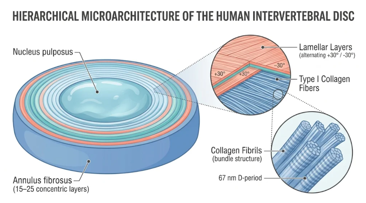
Figure 1. Hierarchy of human intervertebral disc microarchitecture. Schematic cross section of the central nucleus pulposus and surrounding annulus fibrosus lamellae. Inset: ∼30° orientation of type I collagen in lamellae and D-period (67 nm) of collagen fibrils.
The intact intervertebral disc behaves as a biphasic pressure vessel that transforms compressive axial forces into circumferential tensile stresses. Here the shape of the hydrated nucleus pulposus is preserved with its intracavitary volume maintained under axial compressive forces of apposed vertebral bodies in standing, walking, and lifting. The annular ring and cartilaginous endplates provide rigid boundaries to the swelling, creating high hydrostatic pressure within the nuclear cavity(Bhattacharjee et al., 2012).
This hydrostatic pressurization is transmitted to the inner annular ring and also disperses the cartilaginous endplates cranially and caudally. This multiaxial internal pressurization is converted by the annulus fibrosus to high circumferential tensile stress (hoop stress). Individual type I collagen bundles are well adapted to resist this stretch because of their high tensile stiffness and low compressive rigidity(Newham et al., 2026).
Hydrostatic pressure causes bulging and stretching of the alternate oblique collagen fibers in a radial direction. At low strain, individual fibrils de-furl from slightly tilted/helical configurations, exploiting their major molecular backbone. With further loading, the fibers partially reorient toward an angle of the circumferential axis to accommodate any lateral expansion of the disc(Bader et al., 2025). This transfer of stress is facilitated by radial gradients in tissue composition: the soft, proteoglycan-rich inner annulus cushions the sharp mechanical transition from hydrogel to solid tissue, and the stiff, type I collagen-rich outer lamellae support the peak tensile hoop stresses. Hence, the outer annulus is crucial to internal nuclear pressurization, as a failing outer wall will equate to mechanical decompression of the motion segment itself(Torre, Mroz, Bartelstein, Huang, & Iatridis, 2019).
The spinal disc must accommodate synkinetic lateral flexion/extension and axial rotation for spinal kinematics. Physiologic motions of the spine are not simple axial compression, and the shearing forces from dynamic multi-planar forces produce large and complex shear strains across the disc space. The alternating layers of collagen armature of the annulus are capable of resisting such forces and stabilizing the spine(Gregory, Bae, Sah, & Masuda, 2014; Mohd Isa et al., 2022).
When the spinal column rotates or twists about its axis, shear stresses act in circumferential and radial planes of the disc. The lamellae are angled in opposite directions to one another, so that during unilateral axial rotation of the spine, about half of the collagen fibers of the annulus fibrosus are taut, resisting the rotation, and the other half slack or mechanically unloaded. Reversing the twist tightens the slack fibers and slackens the other set. This alternating recruitment of fibers prevents hyper-extension of the fibers, but allows rotational stiffening in either of the mobility directions of the spine(Chu et al., 2018; Mohd Isa et al., 2022).
The lamellar sheets are not independent. The interlamellar matrix (less than 30 μm) between neighboring lamellar sheets is a specialized matrix that contains no collagen. It consists of an orthotropic network (+/- 45 degree to the principal collagen bundles) of thin and thick elastic fibers embedded in a low-fiber-lubricating ground substance rich in decorin, biglycan, lubricin, and water(Tavakoli et al., 2020).
This inter-lamellar matrix contains trans-lamellar cross-bridges (fine branching fibrous interlayer cross-bridges) (figure 2). These cross-bridges inhibit excessive adjacent sheet sliding during pivot rocking and torsional rocking(Newham et al., 2026). And the net of elastic fibers endows the lamellae with the viscoelastic rebound needed to restore the lamellae to the original place when the torsional loading is released. These stiff lamellae of type I collagen in an elastic sheath of inter-lamellar compliance bridged between the lamellae by trabeculae constitute a highly dynamic, shear-resistant composite that can withstand decades of repetitive torsion(Bader et al., 2025; Tavakoli et al., 2020).
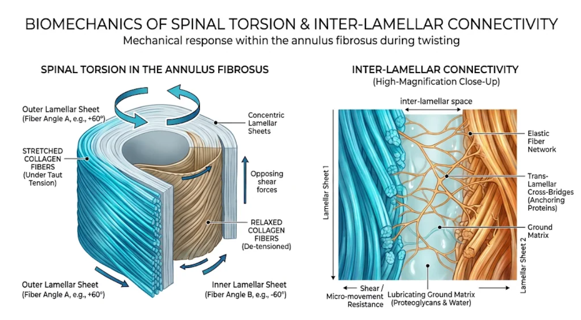
Figure 2. Torsional mechanical properties of annulus fibrosus. Macro- and micro-structural responses of the annulus fibrosus during spinal torsion, highlighting selective collagen fiber tension and inter-lamellar shear resistance.
Thus, an aberrant and mechanistically destructive catabolic cycle combine within the annulus fibrosus the mechanical and anatomic balance of DDD. Matrix ion nutrition, micro-trauma, rotation/twisting, and mechanical overload that is inappropriate modify the synthetic potential of resident disc cells. Disc cells change from an anabolic to an aggressive catabolic state in previous observations of disc degeneration when they starve and acidify the cell microenvironment(Chu et al., 2018; Peng, 2025).
Thus, the resident fibrochondrocytes in the injured annulus will normally upregulate expression/production of matrix-degrading enzymes such as matrix metalloproteinases (collagenase/MMP-1, MMP-8, MMP-13), gelatinase, and stromelysins (MMP-2, MMP-3, MMP-9, MMP-14). The processes are further enhanced by the invasion or senescence of immune cells and their production of cytokines such as interleukin-1β or tumor necrosis factor-α, which impede physiological extracellular-matrix synthesis(Cambria et al., 2020; Chang, Chen, & Huang, 2022).
Collagen synthesis becomes grossly uncoupled. Type I collagen fibrils are tightly ordered in healthy outer lamellae, whereas severe disc degeneration is characterized by aberrant gene expression, down-regulation of native organizing factors such as cartilage oligomeric matrix protein and SOX9, and localized fibrotic over-expression of unstructured collagen(Mohd Isa et al., 2022). However, the lack of physiological tensile pre-strain within native annulus predisposes to enzymatic damage by MMP-1, as shown by in vivo and in vitro experiments. Conversely, physiological tension protects annular collagen fibrils from rapid proteolytic degradation. Loss of disc height and internal hydrostatic pressurization removes this tensile state from annular lamellae and results in rapid disruption of collagen microarchitecture(Lotz, Hadi, Bratton, Reiser, & Hsieh, 2008).
Microstructural studies have revealed disorganization of fibril alignment, swollen and irregular lamellar boundaries, and loss of birefringence in degenerate fibrous annulus tissues and softer structures (by optical coherence tomography and second-harmonic generation microscopy). This is consistent with degenerative changes such as altered tissue geometry with lamellar compartment thickening, disruption of tissue-optimal fiber micromechanics, and damage of interlamellar cross-linking networks such as matrix protein-linking enzyme transglutaminase-2(Bader et al., 2025).
Microstructural unraveling of the annulus fibrosus leads to catastrophic mechanical failure of the spinal motion segment. Mechanically tested degenerate outer annular tissue has dramatically reduced simple shear moduli. Static shear moduli (radially and circumferentially) are significantly lower in symptomatic degenerate discs compared to non-degenerative controls. This loss in shear modulus inversely correlates with increased lamellar thicknesses and Pfirrmann degeneration grade. Moreover, dynamic mechanical analysis by St-Pierre et al. confirms decreases in tissue hysteresis, storage modulus, and stress relaxation, indicating the inability of degenerate annulus to prevent the attenuation of mechanical energy and dampening of rotational forces(Bader et al., 2025).
Degeneration of inter-lamellar matrix components results in massive loss of lamellar adhesion. Peel tests on animal discs demonstrate that needle puncture and matrix degradation will decrease inter-lamellar cohesiveness by as much as 30%. This perceived loss of interlamellar integrity predisposes the annular delamination or concentric sheets peeling from each other during torsion and compressive loading(Gregory et al., 2014).
Additionally, tears of the annulus fibrosus architecture (figure 3) may be divided into 3 types: (A) circumferential tears/delaminations; (B) rim tears (at the insertion to the vertebrae); and (C) radial fissures through the wall of the lamellae. -a radial fissure at the lamellar edge due to increased shear strain (susceptible to failure) over the trans-lamellar cross-bridges. Annular rim tears occur at the site of entrenchment of Sharpey’s fibers into the vertebral ring apophysis(Peng, 2025).
The most common and clinically important of these disruptions are “radial fissures,” which run horizontally (perpendicular) across successive lamellar sheets from the inner annulus (nucleus) to the outer rim. These form fissures that cavitate outward as fractious, dry core contents are expelled by the internal pressures from the nucleus, disrupting the actual ‘walls’ of the intervertebral disc(Adams & Roughley, 2006).
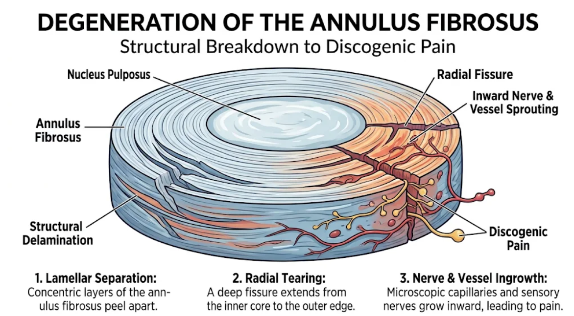
Figure 3. The pathophysiology of discogenic pain. Cross-sectional schematic of annular fibrosus degeneration. Structural delamination causes pain (discogenic), budding into splay inwards because of the formation of radial fissures (letting buds of sensory nerves and microscopic capillaries).
Translating microarchitecture in mechanics into clinical symptoms places the annulus fibrosus at the center of the pathogenesis of discogenic low back pain. The disc is essentially aneural and avascular in the normal adult spine, with sensory nerve endings and capillaries extending only to the outer 2-3 mm of the annulus fibrosis. The absence of nerves is mediated by high tissue density, compressive hydrostatic pressure, and intact extracelluar matrix components that serve as a barrier(Mohd Isa et al., 2022; Peng, 2025).
However, if the mechanical damage results in full-thickness radial disruptions fracturing of the outer annular lamellae, the local microenvironment changes profoundly. Full-thickness radial tears in the outer annulus trigger a delayed and abnormal wound healing response. Unlike vascularized tissues, annulus repair originates peripherally (on the fissure edges). The small number of disc cells dooms physiological healing, as these metabolically inactive cells cannot rapidly replace the organized bundles of type I collagen. Instead, disorganized, vascularized granulation tissue invades the fissure(Peng, 2025; Stich et al., 2020).
In these conditions of stress, disc cells produce proinflammatory cytokines, such as interleukin-1, interleukin-6, and tumor necrosis factor-α, to promote vascular endothelial growth factor and its receptor and nerve growth factor production(Guo et al., 2025). This neurotrophic signal causes the ingrowth of proprioceptive nociceptive nerve fibers into the normally aneural inner lamellae and central nucleus pulposus. These sprouted unmyelinated fibers display elevated nociceptive neuropeptides (e.g., substance P, calcitonin gene-related peptide) and markers of axonogenesis (e.g., growth-associated protein 43). Subsequently, the sensitized, peptidergic nociceptors are susceptible to mechanical stimulation by the delaminated lamellae during activities of daily living, leading to persistent discogenic low back pain. Such inflamed, vascularized annular tears frequently have focal high-intensity zones on T2-weighted magnetic resonance imaging scans, a hallmark of painful internal disc disruption(Mohd Isa et al., 2022; Peng, 2025).
The anatomical structure of the lumbar spine explains the unusual clustering of these mechanical failures in specific areas. Experimental and clinical evidence suggest that the posterior, and particularly the posterolateral, annulus fibrosus is much less peel resistant and prone to delamination than the anterior annulus. Finally, the posterior wall is thinner and experiences higher flexion and torsion and larger angular deformation of collagen fibers during axial compression. Low interlamellar adhesion and high local strain lead to structural overload at the posterior-lateral annulus, which can lead to failure, herniation, and nerve root compression(Peng, 2025).
These biomechanical observations have clinical implications for current and future therapy. Conventional clinical approaches have involved nucleus pulposus rehydration, nucleotomy, or hydrogel injection, yet clinical evidence confirms that nucleus repair is futile without effective sealing at the periphery. Yet, injected hydrogels or cell-embedded biomaterials will extrude from a delaminated still unrepaired annulus, and mechanical instability will lead to chronic herniation. Therefore, treatment of degenerative disc disease should also entail reconstructive surgery to realign transected lamellae of the annulus fibrosus to reestablish laminated tensile architecture, inter-lamellar intermolecular cohesion, and seal radial tears to abate pathological nerve root stimulation(Chu et al., 2018).
The housing of an intervertebral disc: the annulus fibrosus. Its peculiar microstructure (15-25 concentric lamellae of angle-ply type I collagen fibers with elastic network between lamellae and trans-lamellar cross-bridges) transforms the nucleus pulposus hydrostatic pressure to the circumferential hoop stress providing global multiaxial rotational stability. In degenerative disc disease, excessive mechanical loading, loss of protective pre-strain, and catabolic matrix degenerations cause the unraveling of collagen fibers, flaking of inter-lamellar delaminations, and progressive radial fissuring. Such loss of shear resistance and barrier function permits inward migration of nociceptive nerve fibers and inflammatory vessel sprouting and leads to chronic discogenic back pain. Ultimately, the prevention of spinal degeneration will require clinical strategies that maintain annular microarchitecture, restore interlamellar bond strength, and functionally restore the outer fibrous ring.
Adams, M. A., & Roughley, P. J. (2006). What is intervertebral disc degeneration, and what causes it? Spine (Phila Pa 1976), 31(18), 2151-2161. doi:10.1097/01.brs.0000231761.73859.2c
Bader, T. J., Dhiman, M., Vercio, L. L., Bouchard, J., Nicholls, F., Evaniew, N., . . . Swamy, G. (2025). Reduced Shear Modulus and Altered Lamellar Morphology of the Outer Annulus Fibrosus in Painful Intervertebral Disc Degeneration Compared With Tissue From Non-Surgical Controls. JOR Spine, 8(4), e70123. doi:10.1002/jsp2.70123
Bhattacharjee, M., Miot, S., Gorecka, A., Singha, K., Loparic, M., Dickinson, S., . . . Ghosh, S. (2012). Oriented lamellar silk fibrous scaffolds to drive cartilage matrix orientation: towards annulus fibrosus tissue engineering. Acta Biomater, 8(9), 3313-3325. doi:10.1016/j.actbio.2012.05.023
Cambria, E., Arlt, M. J. E., Wandel, S., Krupkova, O., Hitzl, W., Passini, F. S., . . . Wuertz-Kozak, K. (2020). TRPV4 Inhibition and CRISPR-Cas9 Knockout Reduce Inflammation Induced by Hyperphysiological Stretching in Human Annulus Fibrosus Cells. Cells, 9(7). doi:10.3390/cells9071736
Chang, H. I., Chen, C. N., & Huang, K. Y. (2022). Mechanical Stretch-Induced NLRP3 Inflammasome Expression on Human Annulus Fibrosus Cells Modulated by Endoplasmic Reticulum Stress. Int J Mol Sci, 23(14). doi:10.3390/ijms23147951
Chu, G., Shi, C., Wang, H., Zhang, W., Yang, H., & Li, B. (2018). Strategies for Annulus Fibrosus Regeneration: From Biological Therapies to Tissue Engineering. Front Bioeng Biotechnol, 6, 90. doi:10.3389/fbioe.2018.00090
Gregory, D. E., Bae, W. C., Sah, R. L., & Masuda, K. (2014). Disc degeneration reduces the delamination strength of the annulus fibrosus in the rabbit annular disc puncture model. Spine J, 14(7), 1265-1271. doi:10.1016/j.spinee.2013.07.489
Guo, X., Lu, Z., Xiao, W., Huang, H., Wu, J., Zou, F., . . . Jiang, J. (2025). Exploring the Causes of Intervertebral Disc Annulus Fibrosus Impairment. Cell Mol Bioeng, 18(2), 109-121. doi:10.1007/s12195-025-00844-3
Li, J., Liu, C., Guo, Q., Yang, H., & Li, B. (2014). Regional variations in the cellular, biochemical, and biomechanical characteristics of rabbit annulus fibrosus. PLoS One, 9(3), e91799. doi:10.1371/journal.pone.0091799
Lotz, J. C., Hadi, T., Bratton, C., Reiser, K. M., & Hsieh, A. H. (2008). Anulus fibrosus tension inhibits degenerative structural changes in lamellar collagen. Eur Spine J, 17(9), 1149-1159. doi:10.1007/s00586-008-0721-y
Mohd Isa, I. L., Teoh, S. L., Mohd Nor, N. H., & Mokhtar, S. A. (2022). Discogenic Low Back Pain: Anatomy, Pathophysiology and Treatments of Intervertebral Disc Degeneration. Int J Mol Sci, 24(1). doi:10.3390/ijms24010208
Newham, E., Parmenter, A. L., Chen, J., Disney, C. M., Snow, T., Brunet, J., . . . Gupta, H. S. (2026). Multimodal X-ray imaging reveals hierarchical fibre mechanics. Nature, 659(8139), 91-99. doi:10.1038/s41586-026-11148-5
Peng, B. G. (2025). Fundamentals of intervertebral disc degeneration and related discogenic pain. World J Orthop, 16(1), 102119. doi:10.5312/wjo.v16.i1.102119
Stich, S., Jagielski, M., Fleischmann, A., Meier, C., Bussmann, P., Kohl, B., . . . Sittinger, M. (2020). Degeneration of Lumbar Intervertebral Discs: Characterization of Anulus Fibrosus Tissue and Cells of Different Degeneration Grades. Int J Mol Sci, 21(6). doi:10.3390/ijms21062165
Tavakoli, J., Diwan, A. D., & Tipper, J. L. (2020). Advanced Strategies for the Regeneration of Lumbar Disc Annulus Fibrosus. Int J Mol Sci, 21(14). doi:10.3390/ijms21144889
Torre, O. M., Mroz, V., Bartelstein, M. K., Huang, A. H., & Iatridis, J. C. (2019). Annulus fibrosus cell phenotypes in homeostasis and injury: implications for regenerative strategies. Ann N Y Acad Sci, 1442(1), 61-78. doi:10.1111/nyas.13964