Our Services
Others
- DFPP
- Shockwave
- IV Drip


“This literature review investigates the biomechanical breakdown in meniscus tears, where the meniscus extrudes beyond the tibial margin due to a loss of circumferential hoop tension, neutralizing its shock-absorbing capacity and overloading knee articular cartilage.”
Knee menisci are specialized fibrocartilaginous structures that play essential roles in knee joint maintenance, load distribution, and shock absorption. These protective functions are primarily achieved by the continuous circular collagen bundles tightly attached to the tibial plateau, which mechanically convert the axial tibiofemoral compressive load into circumferential hoop tension. Structural tears, such as radial tears and posterior meniscal root tears, sever this continuous mechanical loop and remove the resistance to hoop strain. Loss of circumferential tensile containment causes the wedge-shaped meniscus to buckle under physiologic axial loads and herniate over the edge of the tibial plateau (meniscal extrusion). Clinical, biomechanical, and imaging evidence of meniscal hoop tension transmission preventing extrusion and preserving shock attenuation are synthesized in this review. Mechanobiomechanically, the contact area decreases by 30% to >50% at high peak loading, resulting in point loading of the joint surface rather than diffuse loading, thus accelerating the progressive degeneration of articular cartilage, subchondral bone marrow lesions, and the premature deformation of osteoarthritis (OA) by “point” decoupling of load transmission. Therapeutic meniscal centering to restore hoop tension can halt this self-perpetuating degenerative cascade.
Keywords: Knee biomechanics, Meniscal extrusion, Mechanotransduction, Meniscal tears, Hoop tension, Shock absorption, Posterior root tear, Contact mechanics
During normal daily living activities such as ambulating about, climbing stairs, and even squatting down, the human knee is subjected to repeated multi-axial forces of multiples of body weight(Lee, Ha, & Kim, 2014). For this joint to function properly, mechanical congruency and balance must be present between the rounded or convex femoral condyles and the relatively flat or gently sloping proximal tibial plateaus. The medial and lateral menisci, cresent-shaped fibrocartilages, course along their respective joint spaces, increasing the area of contact between the two articular cartilages and thus diminishing the surface pressure. The menisci do more than increase the distance between articular surfaces; they contribute to load and shock absorption, transmitting 40 to 70% of the load across the joint at full extension and 90% of the load at greater degrees of flexion(M. S. Kim, In, Kim, Jeong, & Sohn, 2025; Langhans, Lamba, Saris, Smith, & Krych, 2023).
The key feature of meniscal function is maintenance of internal circumferential hoop tension. Due to its wedge cross-section, the meniscus can displace radially outward in response to axial compressive joint loads. This outward excursion is typically resisted by the density and continuity of circumferential type I collagen fibers, oriented anterior to posterior, and anchored in a bony Root insertion. This juxtapositioning harnesses biaxiality, converting radially evoked expansive forces into circumferential tension, thereby generating hoop tension(Bonasia, Pellegrino, D’Amelio, Cottino, & Rossi, 2015; Fox, Bedi, & Rodeo, 2012).
However, this hoop tension fails with certain meniscal injuries, such as a complete radial tear of the meniscus or a tear of the posterior meniscal root. In the absence of hoop tension, the meniscus cannot be prevented from moving radially during physiologic load. Meniscal extrusion is the mean radial meniscal displacement measured from the margin of the tibial plateau on imaging. This “foreign” meniscal anchor may alter known knee kinematics and tibiofemoral joint load(Langhans et al., 2023; Pache et al., 2018). We discuss here the mechanical mechanisms for loss of hoop tension and complete loss of knee joint shock absorption induced by meniscal extrusion and progressive cycle of cartilage destruction and osteoarthritis(M. S. Kim et al., 2025).
Its meniscus is populated with a unique proteoglycan-rich extracellular matrix that exhibits a multilayered linear compression and can bear high weight loads. In a histologically normal meniscus, this organic solid matrix consists of 25 to 30% and 70 to 75% water, mainly (over 75%) as dense type 1 collagen. The collagen matrix is further organized into 3 zones, providing directionality to loads(Andrews et al., 2014; M. S. Kim et al., 2025):
1. Superficial Articular: Thin random weave layer of fine collagen fibrils on femoral and tibial sides provides frictionless gliding surface(Melrose, Smith, Cake, Read, & Whitelock, 2005).
2.Intermediate lamellar layer/interzonal layer: Beneath the superficial coat: the horns are generally arranged radially by the collagen bundles with inclusion of deep ones(Andrews et al., 2014).
3. Deep Central Core: Most of the mechanical support tissue of large, vertical cords of type I collagen, which form a continuous stream, arranged circumferentially around the entire arc of the meniscus. These are interspersed with radial tie fibers that bind the circumferential loops together, imparting cohesion and preventing cleavage/delamination of the tissue under longitudinal tensile strain(Langhans et al., 2023; Melrose et al., 2005).
This intricate structural framework is depicted in Figure 1, as shown in the figure where the continuous circumferential collagen loops convert axial compression into tensile containment forces. When exposed to axial compressive loads, the convex femoral condyle forces the wedge-shaped meniscus inferiorly, and this vector also creates a radial vector that tends to extrude the fibrocartilage radially out of the joint(Langhans et al., 2023). The anterior and posterior horns are anchored to the subchondral bone of the tibial plateau by a robust fibrocartilaginous root enthesis that resists radial extrusion. This radial force generates a circumferential tension, the ‘hoop tension’ of the ‘steel hoops’ of a wooden barrel. The lysyl oxidase enzymatic intermolecular cross-linking of collagen further increases the tensile modulus and eliminates the slip/breakage of collagen fibrils under high loads(Bonasia et al., 2015; M. S. Kim et al., 2025).
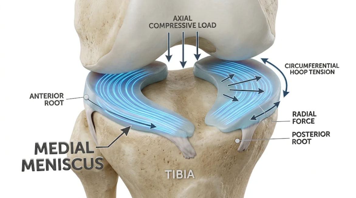
Figure 1. Biomechanics of Meniscal Hoop Tension. Conversion of axial compressive loads into circumferential hoop tension within the medial meniscus to resist radial displacement.
Structural lesions break the continuity of circumferential collagen fibers and upset the delicate biomechanical balance. Pathological extrusion of the meniscus Coronal images reveal extrusion of at least 3 mm from the margin of the tibial plateau. Normal subjects may have physiological extrusion, up to 1-2 mm and especially during dynamic weight bearing, but pathological extrusion is loss of normal internal mechanical restraint to the meniscus(Langhans et al., 2023).
Mechanisms of Failure of Hoop Tension Based on the Tear Morphology:
Posterior root tears are a final failure in hoop mechanics. In a radial or bony avulsion tear, 9-10 mm from the tibial root, most of the load across the posterior horn of the medial meniscus with axial and shear load in flexion leads to complete dissociation of the circumferential hoop from its bony insertion. The tension within the circumferential hoop is lost completely, and the mechanical hoop is abrogated(Lee et al., 2014). Additional weight-bearing pulls the meniscus from the joint line. With chronic physiologic loading in root tear, the gap from the tear enlarges and there is significant meniscal extrusion (>4 to 5 mm)(Langhans et al., 2023).
Radial tears from 50% to 90% of the width of the meniscus cross the circumferential collagen fibers of the body. Radial transverse tears divide the primary load-bearing fibers in half, unlike longitudinal tears that run parallel to collagen bundles. The resistance to hoop strain drastically decreases when the tear depth exceeds 60% of the width, enabling the unanchored tissue halves to spread apart and extrude under axial loads(Walker & Erkman, 1975).
Recent literature highlights that meniscal extrusion is not driven solely by root tears. The meniscal (coronary) ligament anchors the external base of the meniscus to the tibial plateau line. Recent magnetic resonance/cadaveric studies suggest that rupture/chronic stretch of the meniscotibial ligament may occur as an accompaniment to or even in advance of posterior root failure. Without fixation to the coronary ligament, the meniscus extrudes medially, overloading the posterior root beyond normal limits until failure occurs with normal activities(Kelly, Daniel, Smith, & Krych, 2026).
Figure 2. summarizes the spectrum of failure: unopposed division of the circumferential fibers in a posterior root tear allows unopposed extrusion of the meniscal body. These axial loading forces are translated laterally off the surface of the meniscal body in the absence of root anchoring(Langhans et al., 2023).
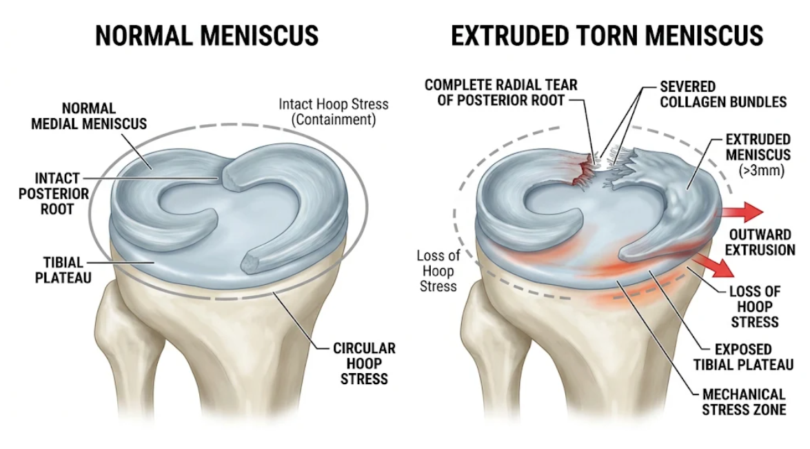
Figure 2. Normal vs. Extruded Meniscus Biomechanics. (Left) Normal medial meniscus preserves circular hoop stress. (Right) Nonfunctional complete radial tear of posterior root with loss of hoop stress, significant (>3 mm) extrusion, and excess mechanics on bare tibial plateau portion.
The shocked-off should remove the hoop tension and no menisci extrusion, so the damper and shock absorber characters of the knee become nonfunctional. Healthy human menisci dissipate mechanical shock through several physiological mechanisms(Lee et al., 2014):
• Viscoelastic Fiber Deformation: The collagen network shows dynamic elongation under load, dissipating kinetic energy(Petersen & Tillmann, 1998).
• Fluid Pressurization and Drag: The compressive forces expel fluid from the low permeable matrix of proteoglycans. Hydraulic pressure and viscous drag of interstitial flow dissipate the energy of impact during heel strike and running(Petersen & Tillmann, 1998).
Again, a loss of the meniscus is accompanied by a reduction in the shock absorption capacity of the knee by 20% to 68%. Extrusion of the meniscus from the tibial rim displaces it from the primary weight-bearing axis. Such extrusion of the wedge-shaped shock absorber catastrophically deranges the joint contact mechanics(M. S. Kim et al., 2025; Lee et al., 2014):
Normal medial menisci would be covering 50% to 75% of the medial plateau and offer broad tibial-femoral osseous contact surface. Biomechanical models of force across cadaveric tibial plateaus in the extruded meniscal state show a decrease in femoral condyle contact area by 30 to 50% due to extruded menisci. In the >4 mm extruded medial meniscus knee, at flexion angles of interest, contact areas of the femoral condyle relative to nominal tibial plateau areas have shown a significant reduction in contact.(Makiev et al., 2022).
Localization of Compressive Forces into a Small Articular Footprint Decrease in Contact Area This loss of hoop tension manifests as an associated increase in maximum contact stresses of 25% to >100% in root tears in both cadaveric and finite-element studies, with focal contact stresses 3 to 5 times native. The meniscus is biomechanically incompetent and the contact stress profile is identical to that of full meniscectomy(Lee et al., 2014).
Meniscal extrusion increases tissue loads and redistributes tensile joint loading. These redistributions shift the posteromedial and posterocentral peak pressures from dynamic gait simulations to unconditioned cartilage tissues of the tibial plateau to withstand compressive and shear strain. Subjecting these unconditioned cartilage regions to mechanical overload(M. S. Kim et al., 2025).
Figure 3. Altered tibiofemoral contact mechanics: wide contact/low pressure in the intact knee vs. reduced contact area/severe peak localized stress caused by meniscal extrusion.
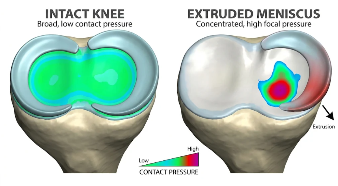
Figure 3. Tibial Contact Pressure due to Meniscal Extrusion. Tibial Contact Pressure Effect of Meniscal Extrusion. Distribution of contact pressure in 3D biomechanical heat-map. Low loading areas with broad distribution in an intact knee (left) vs. selective high focal elevation of pressure on tibial plateau with meniscal extrusion (right).
The extruded meniscus is left behind, and the loss of hoop stress results in a degenerative cascade in the compartment:
1. Delamination and fibrillation of cartilage: The hyaline cartilage cannot withstand the columnar compressive load due to excessive focal loading. It leads to the destruction of chondrocytes and rupture of the superficial proteoglycan-collagen meshwork, thereby speaking to articular cartilage dose to wear and tear and reduction of the joint space (Langhans et al., 2023).
2. Overload in BML and subchondral bone: In the absence of the meniscus, shock waves are transmitted to the subchondral bone plate (which is unable to dissipate kinetic energy). This chronic mechanical overload results in microtumors, which are typically visible on an MRI as the edema of the medullar cave and the cysts of the deep bone(Langhans et al., 2023).
3. Spontaneous osteonecrosis and insufficiency fractures: Trabecular microfractures due to overloaded subchondral bone. Spontaneous Osteonecrosis of the Knee (SONK or SIFK): This is not so much vascular avascular necrosis but a fracture through mechanical overload since up to 80% of SONK patients have a posterior root tear of the meniscus with meniscal extrusion(Langhans et al., 2023).
4. Marginal osteophytosis and mechanical malalignment: The proximal rim of tibia develops the marginal osteophytes to increase the load-bearing area to stabilize the mechanical maladjustment developed due to the change by blocking the stress exerted. In longitudinal cohort studies, degree of meniscal extrusion has been shown to be associated with osteophyte size and radiographic progression of OA (Kellgren-Lawrence grade). In addition, medial joint space narrowing results in progressive varus deformity and lateral displacement of the mechanical line of force, thereby encouraging further extrusion by a continuing downward spiral(D. H. Kim, Lee, Kim, & Cha, 2020; Langhans et al., 2023).
To summarize, as noted by Makiev et al. 2022, the literature states that meniscal extrusion should be seen as a visual sign of monumental structural failure and not a benign incidental finding in imaging(Makiev et al., 2022). Only simple meniscectomy or debridement has been considered in terms of meniscal tear morphology. However, in longer-look biomechanical studies, cellulitis root tear resective debridement resulted in the restoration of hoop tension and the failure to reduce contact pressures but accelerated TKA (total knee arthroplasty)(Ebata et al., 2025; Lee et al., 2014).
Preservation of the joint begins with restoring the anatomic mechanistic capacity of the meniscus (see following). Biomechanical studies have demonstrated that anatomic transtibial repair of the posterior root pattern tear restores normal tibiofemoral pressures and circumferential hoop stresses. This is also a major clinical paradox, as even patients who undergo anatomic posterior root reconstruction can have a significant proportion of progressively evolved meniscal extrusions (into 60% in some series) on MRI despite complete healing to bone(Langhans et al., 2023; Lee et al., 2014).
This clinical finding is frequently indicative of insufficient hoop tensioning to compress the chronically displaced and contracted meniscal body across the tibial plateau. The subluxation of the meniscal rim over the articular margin is allowed by the rupture and stretching of the peripheral restraints (meniscotibial ligament)(Kelly et al., 2026). Therefore, surgical approaches to the joint have focused on combined reparative techniques. At present, the techniques for meniscal centralization utilize the extrusional fixation of the extruded meniscal body to the rim of the tibial plateau with the use of suture anchors. Initial studies on cadaveric and clinical practice show that meniscal centralization with root repair significantly reduces extrusion, restores contact mechanics, and attenuates the onset of osteoarthritis compared with root repair alone. Finally, high tibia osteotomy for limb malalignment (E-Variable) remains an important consideration in patients with pronounced associated Varus malalignment resulting in increased contact load in the medial compartment.(Ebata et al., 2025; Langhans et al., 2023; Makiev et al., 2022).
Meniscal extrusion: a key clinical and biomechanical indicator of compromised hoop tension in meniscal tears. Posterior root tears, large radial tears, or meniscotibial ligament rupture disrupt the continuity of the circumferential collagen and prevent the meniscus from resisting axial compression by compression tension. Both radial and root tears lead to extrusion of tissue radially from the weight-bearing corridor, resulting in loss of this biologic shock absorber of the knee. This results in up to 50% reductions in tibiofemoral contact area and multiple increases in local peak contact stresses, provoking deleterious point-loading similar to total meniscectomy. This mechanical failure induces cartilage fibrillation, subchondral bone marrow edema, insufficiency fractures, and rapidly progressive osteoarthritis. Today’s clinical management should emphasize therapies aimed at arresting the destructive biomechanical cascade and preserving the native knee joint by restoring hoop tension continuity and centralizing the meniscus anatomically.
Andrews, S. H., Rattner, J. B., Abusara, Z., Adesida, A., Shrive, N. G., & Ronsky, J. L. (2014). Tie-fibre structure and organization in the knee menisci. J Anat, 224(5), 531-537. doi:10.1111/joa.12170
Bonasia, D. E., Pellegrino, P., D’Amelio, A., Cottino, U., & Rossi, R. (2015). Meniscal Root Tear Repair: Why, When and How? Orthop Rev (Pavia), 7(2), 5792. doi:10.4081/or.2015.5792
Ebata, T., Iwasaki, K., Sato, T., Hosokawa, Y., Hamasaki, M., Sato, D., . . . Iwasaki, N. (2025). The Relationship of Meniscal Extrusion to Subchondral Bone Density in Medial Knee Osteoarthritis. Orthop J Sports Med, 13(8), 23259671251366424. doi:10.1177/23259671251366424
Fox, A. J., Bedi, A., & Rodeo, S. A. (2012). The basic science of human knee menisci: structure, composition, and function. Sports Health, 4(4), 340-351. doi:10.1177/1941738111429419
Kelly, J. J., Daniel, A. V., Smith, P. A., & Krych, A. J. (2026). Meniscal Extrusion: A Key Modifiable Driver of Meniscal and Cartilage Degeneration. Curr Rev Musculoskelet Med, 19(1), 16. doi:10.1007/s12178-026-10006-1
Kim, D. H., Lee, G. C., Kim, H. H., & Cha, D. H. (2020). Correlation between meniscal extrusion and symptom duration, alignment, and arthritic changes in medial meniscus posterior root tear: research article. Knee Surg Relat Res, 32(1), 2. doi:10.1186/s43019-019-0019-x
Kim, M. S., In, Y., Kim, H., Jeong, J., & Sohn, S. (2025). Why Hoop Tension Matters: A Biomechanical Perspective on Medial Meniscus Posterior Root Tears-A Narrative Review. Bioengineering (Basel), 12(6). doi:10.3390/bioengineering12060638
Langhans, M. T., Lamba, A., Saris, D. B. F., Smith, P., & Krych, A. J. (2023). Meniscal Extrusion: Diagnosis, Etiology, and Treatment Options. Curr Rev Musculoskelet Med, 16(7), 316-327. doi:10.1007/s12178-023-09840-4
Lee, D. W., Ha, J. K., & Kim, J. G. (2014). Medial meniscus posterior root tear: a comprehensive review. Knee Surg Relat Res, 26(3), 125-134. doi:10.5792/ksrr.2014.26.3.125
Makiev, K. G., Vasios, I. S., Georgoulas, P., Tilkeridis, K., Drosos, G., & Ververidis, A. (2022). Clinical significance and management of meniscal extrusion in different knee pathologies: a comprehensive review of the literature and treatment algorithm. Knee Surg Relat Res, 34(1), 35. doi:10.1186/s43019-022-00163-1
Melrose, J., Smith, S., Cake, M., Read, R., & Whitelock, J. (2005). Comparative spatial and temporal localisation of perlecan, aggrecan and type I, II and IV collagen in the ovine meniscus: an ageing study. Histochem Cell Biol, 124(3-4), 225-235. doi:10.1007/s00418-005-0005-0
Pache, S., Aman, Z. S., Kennedy, M., Nakama, G. Y., Moatshe, G., Ziegler, C., & LaPrade, R. F. (2018). Meniscal Root Tears: Current Concepts Review. Arch Bone Jt Surg, 6(4), 250-259.
Petersen, W., & Tillmann, B. (1998). Collagenous fibril texture of the human knee joint menisci. Anat Embryol (Berl), 197(4), 317-324. doi:10.1007/s004290050141
Walker, P. S., & Erkman, M. J. (1975). The role of the menisci in force transmission across the knee. Clin Orthop Relat Res(109), 184-192. doi:10.1097/00003086-197506000-00027