Our Services
Others
- DFPP
- Shockwave
- IV Drip


“This literature review discusses the pathophysiological transition from compensated to decompensated liver cirrhosis. The review focuses on the breakdown of the microvascular architecture, the loss of metabolic functional reserve, mitochondrial energy collapse and the systemic detoxification failure.”
Liver cirrhosis is the end-stage of chronic liver injury characterized by diffuse parenchymal reorganization, fibrotic septa and regenerative nodule formation. Clinically, cirrhosis progresses through a dynamic spectrum, which is subdivided into two distinct functional phases: compensated liver cirrhosis and decompensated liver cirrhosis. Compensated cirrhosis is generally asymptomatic owing to the remarkable functional reserve of the hepatic parenchyma and compensatory microvascular remodeling. However, continued cellular injury, architectural disruption and persistent systemic inflammation eventually exceed this compensatory capacity resulting in frank clinical decompensation with the development of ascites, variceal bleeding, hepatic encephalopathy and jaundice. This transition represents a major prognostic watershed with a dramatic decline in median survival from more than twelve years in compensated disease to less than two years after the initial decompensating event. This review of the literature combines current pathophysiological concepts of the functional transition from compensated to decompensated liver cirrhosis. In particular, this review emphasizes the structural disruption of liver microcirculation, the progressive increase in intrahepatic vascular resistance, the shift from aerobic mitochondrial oxidative phosphorylation to extramitochondrial glycolysis, and the profound disruption of hepatic metabolic homeostasis and systemic detoxification. Furthermore, this review discusses the aggravation of metabolic toxicity by systemic inflammation, gut-liver axis dysbiosis and muscle wasting, and emerging evidence on possible hepatic recompensation after etiological control.
Chronic liver disease and cirrhosis are major causes of morbidity and mortality globally, with global epidemiological estimates indicating that they result in over 1 million deaths per year(Lee & Saffo, 2023). The major causes of chronic liver injury are alcohol related liver disease, chronic viral hepatitis infection, and metabolic dysfunction-associated steatotic liver disease. Regardless of the etiology, chronic necroinflammation triggers a continuous wound-healing process characterized by excess production of extracellular matrix, activation of hepatic stellate cells, and disorganization of the parenchymal architecture(Perez, Bolte, Bigelow, Dickson, & Shah, 2021).
Traditionally, cirrhosis has been viewed as a static, irreversible end-stage disease. However in contemporary hepatology, cirrhosis is seen as a highly dynamic spectrum modulated by the pathophysiologic interplay of hepatic vascular resistance, metabolic reserve, organ crosstalk and systemic immune activation(Garcia-Tsao, Friedman, Iredale, & Pinzani, 2010). The most relevant prognostic milestone of this clinical continuum is the difference between compensated liver cirrhosis and decompensated liver cirrhosis(Kumar, Kumar, & Prakash, 2023).
In compensated cirrhosis there is histological scarring but preservation of essential synthetic, metabolic and clearance functions. The high regenerative potential and functional redundancy of the hepatic parenchyma preserve systemic homeostasis at early stages of the disease(Perez et al., 2021). Structural microvascular changes are initially compensated for by intrinsic neurohumoral mechanisms. It is estimated that 5-7% of patients with compensated cirrhosis develop decompensated cirrhosis per year. At this critical inflection point, physiologic compensation is not sufficient to compensate fully resulting in obvious clinical complications such as ascites, variceal bleeding, hepatic encephalopathy and acute kidney injury(Perez et al., 2021).
The exact pathophysiological mechanisms that regulate this transition need to be understood. Decompensated state is not a simple progression of fibrosis but a systemic syndrome of cardiovascular dysfunction, severe metabolic failure, loss of detoxification capacity and widespread systemic inflammation(Engelmann, Clària, Szabo, Bosch, & Bernardi, 2021; Garcia-Tsao et al., 2010). This review offers an in-depth synthesis of the cellular, hemodynamic, metabolic and biochemical mechanisms involved in the loss of hepatic compensation with emphasis on the central role of metabolic and clearance failure in the clinical course of decompensated liver cirrhosis.
The transition from compensated to decompensated cirrhosis is due to the progressive loss of functional parenchymal mass and the failure of homeostatic adaptations. In early compensated cirrhosis the liver maintains systemic metabolic stability by hepatocyte hyperplasia and compensatory hyper-perfusion of surviving hepatic lobules(Perez et al., 2021; Wang, Yi, Li, Xu, & Tang, 2025). However, progressive parenchymal extinction ensues due to continued injury where microvascular thrombosis and ischemia result in localized parenchymal collapse, replacement by fibrous tissue, and loss of functional hepatocytes(Engelmann et al., 2021).
The transition revolves around the evolution of intrahepatic vascular resistance. Intrahepatic resistance is comprised of two major parts: a fixed structural component and a dynamic functional component. The structural component is due to collagen deposition in the Space of Disse, sinusoidal capillarization, regenerative nodular compression and vascular occlusion(Garcia-Tsao et al., 2010; Iwakiri & Groszmann, 2007). The dynamic component is due to active contraction of activated hepatic stellate cells and vascular smooth muscle cells surrounding sinusoids mediated by imbalance of reduced intrahepatic vasodilators mainly nitric oxide and increased intrahepatic vasoconstrictors like endothelin-1, norepinephrine, and thromboxane A2(Kumar et al., 2023).
The rising intrahepatic resistance results in clinically relevant portal hypertension, which is generally defined as a hepatic venous pressure gradient >10 mmHg(Figure 1)(Engelmann et al., 2021). Clinically significant portal hypertension is the main pathophysiological mechanism of decompensation. When the pressure gradient rises above this physiologic threshold, portosystemic collateral vessels form and shunt portal blood flow away from the functional hepatic parenchyma into the systemic circulation(Romero-Gómez et al., 2004). Portosystemic shunting reduces portal pressure locally, but at the cost of severely impeding the liver’s ability to deal with the nutrient-rich and toxin-rich blood from the gastrointestinal tract. Portosystemic shunting thus accelerates the systemic detoxification failure and metabolic chaos(Garcia-Tsao et al., 2010).
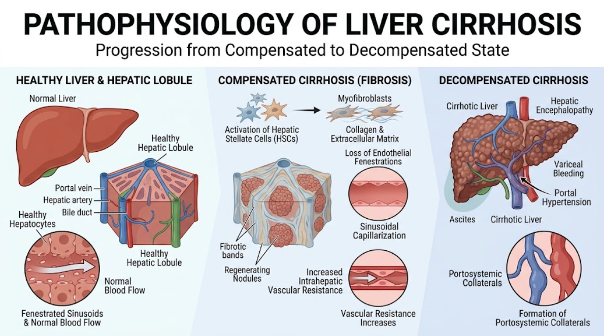
Figure 1. Pathophysiological progression of liver cirrhosis from a compensated to a decompensated state. Scheme of the development from normal liver and hepatic lobule to compensated and decompensated cirrhosis. The transition (left to right) illustrates key pathophysiological mechanisms including activation of hepatic stellate cells (HSCs) into myofibroblasts, collagen deposition and sinusoidal capillarization with loss of endothelial fenestrations (middle). These changes result in increased intrahepatic vascular resistance and fibrotic band formation leading to decompensated cirrhosis (right) with portal hypertension, ascites, hepatic encephalopathy and portosystemic collaterals.
The structural remodeling of hepatic microcirculation affects the delicate transport interface between sinusoidal blood flow and hepatocyte microvilli. In normal liver tissue, the endothelial cells of the sinusoids are fenestrated, allowing rapid exchange of proteins, metabolites and toxins(Cerini et al., 2016). Sinusoidal capillarization occurs in cirrhosis, in which endothelial cells lose fenestrations, develop a continuous basement membrane, and are surrounded by dense collagen matrices in the Space of Disse. This barrier represents a serious hindrance to passive molecular transport, which limits the access of oxygen and essential substrates to hepatocytes and the exit of metabolic clearance products into the bloodstream(Moreau et al., 2020).
At the same time, chronic portal hypertension modifies extrahepatic cardiovascular hemodynamics. Clinically relevant portal hypertension leads to increased production of endothelial nitric oxide synthase and vascular endothelial growth factor in the splanchnic arterial bed. Splanchnic vasodilation results in severe arterial hypotension and a reduction in effective arterial blood volume(Garcia-Tsao et al., 2010). In compensated cirrhosis the body compensates for splanchnic vasodilation by activation of compensatory systemic neurohumoral pathways such as the renin-angiotensin-aldosterone system, the sympathetic nervous system and non-osmotic vasopressin release. These systemic reflexes preserve arterial blood pressure and renal perfusion in the early stages of the disease(Garcia-Tsao et al., 2010).
As liver disease progresses to decompensation, however, splanchnic pooling becomes so severe that compensatory vasoconstrictor mechanisms are unable to maintain effective blood volume. The hyperdynamic circulatory state follows with increased heart rate and increased cardiac output(Nazar et al., 2013). Eventually cirrhotic cardiomyopathy develops reducing cardiac response to stress. This is the result of a combination of unopposed arterial vasodilation, cardiac exhaustion and systemic microvascular constriction leading to systemic organ hypoperfusion, renal dysfunction and fluid retention (ascites, hepatorenal syndrome)(Garcia-Tsao et al., 2010).
Cellularly, the transition to decompensated cirrhosis is marked by a global collapse of hepatic energy metabolism. The healthy liver is a metabolic hub of the body, orchestrating carbohydrate storage, gluconeogenesis, lipid β-oxidation, protein synthesis, and oxidative phosphorylation(Moreau et al., 2020). Decompensated liver disease is marked by acute and chronic parenchymal injury with profound mitochondrial dysfunction and failure to maintain systemic energy homeostasis(Regev et al., 2002).
In normal liver tissue, cellular adenosine triphosphate is mainly produced by mitochondrial oxidative phosphorylation involving the electron transport chain. Chronic exposure to reactive oxygen species, damage associated molecular patterns and pro-inflammatory cytokines in decompensated cirrhosis culminates in structural damage of mitochondrial membranes and respiratory chain complexes(Engelmann et al., 2021). The impaired oxidative phosphorylation leads to a dramatic decrease in cellular ATP production, forcing hepatocytes and extrahepatic tissues to undergo a metabolic shift to extramitochondrial glycolysis. This metabolic re-routing is highly inefficient and gives far less ATP per glucose molecule, similar to the Warburg effect in neoplastic tissues(Engelmann et al., 2021).
In an effort to meet the enormous systemic energy requirements created by ongoing systemic inflammation and immune cell activation, the body enters a profound hypercatabolic state as shown in figure 2. The liver and the extrahepatic tissues stimulate the peripheral lipolysis in the adipose tissue and proteolysis in the skeletal muscle. Accelerated proteolysis leads to severe muscle wasting and sarcopenia, which is an independent risk factor for clinical decompensation and mortality. Sarcopenia is not only a sign of malnutrition but also directly contributes to the acceleration of metabolic failure(Del Cioppo, Faccioli, & Ridola, 2025). The skeletal muscle is an important alternative organ for temporary ammonia detoxification by the glutamine synthetase. Therefore, loss of muscle mass per se aggravates systemic hyperammonemia and accelerates the transition to overt organ dysfunction(Del Cioppo et al., 2025; Engelmann et al., 2021).
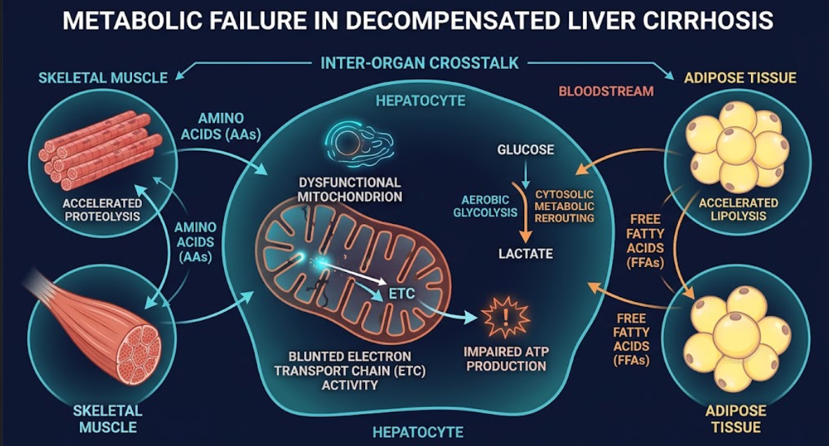
Figure 2. Metabolic Failure in Decompensated Liver Cirrhosis. Diagrammatic representation of inter-organ crosstalk and cellular metabolic dysregulation. Hepatocyte mitochondria dysfunction leads to blunted electron transport chain (ETC) activity, impaired ATP production and cytosolic metabolic rerouting to aerobic glycolysis. Concurrently, other tissues show a compensatory increase in proteolysis in skeletal muscle and lipolysis in adipose tissue.
In addition, hepatic synthetic capacity collapses in decompensation. Production of essential plasma proteins, including albumin, non-antithrombin coagulation factors and transport proteins, decreases sharply. Albumin is not just a regulator of plasma oncotic pressure but has important anti-oxidant, anti-inflammatory and toxin binding properties(Ginés et al., 1998). The serum total albumin level falls in decompensated cirrhosis and the albumin that remains in circulation is heavily damaged by oxidative post-translational modification. This loss of functional albumin results in defective intravascular binding and neutralization of pro-inflammatory lipids, endogenous toxins and circulating pathogen products, which increases systemic toxicity and tissue injury(Engelmann et al., 2021).
The liver is an essential functional cornerstone in the removal and detoxification of endogenous waste products and gut-derived toxins. The evolution pathophysiological to decompensated cirrhosis is characterized by the global failure of hepatic clearance pathways. This failure is due to two synergistic abnormalities; loss of functional hepatocyte enzymatic machinery and massive portosystemic shunting which bypasses the liver altogether(Garcia-Tsao et al., 2010).
Ammonia clearance is the classic paradigm of failure of hepatic detoxification. In healthy states, ammonia generated from the metabolism of intestinal bacteria and systemic amino acid catabolism is detoxified by periportal hepatocytes through the urea cycle and by perivenous hepatocytes through glutamine synthesis(Schuppan & Afdhal, 2008). In decompensated cirrhosis, the activity of the urea cycle enzymes is markedly reduced because of the loss of hepatocytes and structural capillary barriers. At the same time, portal hypertension causes portosystemic shunting, allowing unfiltered intestinal ammonia to enter directly into the systemic arterial circulation(Garcia-Tsao et al., 2010; Weber, Friedman, & Fresard, 1988).
As shown in figure 3, increased circulating ammonia passes the blood-brain barrier and enters cerebral astrocytes. Astrocytes convert ammonia to glutamine via glutamine synthetase. Excess glutamine accumulation within the cell induces an osmotic gradient, which attracts water to astrocytes and results in low-grade cerebral edema, astrocyte swelling, oxidative stress, and neuroinflammation. This cascade clinically manifests as hepatic encephalopathy (HE) ranging from mild cognitive impairment and asterixis to somnolence and deep coma(Engelmann et al., 2021; Garcia-Tsao et al., 2010).
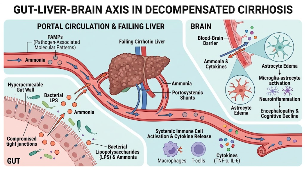
Figure 3. The Gut-Liver-Brain Axis in Decompensated Cirrhosis. Schematic representation of the pathophysiology of hepatic encephalopathy Intestinal permeability and portosystemic shunting increase, allowing ammonia and bacterial endotoxins to bypass the failing liver. These circulating toxins and systemic cytokines traverse the blood brain barrier to induce astrocyte edema, microglia-astrocyte activation, and subsequent neuroinflammation, resulting in cognitive decline.
Detoxification failure is much more than nitrogenous waste products. The cirrhotic liver fails to clear bilirubin, endogenously produced hormones, eicosanoids and circulating microvascular vasoactive substances(D’Amico, Garcia-Tsao, & Pagliaro, 2006). Clinical evidence of accumulation of bilirubin includes scleral icterus and overt jaundice, suggestive of severe hepatic excretory dysfunction. Furthermore, impaired lipid clearance leads to the accumulation of toxic acylcarnitines, free fatty acids and altered sphingolipids, which fuels systemic inflammation, mitochondrial uncoupling and endothelial damage(Engelmann et al., 2021).
Adding to the failure of detoxification is gut-liver axis dysbiosis and immune dysfunction associated with cirrhosis. Intestinal hyperpermeability, also called a leaky gut, in advanced cirrhosis portal hypertensive enteropathy is caused by mucosal ischemia and altered tight junction proteins(Kumar et al., 2023). Intestinal bacterial overgrowth and dysbiosis lead to widespread translocation of viable bacteria, bacterial fragments and pathogen-associated molecular patterns such as lipopolysaccharide, flagellin and bacterial DNA into the portal and systemic circulations(Betrapally, Gillevet, & Bajaj, 2017; Shao et al., 2018).
At the same time, the cirrhotic liver is associated with profound immune dysfunction, with a paradoxical combination of systemic immune activation and functional immune paralysis(Kumar et al., 2023). Translocated pathogen-associated molecular patterns and endogenously released damage-associated molecular patterns bind to Toll-like receptors located on Kupffer cells, circulating monocytes, and neutrophils. Binding induces activation of nuclear factor kappa B (NF- κB) and inflammasome pathways leading to massive release of pro-inflammatory cytokines including tumor necrosis factor-alpha (TNF-α), interleukin-1β, interleukin-6 and interleukin-8(Bukong et al., 2018). This ongoing systemic inflammatory cascade leads to systemic oxidative stress, endothelial dysfunction and generalized tissue injury. This leads the patient to progress from compensated cirrhosis to acute decompensation or acute-on-chronic liver failure(Garcia-Tsao et al., 2010).
For decades, the transition from compensated to decompensated cirrhosis was viewed as a one-way street, an irreversible pathway to liver transplantation or death(Kumar et al., 2023). However, the concept of hepatic recompensation has been introduced by clinical data and consensus statements, particularly the Baveno VII consensus. Recompensation has been defined as the sustained partial or complete reversal of structural and functional abnormalities of cirrhosis after effective and long-term elimination of the primary underlying etiology(Wang et al., 2025).
Clinical studies have shown that successful etiological control (eg, sustained viral suppression in chronic hepatitis B, sustained virological response in hepatitis C, or durable alcohol abstinence in alcohol-related cirrhosis) can halt ongoing necroinflammation and reverse hepatic functional reserve(Wang et al., 2025). Formal recompensation per the Baveno VII consensus criteria requires the presence of three core criteria for at least 12 months: (1) sustained suppression or full clearance of the primary etiology; (2) sustained resolution of clinical ascites (off diuretics), encephalopathy (off lactulose/rifaximin), and no variceal rebleeding; and (3) stable, marked improvement in synthetic liver function markers such as serum albumin, bilirubin, and prothrombin time/INR(de Franchis, Bosch, Garcia-Tsao, Reiberger, & Ripoll, 2022).
At cellular and histological levels, recompensation is mediated by three interconnected processes of hepatic tissue repair: active degradation of extracellular matrix by matrix metalloproteinases, vigorous regeneration of hepatocytes and reconstitution of functional architecture of liver lobules(Wang et al., 2025). In the absence of persistent necroinflammation, collagen degradation outpaces fibrogenesis, thick acellular scarring thins, and intrahepatic microvascular tone decreases(Kumar et al., 2023). Better translobular perfusion leads to a reduction of portal pressure and allows repopulation of collapsed areas by new hepatocytes, which partially restores urea cycle capacity, mitochondrial oxidative phosphorylation and systemic clearance mechanisms(Wang et al., 2025).
While the potential for recompensation is promising, clinical studies point out the definite physiological limits of structural reversal. In advanced stages of extensive vascular occlusion, severe parenchymal extinction and dense acellular collagen cross-linking, structural basement scaffold is lost irreversibly(Perez et al., 2021). In such cases, the regeneration of the hepatocyte results in isolated unorganized nodules which continue to compress the surrounding venous outlets leading to perpetuation of portal hypertension despite the control of the etiology. Furthermore, clinically significant portal hypertension and risk of hepatocellular carcinoma might persist even after clinical recompensation, requiring lifelong hepatological surveillance and guidance-directed therapy(Ferrarese et al., 2026; Sharma & Roy, 2023).
The literature synthesis presented in this review highlights a profound paradigm change in hepatology: cirrhosis is not a uniform static stage but a highly dynamic biological spectrum with complex metabolic, cardiovascular and immunological interactions. The old paradigm was almost exclusively mechanical portal pressure and gross histological scar tissue(Garcia-Tsao et al., 2010). While portal hypertension remains the underlying hemodynamic driver of portosystemic collateral formation and fluid retention, novel data suggests that metabolic failure, mitochondrial failure and systemic inflammation are important equal drivers of clinical decompensation and multi-organ failure(Regev et al., 2002).
The tipping point from compensated to decompensated disease is systemic(Perez et al., 2021). In compensated phase, the body is in a functional equilibrium. Hepatocyte reserve compensates synthetic demands, neurohumoral reflexes support arterial blood pressure, and skeletal muscle counteracts impaired hepatic clearance(Engelmann et al., 2021). Decompensation is when these inter-organ compensatory mechanisms are overwhelmed at the same time. The decline in mitochondrial oxidative activity forces the tissues into anaerobic glycolysis. Systemic catabolism destroys muscle mass needed for alternative ammonia clearance. Gut-derived bacterial translocation triggers systemic inflammatory cascades that paralyze immune function and induce microvascular shock(Engelmann et al., 2021).
Decompensation as a multisystem metabolic and inflammatory syndrome is an important clue for therapeutic intervention(Lee & Saffo, 2023). The old way of managing was by treating the symptoms, with diuretics for ascites, lactulose for encephalopathy and endoscopic banding for varices. These measures are still important, but new disease-modifying strategies are aimed at preserving or restoring the compensatory threshold. Non-selective β blockers reduce portal inflow and attenuate bacterial translocation and systemic inflammation(Iwakiri & Groszmann, 2007). Emerging pharmacotherapies such as statins for improving sinusoidal endothelial function, SGLT2 inhibitors for altering hemodynamics, and long-term albumin infusions for restoring oncotic and anti-inflammatory capacity represent novel multimodal approaches to prevent initial decompensation(Garcia-Tsao et al., 2010; Lee & Saffo, 2023).
Finally, the validation of hepatic recompensation confirms the principle that early intervention can affect the natural history of end-stage liver disease(Wang et al., 2025). But major diagnostic gaps are present. Prognostic scoring systems, including the Child-Pugh and MELD scores, are mainly based on static biological parameters and do not take into account metabolic reserve, mitochondrial viability, sarcopenia, or subclinical systemic inflammation(Del Cioppo et al., 2025). Identification of accurate molecular markers of liver regeneration and refined non-invasive biomarkers including body composition analysis are unmet needs to improve patient risk stratification and personalize therapy in chronic liver disease(Garcia-Tsao et al., 2010).
Transition from compensated to decompensated liver cirrhosis is a major prognostic watershed characterized by collapse of hepatic functional reserve, severe portal hypertension, and systemic failure that is widespread. Compensation loss stems from intricate intrahepatic microvascular remodeling, sinusoidal capillarization, mitochondrial oxidative failure, and disruption of systemic detoxification pathways. This leads to an accumulation of ammonia, gut-derived endotoxins and pro-inflammatory cytokines, resulting in a systemic inflammatory state that causes ascites formation, variceal bleeding, hepatic encephalopathy and secondary organ failure. Modern etiological therapy has shown that partial structural and functional recompensation is possible, but the main clinical goal is to prevent the first transition. Further studies of the cellular mechanisms that drive microvascular remodeling, mitochondrial recovery, and immune dysfunction are critical for developing novel chemopreventive strategies to maintain metabolic reserve and improve transplant-free survival in patients with chronic liver disease.
Betrapally, N. S., Gillevet, P. M., & Bajaj, J. S. (2017). Gut microbiome and liver disease. Transl Res, 179, 49-59. doi:10.1016/j.trsl.2016.07.005
Bukong, T. N., Cho, Y., Iracheta-Vellve, A., Saha, B., Lowe, P., Adejumo, A., . . . Szabo, G. (2018). Abnormal neutrophil traps and impaired efferocytosis contribute to liver injury and sepsis severity after binge alcohol use. J Hepatol, 69(5), 1145-1154. doi:10.1016/j.jhep.2018.07.005
Cerini, F., Vilaseca, M., Lafoz, E., García-Irigoyen, O., García-Calderó, H., Tripathi, D. M., . . . García-Pagán, J. C. (2016). Enoxaparin reduces hepatic vascular resistance and portal pressure in cirrhotic rats. J Hepatol, 64(4), 834-842. doi:10.1016/j.jhep.2015.12.003
D’Amico, G., Garcia-Tsao, G., & Pagliaro, L. (2006). Natural history and prognostic indicators of survival in cirrhosis: a systematic review of 118 studies. J Hepatol, 44(1), 217-231. doi:10.1016/j.jhep.2005.10.013
de Franchis, R., Bosch, J., Garcia-Tsao, G., Reiberger, T., & Ripoll, C. (2022). Baveno VII – Renewing consensus in portal hypertension. J Hepatol, 76(4), 959-974. doi:10.1016/j.jhep.2021.12.022
Del Cioppo, S., Faccioli, J., & Ridola, L. (2025). Hepatic cirrhosis and decompensation: Key indicators for predicting mortality risk. World J Hepatol, 17(3), 104580. doi:10.4254/wjh.v17.i3.104580
Engelmann, C., Clària, J., Szabo, G., Bosch, J., & Bernardi, M. (2021). Pathophysiology of decompensated cirrhosis: Portal hypertension, circulatory dysfunction, inflammation, metabolism and mitochondrial dysfunction. J Hepatol, 75 Suppl 1(Suppl 1), S49-s66. doi:10.1016/j.jhep.2021.01.002
Ferrarese, A., Perricone, G., Vivian, L. M., Zanetto, A., Viel, G., Burra, P., . . . Germani, G. (2026). Liver Transplantation for Alcohol-Related Liver Disease in 2026: Phenotypes, Selection, and Post-Transplant Care. Journal of Clinical Medicine, 15(20), 7840. Retrieved from doi:10.3390/jcm15207840
Garcia-Tsao, G., Friedman, S., Iredale, J., & Pinzani, M. (2010). Now there are many (stages) where before there was one: In search of a pathophysiological classification of cirrhosis. Hepatology, 51(4), 1445-1449. doi:10.1002/hep.23478
Ginés, P., Berl, T., Bernardi, M., Bichet, D. G., Hamon, G., Jiménez, W., . . . Schrier, R. W. (1998). Hyponatremia in cirrhosis: from pathogenesis to treatment. Hepatology, 28(3), 851-864. doi:10.1002/hep.510280337
Iwakiri, Y., & Groszmann, R. J. (2007). Vascular endothelial dysfunction in cirrhosis. J Hepatol, 46(5), 927-934. doi:10.1016/j.jhep.2007.02.006
Kumar, R., Kumar, S., & Prakash, S. S. (2023). Compensated liver cirrhosis: Natural course and disease-modifying strategies. World J Methodol, 13(4), 179-193. doi:10.5662/wjm.v13.i4.179
Lee, S., & Saffo, S. (2023). Evolution of care in cirrhosis: Preventing hepatic decompensation through pharmacotherapy. World J Gastroenterol, 29(1), 61-74. doi:10.3748/wjg.v29.i1.61
Moreau, R., Clària, J., Aguilar, F., Fenaille, F., Lozano, J. J., Junot, C., . . . Angeli, P. (2020). Blood metabolomics uncovers inflammation-associated mitochondrial dysfunction as a potential mechanism underlying ACLF. J Hepatol, 72(4), 688-701. doi:10.1016/j.jhep.2019.11.009
Nazar, A., Guevara, M., Sitges, M., Terra, C., Solà, E., Guigou, C., . . . Ginès, P. (2013). LEFT ventricular function assessed by echocardiography in cirrhosis: relationship to systemic hemodynamics and renal dysfunction. J Hepatol, 58(1), 51-57. doi:10.1016/j.jhep.2012.08.027
Perez, I. C., Bolte, F. J., Bigelow, W., Dickson, Z., & Shah, N. L. (2021). Step by Step: Managing the Complications of Cirrhosis. Hepat Med, 13, 45-57. doi:10.2147/hmer.S278032
Regev, A., Berho, M., Jeffers, L. J., Milikowski, C., Molina, E. G., Pyrsopoulos, N. T., . . . Schiff, E. R. (2002). Sampling error and intraobserver variation in liver biopsy in patients with chronic HCV infection. Am J Gastroenterol, 97(10), 2614-2618. doi:10.1111/j.1572-0241.2002.06038.x
Romero-Gómez, M., Ramos-Guerrero, R., Grande, L., de Terán, L. C., Corpas, R., Camacho, I., & Bautista, J. D. (2004). Intestinal glutaminase activity is increased in liver cirrhosis and correlates with minimal hepatic encephalopathy. J Hepatol, 41(1), 49-54. doi:10.1016/j.jhep.2004.03.021
Schuppan, D., & Afdhal, N. H. (2008). Liver cirrhosis. Lancet, 371(9615), 838-851. doi:10.1016/s0140-6736(08)60383-9
Shao, L., Ling, Z., Chen, D., Liu, Y., Yang, F., & Li, L. (2018). Disorganized Gut Microbiome Contributed to Liver Cirrhosis Progression: A Meta-Omics-Based Study. Front Microbiol, 9, 3166. doi:10.3389/fmicb.2018.03166
Sharma, S., & Roy, A. (2023). Recompensation in Cirrhosis: Current Evidence and Future Directions. J Clin Exp Hepatol, 13(2), 329-334. doi:10.1016/j.jceh.2022.08.002
Wang, J. Y., Yi, B., Li, C. Y., Xu, H. Q., & Tang, S. H. (2025). Pay attention to the value of liver regeneration in the re-compensation of decompensated cirrhosis. World J Gastroenterol, 31(24), 106564. doi:10.3748/wjg.v31.i24.106564
Weber, F. L., Jr., Friedman, D. W., & Fresard, K. M. (1988). Ammonia production from intraluminal amino acids in canine jejunum. Am J Physiol, 254(2 Pt 1), G264-268. doi:10.1152/ajpgi.1988.254.2.G264