Our Services
Others
- DFPP
- Shockwave
- IV Drip


“In this literature review, we discuss how a slow wave of cortical electrical depolarization causes transient microvascular changes and sensory aura phenomena in chronic migraine.”
Chronic migraine is a debilitating neurological disorder with recurrent severe headaches and widespread sensory disturbances. In some individuals, especially migraine with aura, transient neurological deficits precede the attacks, most commonly visual symptoms such as scintillating scotomas, fortification spectra and localized blurriness. The major neurophysiological substrate for these aura manifestations is cortical spreading depression (CSD). CSD is a self-propagating wave of massive neuronal and glial depolarization that moves slowly across the cerebral cortex at a speed of two to five millimeters per minute. This large electrophysiological event is defined by loss of ion homeostasis across cell membranes, prominent efflux of intracellular potassium accompanied by massive influx of sodium and calcium, and widespread release of excitatory amino acids, in particular glutamate, into the extracellular milieu. Following the initial wave of intense cellular hyperexcitability, CSD leads to a prolonged period of bioelectrical silence, or cortical depression. Importantly, CSD causes profound changes in cerebral hemodynamics via neurovascular coupling, which is characterized by a transient period of focal hyperemia followed by a prolonged wave of cortical hypoperfusion or oligemia due to transient microvascular constriction. This review summarizes the current literature on molecular triggers, ionic shifts, propagation kinetics and neurovascular consequences of CSD in chronic migraine. Here we extend our previous work characterizing trigeminal nerve sensitization and neuroimmune cascades and further explore intrinsic cortical mechanisms that initiate and sustain CSD, and how transient vascular changes directly cause visual and sensory aura symptoms.
Migraine is one of the most common and disabling neurological disorders in the world, and places a huge personal and economic burden on millions of people(Kitamura & Imai, 2024). In clinical terms migraine is classified into episodic and chronic migraine, the latter being defined as 15 or more days of headache per month for at least 3 months. In this population, approximately one-third of individuals experience a discrete stage of transient neurological symptoms referred to as migraine aura. Symptoms build up over a period of five to twenty minutes, usually lasting less than sixty minutes, and most commonly present as visual disturbances, such as shimmering lights, expanding blind spots, blurred vision, and geometrical fortification spectra(Cui, Kataoka, & Watanabe, 2014; Mehnert, Fischer-Schulte, & May, 2023).
The origin of migraine aura has been debated for decades, between purely vascular models, attributing symptoms to primary cerebral vasospasm, and neural models emphasizing intrinsic cortical dysfunction. Modern neuroimaging and electrophysiological research have firmly reconciled these concepts and identified Cortical Spreading Depression (CSD) as the unifying neurobiological mechanism underlying aura phenomena(Tfelt-Hansen, 2010). CSD was first described by Aristides Leao in 1944 and was later associated with visual scotomas by Karl Lashley and Peter Milner. CSD is a slowly propagated wave of massive cellular depolarization that spreads across the cortical gray matter(Close, Eftekhari, Wang, Charles, & Russo, 2019; Lashley, 1941; A. A. P. Leao, 1944).
As discussed in our previous review on the activation and sensitization of the trigeminovascular system, cranial pain and persistent hypersensitivity are due to complex vessel-to-neuron signaling and neuroimmune crosstalk(Pietrobon & Striessnig, 2003). Building on what was described in our previous work, the present review shifts the focus from peripheral nociceptive pathways to the intrinsic cortical events preceding headache onset(Messlinger, 2009). In our previous review we discussed how dural afferents become sensitized by inflammatory mediators, and the present literature review extends existing knowledge by dissecting the precise electrophysiological and cellular events of CSD. This paper specifically discusses how the convergence of local ionic failure, glutamate excitotoxicity and microvascular constriction leads to the characteristic visual aura of chronic migraine(Close et al., 2019).
Cortical Spreading Depression is fundamentally a catastrophic breakdown of cellular membrane potential in a dense population of cortical neurons and glial cells. Under physiological resting conditions the brain actively maintains steep electrochemical gradients across neuronal membranes by continuous activity of energy-dependent ion pumps, particularly the sodium-potassium ATPase. CSD is initiated when the local metabolic stress, excessive focal excitation, or impaired ion clearance lead to extracellular potassium concentrations above a critical threshold, usually between twelve and fifteen millimolar(Close et al., 2019; Hanalioglu et al., 2022).
Once this threshold is crossed voltage gated ion channels open en masse causing a wave of depolarization through the cell. In this phase the intracellular potassium passes into the extracellular interstitial space to maximum concentrations of thirty to sixty millimolar(Lauritzen et al., 2011). At the same time, extracellular sodium, calcium and chloride ions flood rapidly into neurons and glia. The sudden increase in sodium and water causes acute swelling of neurons and beading of dendrites, and shrinks the extracellular space by almost half for a period of time(Lauritzen et al., 2011).
Electrophysiologically, the sudden loss of membrane potential is observed as a dramatic shift in the direct current potential and a complete cessation of spontaneous electrical activity in the brain. Neurons can enter a state called a depolarization block in which they are unable to generate action potentials. This period of electrical silence, or spreading depression, lasts several minutes until ATP-fueled membrane pumps restore ionic homeostasis(Dohmen et al., 2008; A. A. Leao, 1947).
This ionic collapse is driven and sustained by an essential component, glutamate signaling. Neurons depolarize and large amounts of glutamate are released in synaptic and extrasynaptic clefts. Glutamate binds to ionotropic N-methyl-D-aspartate (NMDA) and alpha-amino-3-hydroxy-5-methyl-4-isoxazolepropionic acid (AMPA) receptors on adjacent pyramidal cells, resulting in more calcium and sodium entry and strengthening the depolarizing wave(Kitamura & Imai, 2024). This vesicular release of neurotransmitters is facilitated by voltage-gated calcium channels, especially Cav2.1 P/Q-type channels, resulting in a self-regenerative positive feedback loop(Charles & Brennan, 2009).
CSD moves in space over the cerebral cortex with highly predictable dynamics, contiguous at a rate of two to five millimeters per minute. This slow rate of propagation is qualitatively different from the fast propagation of action potentials or epileptic seizure discharges that travel across neural networks at meters per second. CSD’s slow speed fits precisely the clinical spread of visual aura symptoms across the visual field(Lauritzen et al., 2011).
The anatomical and physiological characteristics of the primary visual cortex, located in the occipital lobe, make it especially susceptible to CSD ignition. The visual cortex has a high packing density of neurons and a relatively low ratio of astrocytic coverage compared to other cortical areas(Lauritzen, 1994). Astrocytes are the main cells that clear extracellular potassium and glutamate. The sparse astrocytic network in layer two and three of the visual cortex limits local buffering capacity, allowing ion concentrations to reach depolarization thresholds more easily(Charles & Brennan, 2009).
As depicted in figure 1, a major role in modulating the CSD wave front is played by astrocytes and microglial cells. As neurons depolarize to produce an electrical signal, astrocytes also undergo intracellular calcium waves that spread through the astrocytic syncytium by means of gap junctions(Charles, 1998). These astrocytic calcium waves induce the release of adenosine triphosphate (ATP), epoxyeicosatrienoic acids and additional potassium into the extracellular space(Haydon & Carmignoto, 2006). Although astrocytic calcium waves are not the propagating wave of CSD, they locally modulate tissue excitability and neurovascular responses. Microglia are also rapidly activated during repeated CSD events, releasing pro-inflammatory cytokines such as interleukin-1β and tumor necrosis factor-alpha (TNF-α) which lower the CSD threshold and promote recurrent attacks in chronic migraineurs(Kitamura & Imai, 2024).
Important knowledge has been gained using genetically modified animal models about the molecular determinants controlling susceptibility to CSD. Mutations in genes encoding ion channels and membrane pumps, such as CACNA1A (encoding Cav2.1 calcium channels), ATP1A2 (encoding the α-2 subunit of sodium-potassium ATPase) and SCN1A (encoding Nav1.1 sodium channels), substantially reduce the electrical threshold to trigger CSD(Charles & Brennan, 2009). These mutations cause either an excessive release of glutamate or a defective reuptake of potassium, establishing increased cortical excitability as a core biological determinant of aura susceptibility(Hanalioglu et al., 2022).
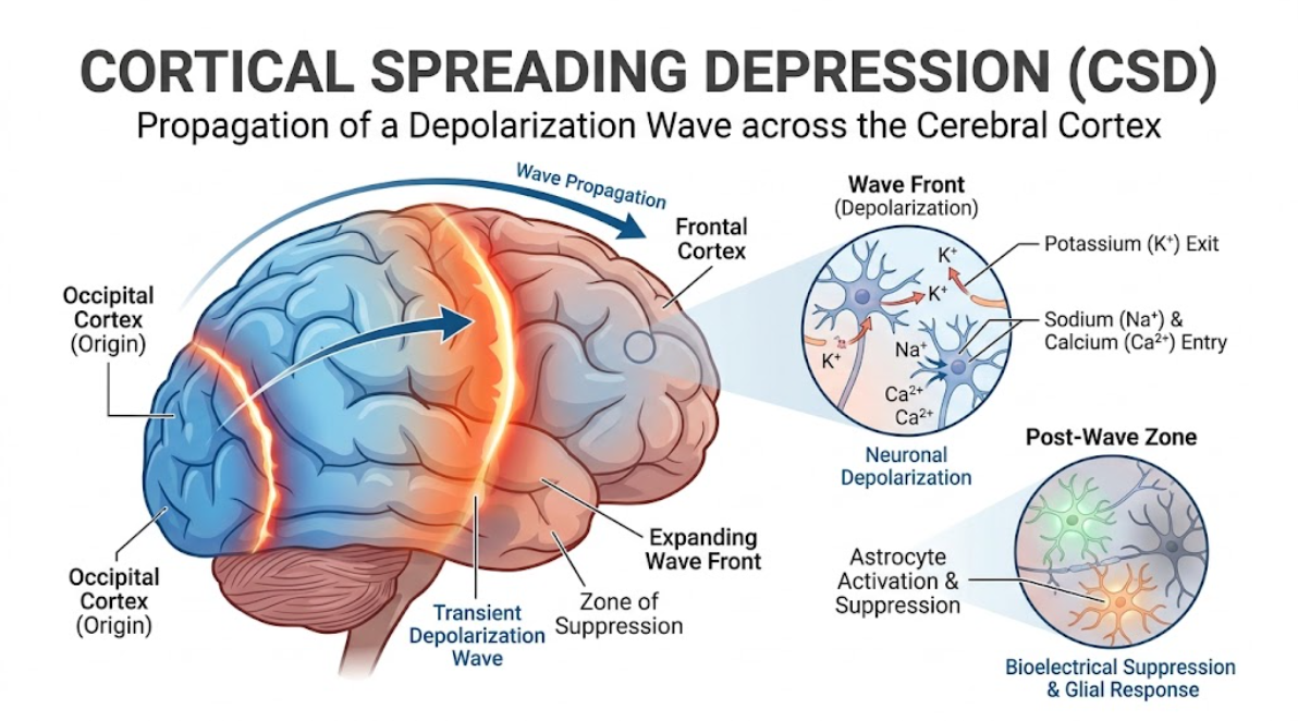
Figure 1. Progression and Cellular Mechanisms of Cortical Spreading Depression (CSD). Schematic representation of CSD wave propagation from the occipital cortex moving anteriorly across the cerebral cortex. The enlarged views emphasize the transient wave front of neuronal depolarization with potassium (K⁺) efflux and sodium/calcium (Na⁺/Ca²⁺) influx and a post-wave zone of bioelectrical suppression and astrocyte activation.
Cortical Spreading Depression induces profound hemodynamic changes throughout the cortical microvasculature demonstrating the complex coupling between neuronal electrical activity and local blood flow. The vascular response after a CSD wave is usually biphasic with an initial transient hyperemic phase followed by a prolonged period of hypoperfusion or oligemia(Close et al., 2019; Tfelt-Hansen, 2010).
When the depolarization wave reaches the arterioles, the high metabolic demand and rapid liberation of vasoactive substances (e.g. nitric oxide, calcitonin gene-related peptide (CGRP), and potassium) result in a short but intense dilation of the arteries. Regional cerebral blood flow is markedly increased above baseline for 1-2 minutes. However, as the intense depolarization subsides and sustained extracellular ionic shifts become established, perivascular smooth muscle cells contract and microvascular constriction becomes quite pronounced(Lauritzen et al., 2011).
This secondary vascular constriction results in a prolonged reduction in regional cerebral blood flow, with local perfusion decreased twenty to thirty percent below resting levels. This stage of propagating oligemia continues for several hours, long after neuronal membrane potentials have normalized. Critically, these same hemodynamic changes have been verified in fMRI studies measuring blood oxygen level-dependent (BOLD) signals in humans experiencing spontaneous visual aura(Lauritzen et al., 2011).
Early fMRI studies demonstrated a focal increase of BOLD signal in the extrastriate visual cortex (area V3A) corresponding to the early hyperemic vasodilation. This progressed slowly across the occipital cortex at a rate of three to three and a half millimeters per minute, exactly the speed of CSD. A prolonged drop in BOLD signal was observed following the leading hyperemic signal, consistent with protracted cortical oligemia and microvascular constriction(Kitamura & Imai, 2024).
It is important to emphasize that, despite the profound reduction in cerebral blood flow during cortical oligemia, tissue perfusion is still above the critical level for ischemic cell death. Uncomplicated migraine aura preserves high energy phosphate stores (e.g. ATP, phosphocreatine). Therefore, the vascular vasoconstriction observed in CSD is a functional stroke or a permanent ischemic injury, not a structural one(Lauritzen et al., 2011; Tfelt-Hansen, 2010).
The clinical manifestations of migraine aura are direct, perceptual representations of the underlying CSD wave passing over retinotopically organized cortical tissue. Because the primary visual cortex (V1) is organized in an orderly spatial fashion that represents the contralateral visual field, the physical movement of the depolarization wave across the cortical surface directly maps to the perceived motion of visual disturbances(Nozari et al., 2010).
The initial phase of CSD, characterized by pronounced neuronal excitation, enhanced release of glutamate and a short-lived dilation of the microvasculature, corresponds to the positive visual aura phenomena. The transient hyperexcitability of local visual neurons as the front of the CSD wave enters a particular region of the visual cortex produces scintillations, shimmering zig-zag lines, bright flashes and geometric fortification spectra in the corresponding visual field(Mehnert et al., 2023; Silberstein, 2004).
This wave front propagates a region of profound neuronal electrical silence, a state of depolarization block and persistent microvascular constriction (oligemia) as the CSD wave propagates. Such localized cortical suppression is the direct physiological correlate of the negative visual phenomena of aura. The patients see the expanding scotoma, the central or peripheral blind spot, or the zones of blurred vision which immediately follow behind the advancing scintillating border(Mehnert et al., 2023; Silberstein, 2004).
The slow expansion of the scintillating scotoma across the visual field over 20 to 30 minutes (figure 2) is an exact representation of the two to five millimeter per minute progression of CSD across the occipital lobe. If CSD occurs near the occipital pole, symptoms begin in the center of the visual field and spread outward toward the periphery. If CSD starts in anterior occipital regions, the visual aura is experienced in the peripheral visual field and travels centripetally(Mehnert et al., 2023; Silberstein, 2004).
In complex aura presentations, CSD is not limited to the occipital cortex, but propagates slowly into neighboring cortical structures(Petrusic & Zidverc-Trajkovic, 2014). The wave spreads anteriorly over the parietal and temporal lobes. Extension to the primary somatosensory cortex results in sensory aura symptoms, usually with paresthesias or “pins and needles” sensations in the hand, arm and face. After the excitation wave, there is numbness and loss of sensation with the depolarization block. CSD affecting speech areas of the dominant hemisphere such as Broca’s or Wernicke’s areas results in transient dysphasia, difficulty in word finding and paraphasias(Mehnert et al., 2023).
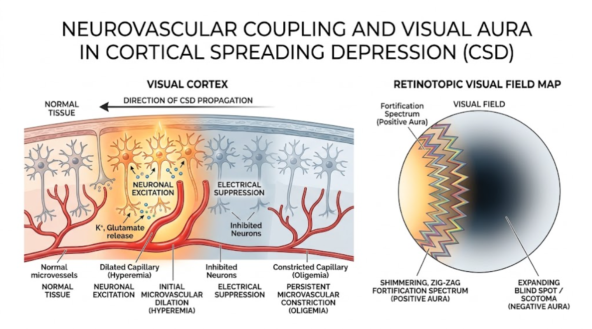
Figure 2. Neurovascular Coupling and Visual Aura in Cortical Spreading Depression (CSD). CSD wave (right to left) propagating in a cross-section of the visual cortex (left). Initial neuronal excitation and K+/glutamate release leading to microvascular dilation (hyperemia) followed by electrical suppression (inhibited neurons) and persistent constriction (oligemia). On the right, the corresponding retinotopic visual field map shows how this cortical event gives rise to an advancing shimmering zig-zag fortification spectrum (positive aura) followed by an expanding blind spot/scotoma (negative aura).
The question in headache medicine for decades has been: how does a neuroelectrical event in the brain cortex translate into the severe, throbbing head pain characteristic of chronic migraine? Recent neurobiological studies have demonstrated that CSD is a powerful endogenous trigger for the activation of the trigeminovascular system, establishing a direct functional connection between cortical aura and cranial nociception(Cui et al., 2014).
The passage of a CSD wave leads to a massive efflux of intracellular ions and neurochemicals, resulting in an inflammatory milieu in the cortical parenchyma and subarachnoid space. High concentrations of extracellular potassium, hydrogen ions, ATP, glutamate and arachidonic acid metabolites diffuse toward pial and meningeal blood vessels(Kunkler, Hulse, Schmitt, Nicholson, & Kraig, 2005). Furthermore, CSD induces the opening of neuronal Pannexin-1 megachannels resulting in the activation of intracellular caspase-1 and release of pro-inflammatory cytokines such as interleukin-1β and high-mobility group box 1 (HMGB1)(Kitamura & Imai, 2024).
These parenchymal inflammatory mediators activate the trigeminal nerve’s primary unmyelinated C-fibers and thinly myelinated A-δ fibers, which innervate pial and dural blood vessels. This parenchymal-to-meningeal signaling cascade results in sterile neurogenic inflammation, dural mast cell degranulation and the release of vasoactive neuropeptides, especially CGRP, into the perivascular space(Cui et al., 2014).
There is an apparent physiological latency of 15–30 min from the onset of CSD until full activation of first-order trigeminal ganglion neurons. This physiological delay agrees with the clinical observation that migraine headache usually begins as the aura symptoms are beginning to resolve(Close et al., 2019; Mehnert et al., 2023). Repeated episodes of CSD lower the threshold for activation of central trigeminovascular neurons in the trigeminocervical complex and thalamus in chronic migraineurs. This re-stimulation perpetuates central sensitization, cutaneous allodynia and chronification of headache(Kitamura & Imai, 2024).
Here is presented a comprehensive literature review which clarifies the key role of Cortical Spreading Depression as the electrophysiological generator of migraine aura and as a critical upstream trigger for trigeminovascular pain activation. This review tries to provide a unifying framework for the complex clinical features of chronic migraine patients with aura by looking at the detailed cellular cascades, ionic shifts and microvascular responses(Close et al., 2019).
A major strength of the CSD concept is its capacity to reconcile historical disputes between primary neural and primary vascular theories of migraine. CSD is primarily a neuroglial event that secondarily drives microvascular behavior through potent neurovascular coupling. Transient initial hyperemia followed by prolonged cortical oligemia suggests that microvascular constriction is an intrinsic secondary consequence of widespread cortical depolarization rather than an isolated primary arterial spasm(Close et al., 2019; Tfelt-Hansen, 2010).
When CSD is considered in the special context of chronic migraine, important pathophysiological differences to episodic forms emerge. Chronic migraineurs are characterized by a state of chronic cortical hyperexcitability and defective sensory habituation in the interictal state. This increased baseline susceptibility lowers the threshold for physiological triggers (such as intense light stimulation, metabolic stress, sleep deprivation) to induce CSD(Hanalioglu et al., 2022). In addition, recurrent CSD waves cause epigenetic changes, chronic microglial activation and structural reorganization in cortical and subcortical pain networks, creating a self-propagating cycle of disease progression(Kitamura & Imai, 2024).
The clinical relevance of CSD suppression is strongly validated by pharmacological studies. Chronic migraine prophylactic agents like topiramate, valproic acid, propranolol and amitriptyline have been shown to increase the CSD ignition threshold and decrease CSD frequency significantly after chronic administration in animal models(Tfelt-Hansen, 2010). Similarly, novel treatments, including NMDA receptor modulation, CGRP receptor antagonism, and non-invasive vagus nerve stimulation, have been demonstrated to decrease cortical excitability and inhibit CSD wave propagation(Kitamura & Imai, 2024).
But key scientific questions remain about CSD dynamics in human patients. Indirect evidence of CSD-like hemodynamic waves during visual aura was strongly provided by neuroimaging studies using functional MRI and SPECT, but the characteristic DC potential shift is seldom recorded by non-invasive scalp electroencephalography due to skull impedance and focal signal cancelation(Lauritzen et al., 2011). Continued progress in high-density electroencephalography, magnetoencephalography, and ultra-high-field functional imaging will be vital to further characterize CSD in conscious human subjects and to translate these physiological insights into targeted, disease-modifying therapies(Tfelt-Hansen, 2010).
Finally, the basic neurobiological substrate of the aura phenomenon in chronic migraine is the Cortical Spreading Depression. CSD is defined as a wave of profound neuronal and glial depolarization that propagates slowly and disrupts cellular ion homeostasis, leading to massive glutamate release and transient silencing of cortical electrical activity. CSD induces a characteristic biphasic hemodynamic response through neurovascular coupling consisting of a transient initial hyperemic dilation followed by a sustained microvascular constriction and cortical oligemia. This contiguous wave of excitation and subsequent electrical depression across retinotopically organized visual cortex provides a precise explanation of the clinical course of positive scintillations followed by expanding scotomas. CSD also provides an important link between cortical events and headache pain by releasing inflammatory mediators that activate and sensitize the trigeminovascular system. Characterization of the cellular and molecular mechanisms of CSD enhances our basic knowledge of the pathophysiology of chronic migraine and sets out promising targets for novel prophylactic therapies.
Charles, A. (1998). Intercellular calcium waves in glia. Glia, 24(1), 39-49. doi:10.1002/(sici)1098-1136(199809)24:1<39::aid-glia5>3.0.co;2-w
Charles, A., & Brennan, K. (2009). Cortical spreading depression-new insights and persistent questions. Cephalalgia, 29(10), 1115-1124. doi:10.1111/j.1468-2982.2009.01983.x
Close, L. N., Eftekhari, S., Wang, M., Charles, A. C., & Russo, A. F. (2019). Cortical spreading depression as a site of origin for migraine: Role of CGRP. Cephalalgia, 39(3), 428-434. doi:10.1177/0333102418774299
Cui, Y., Kataoka, Y., & Watanabe, Y. (2014). Role of cortical spreading depression in the pathophysiology of migraine. Neuroscience Bulletin, 30(5), 812-822. doi:10.1007/s12264-014-1471-y
Dohmen, C., Sakowitz, O. W., Fabricius, M., Bosche, B., Reithmeier, T., Ernestus, R. I., . . . Graf, R. (2008). Spreading depolarizations occur in human ischemic stroke with high incidence. Ann Neurol, 63(6), 720-728. doi:10.1002/ana.21390
Hanalioglu, S., Taskiran-Sag, A., Karatas, H., Donmez-Demir, B., Yilmaz-Ozcan, S., Eren-Kocak, E., . . . Dalkara, T. (2022). Cortical spreading depression can be triggered by sensory stimulation in primed wild type mouse brain: a mechanistic insight to migraine aura generation. J Headache Pain, 23(1), 107. doi:10.1186/s10194-022-01474-0
Haydon, P. G., & Carmignoto, G. (2006). Astrocyte control of synaptic transmission and neurovascular coupling. Physiol Rev, 86(3), 1009-1031. doi:10.1152/physrev.00049.2005
Kitamura, E., & Imai, N. (2024). Molecular and Cellular Neurobiology of Spreading Depolarization/Depression and Migraine: A Narrative Review. Int J Mol Sci, 25(20). doi:10.3390/ijms252011163
Kunkler, P. E., Hulse, R. E., Schmitt, M. W., Nicholson, C., & Kraig, R. P. (2005). Optical current source density analysis in hippocampal organotypic culture shows that spreading depression occurs with uniquely reversing currents. J Neurosci, 25(15), 3952-3961. doi:10.1523/jneurosci.0491-05.2005
Lashley, K. S. (1941). PATTERNS OF CEREBRAL INTEGRATION INDICATED BY THE SCOTOMAS OF MIGRAINE. Archives of Neurology & Psychiatry, 46(2), 331-339. doi:10.1001/archneurpsyc.1941.02280200137007
Lauritzen, M. (1994). Pathophysiology of the migraine aura. The spreading depression theory. Brain, 117 ( Pt 1), 199-210. doi:10.1093/brain/117.1.199
Lauritzen, M., Dreier, J. P., Fabricius, M., Hartings, J. A., Graf, R., & Strong, A. J. (2011). Clinical relevance of cortical spreading depression in neurological disorders: migraine, malignant stroke, subarachnoid and intracranial hemorrhage, and traumatic brain injury. J Cereb Blood Flow Metab, 31(1), 17-35. doi:10.1038/jcbfm.2010.191
Leao, A. A. (1947). Further observations on the spreading depression of activity in the cerebral cortex. J Neurophysiol, 10(6), 409-414. doi:10.1152/jn.1947.10.6.409
Leao, A. A. P. (1944). SPREADING DEPRESSION OF ACTIVITY IN THE CEREBRAL CORTEX. Journal of Neurophysiology, 7(6), 359-390. doi:10.1152/jn.1944.7.6.359
Mehnert, J., Fischer-Schulte, L., & May, A. (2023). Aura phenomena do not initiate migraine attacks—Findings from neuroimaging. Headache: The Journal of Head and Face Pain, 63(8), 1040-1044. doi:https://doi.org/10.1111/head.14597
Messlinger, K. (2009). Migraine: where and how does the pain originate? Exp Brain Res, 196(1), 179-193. doi:10.1007/s00221-009-1756-y
Nozari, A., Dilekoz, E., Sukhotinsky, I., Stein, T., Eikermann-Haerter, K., Liu, C., . . . Moskowitz, M. A. (2010). Microemboli may link spreading depression, migraine aura, and patent foramen ovale. Ann Neurol, 67(2), 221-229. doi:10.1002/ana.21871
Petrusic, I., & Zidverc-Trajkovic, J. (2014). Cortical spreading depression: origins and paths as inferred from the sequence of events during migraine aura. Funct Neurol, 29(3), 207-212.
Pietrobon, D., & Striessnig, J. (2003). Neurobiology of migraine. Nat Rev Neurosci, 4(5), 386-398. doi:10.1038/nrn1102
Silberstein, S. D. (2004). Migraine. Lancet, 363(9406), 381-391. doi:10.1016/s0140-6736(04)15440-8
Tfelt-Hansen, P. (2010). History of migraine with aura and cortical spreading depression from 1941 and onwards. Cephalalgia, 30(7), 780-792. doi:10.1111/j.1468-2982.2009.02015.x