Our Services
Others
- DFPP
- Shockwave
- IV Drip


This article is for educational purposes only and does not replace consultation with a qualified medical professional.
For patients evaluating regenerative medicine for joint preservation, the clinical efficacy of umbilical cord mesenchymal stem cells (UC-MSCs) relies entirely on objective biological mechanisms, not marketing claims. If you’ve been living with chronic hip pain, you already know the frustrating cycle. You get an X-ray. Your doctor spots cartilage degradation. They offer you physical therapy, hand you a prescription for NSAIDs, and eventually escalate to palliative cortisone injections. When those inevitably stop working, you’re told to wait until the joint is destroyed enough to justify an invasive total hip arthroplasty.
It’s a passive, watch-and-wait approach to structural decay.
This clinical guide examines the peer-reviewed evidence behind stem cell therapy for hip osteoarthritis, analyzing biological mechanisms, realistic success rates, and strict contraindications. We’re going to bypass the hype. By focusing on the transition from cartilage
Stem cell therapy for hip osteoarthritis utilizes UC-MSCs to modulate joint inflammation and promote cartilage repair through paracrine signaling.
Osteoarthritis is the most common form of arthritis, characterized by the progressive breakdown of joint cartilage and underlying bone changes (Centers for Disease Control and Prevention, 2024). In the hip joint, this degeneration initiates a chronic inflammatory response that degrades the extracellular matrix, making early staging critical for determining whether a patient falls within the Regenerative Viability Window for cellular therapy.
To understand how regenerative medicine interacts with a failing joint, we have to look closely at the biological mechanics of hip cartilage loss. Articular cartilage, the smooth tissue cushioning the hip joint, isn’t just an inert shock absorber. It is highly specialized, avascular tissue maintained by cells called chondrocytes.
When a patient develops hip osteoarthritis, the joint undergoes a catastrophic shift in homeostasis. Mechanical stress or prior trauma triggers the release of matrix metalloproteinases (MMPs) and aggrecanases—enzymes that literally digest the cartilage from the inside out.
This isn’t simple wear and tear. It’s an active, biochemically driven joint degeneration. The breakdown of the cartilage matrix releases microscopic debris into the synovial fluid. The joint capsule’s lining (the synovium) detects this debris and mounts a severe immune response, triggering painful synovial inflammation. As the articular cartilage thins, the underlying subchondral bone experiences increased mechanical stress. The bone reacts by thickening (sclerosis) and forming jagged osteophytes (bone spurs) around the edges of the acetabulofemoral joint.
This specific sequence of biological destruction is exactly why the concept of “The Regenerative Viability Window” is so vital. Biological interventions require a baseline cellular matrix to build upon. If you inject cellular biologics into a joint that still has some structural scaffolding and active chondrocytes, they can issue chemical commands to halt the MMPs. If you wait until the joint is entirely destroyed, there are no local cells left to receive those biological instructions.
Orthopedic specialists universally categorize hip osteoarthritis across four distinct stages. Identifying your current stage isn’t just about reading an X-ray; it dictates whether you are a candidate for biological intervention or if you’re firmly in surgical territory.
Stage 1: Minor: Patients experience very little pain. Radiographic findings might show a tiny osteophyte, but the joint space narrowing is barely perceptible.
Stage 2: Mild: This is where symptoms become clinically relevant. You might feel morning stiffness or a sharp catch in your groin after a long walk. Radiographs show definite osteophyte formation, though the cartilage is still largely intact.
Stage 3: Moderate: The joint space is visibly narrowing. The cartilage is fraying and thinning significantly. Hip arthritis symptoms escalate—patients often experience pain radiating down to the knee during weight-bearing activities, and putting on shoes becomes physically restrictive.
Stages 2 and 3 represent the optimal Regenerative Viability Window. The joint is highly inflamed, but the structural scaffolding remains.
Stage 4: Severe.:This is end-stage disease. The joint space is completely gone, resulting in severe bone-on-bone friction. The cartilage has eroded entirely in the load-bearing zones, and large osteophytes physically block range of motion.
At Stage 4, the hip falls outside the efficacy window for cellular therapies. As noted in a comprehensive Cleveland Clinic analysis, end-stage bone-on-bone joint degeneration often involves severe structural deformities (2023). Stem cells cannot physically reshape deformed bone or replace a completely missing articular surface. Mechanical failure requires mechanical intervention.
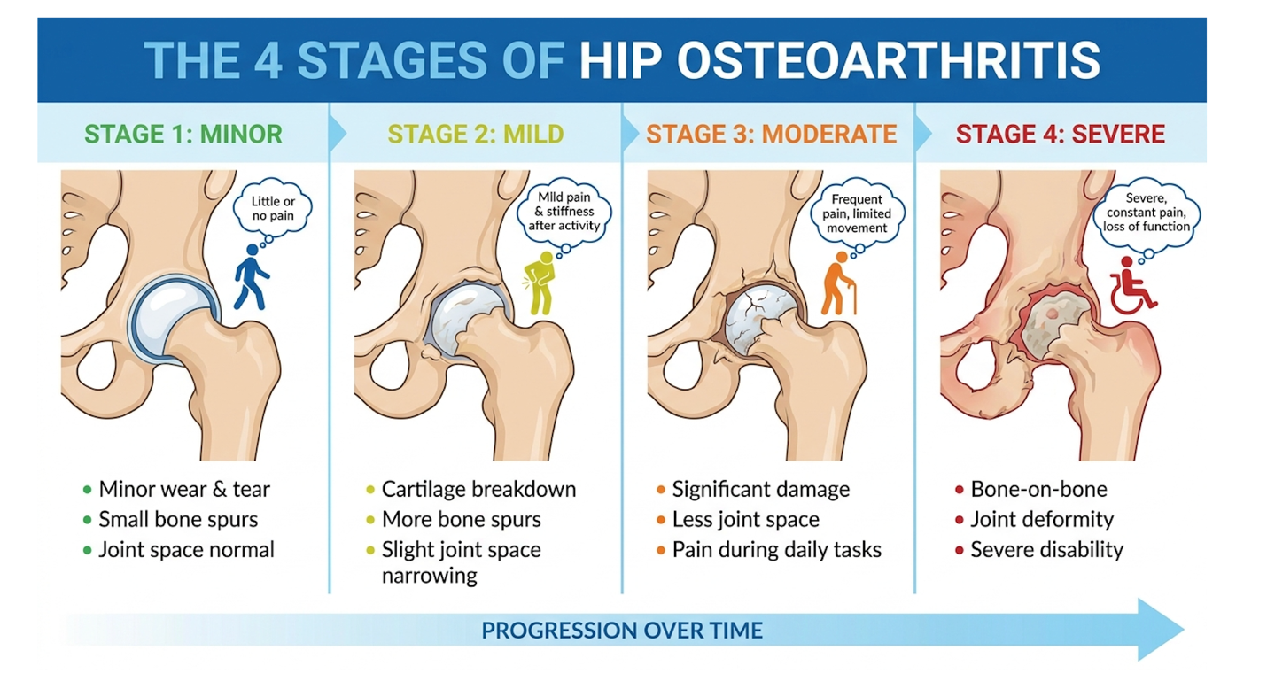
Figure 1: The critical transition between Stage 3 (moderate) and Stage 4 (severe) determines cellular therapy candidacy.
Once the stage of degeneration is identified through MRI or weight-bearing radiographs, clinicians must evaluate the specific biological tools such as UC-MSCs used to combat the inflammation.
Wharton’s jelly-derived MSCs exhibit high proliferative capacity and multi-lineage differentiation potential, allowing them to support joint repair (National Institutes of Health, 2019). These umbilical cord mesenchymal stem cells (UC-MSCs) act as biological signaling centers. Unlike controversial embryonic cells, UC-MSCs are adult-lineage multipotent cells that offer potent anti-inflammatory properties for treating osteoarthritis without ethical complications.
The term “stem cell” is heavily abused in medical marketing. To be scientifically accurate, we are talking strictly about Umbilical Cord Mesenchymal Stem Cells (UC-MSCs).
By definition, MSCs are multipotent stromal cells. This means they possess the biological capability to differentiate into specific mesodermal lineages, specifically osteoblasts (bone cells), chondrocytes (cartilage cells), and adipocytes (fat cells).
But here is where the critical distinction lies: these cells are sourced specifically from Wharton’s jelly, the gelatinous connective tissue found inside the umbilical cord. They are fundamentally distinct from hematopoietic (blood-forming) stem cells, which are primarily extracted from umbilical cord blood. You don’t want blood-forming cells injected into an arthritic joint; you want connective-tissue-forming cells.
Furthermore, I have seen endless patient confusion regarding the ethical nature of these cells. Let’s be absolutely clear. UC-MSCs are completely distinct from embryonic stem cells. Embryonic cells carry deep ethical controversies and a high risk of teratoma (tumor) formation. Wharton’s jelly stem cells are considered adult-lineage multipotent cells. They are safe, highly proliferative, and ethically uncontroversial. A 2021 study published on PubMed explicitly reinforces their strong safety profile and lack of tumorigenicity in orthopedic applications.
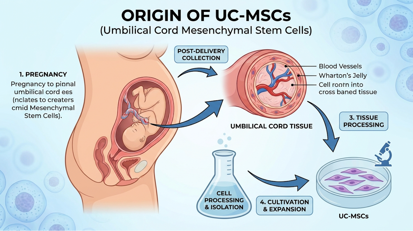
Figure 2: Wharton’s jelly provides a dense, pure population of multipotent mesenchymal stem cells distinct from cord blood.
The unique biological properties of these cells are deeply intertwined with how they are safely and ethically collected from donors.
When you undergo an autologous stem cell procedure—meaning the cells are extracted from your own body—a physician must drill into your iliac crest (pelvis) to aspirate bone marrow, or perform a mini-liposuction to extract adipose (fat) tissue. It’s painful. It involves significant downtime.
Worse, autologous MSC sourcing suffers from the realities of cellular aging. If you are a 65-year-old patient with hip osteoarthritis, your bone marrow stem cells are also 65 years old. They suffer from cellular senescence. Their replication rate is sluggish. Their anti-inflammatory output is heavily diminished compared to younger tissue.
| 📌 If you’re interested in why umbilical cord cells are often preferred over bone marrow or fat-derived cells, we have an interesting article that discusses why UC-MSCs are superior to other stem cell sources, which you can read via the internal link. |
This is why modern regenerative clinics lean heavily toward allogeneic stem cells (cells sourced from a donor).
UC-MSCs are collected from full-term, healthy cesarean section births. The umbilical cord, which is normally discarded as medical waste, is donated with strict maternal consent. The mother undergoes rigorous FDA-aligned infectious disease screening. These are “Day 0” cells. They possess massive proliferative energy and optimal vitality.
And no, your body will not reject them. UC-MSCs are uniquely “immune-privileged.” Because they lack mature Major Histocompatibility Complex (MHC) Class II surface antigens, your immune system’s T-cells do not recognize them as foreign invaders. You can safely receive them without any need for immunosuppressant drugs or tissue matching.
With a clear understanding of their ethical sourcing and immune-privileged status, clinical attention must turn to exactly how these cells alter the disease state within a deteriorating hip joint.
Mesenchymal stem cells promote joint healing primarily through the paracrine secretion of trophic factors that suppress local inflammation (National Library of Medicine, 2017). When injected into a deteriorating hip joint, UC-MSCs rely on a complex mechanism of action involving cell signaling, immune system regulation, and tissue repair to reverse the toxic microenvironment driving osteoarthritis progression.
If you want to stop a joint from destroying itself, you have to shut down the chemical fire inside the synovium. The osteoarthritic hip is essentially trapped in a chronic inflammatory loop. Pro-inflammatory cytokines, specifically Tumor Necrosis Factor-alpha (TNF-alpha) and Interleukin-1 (IL-1), saturate the joint space, signaling the cartilage to undergo accelerated cell death (apoptosis).
This is where the UC-MSC mechanism of action begins.
When UC-MSCs enter this toxic environment, they don’t just passively float around. They actively detect the severe inflammation and begin a process called immunomodulation. The regulatory adjustment of the immune system is arguably their most potent medical capability.
They accomplish this primarily through phenotypic macrophage reprogramming. The hip joint is filled with local immune cells called macrophages. In osteoarthritis, these are predominantly M1 macrophages—aggressive, tissue-destructive cells that sustain the inflammatory loop. UC-MSCs release specific biochemical signals that force these macrophages to switch from the destructive M1 state into the M2 state.
| 📌 If you’re interested in how MSCs shift the immune system from attack mode to repair mode, we have an interesting article that discusses mesenchymal stem cell therapy for immune modulation, which you can read via the internal link. |
M2 macrophages are anti-inflammatory and tissue-repairing. By forcing this shift, the stem cells effectively hijack the joint’s immune response and hit the reset button.
Contrast this biological resetting with non-steroidal anti-inflammatory drugs (NSAIDs) like ibuprofen. NSAIDs merely block pain receptors and temporarily mask the cyclooxygenase (COX) pathways. They do absolutely nothing to alter the behavior of the macrophages destroying the joint. Furthermore, clinical evidence indexed in PubMed indicates that chronic NSAID use can actually impair bone healing and accelerate joint pathology over time.
Once the destructive inflammatory cascade is halted, the cells initiate the second, most critical phase of healing via cell-to-cell communication.
For decades, the mainstream media promoted a severe oversimplification: that stem cells are injected, and they miraculously turn into new cartilage like microscopic building blocks. That is scientifically inaccurate.
The true power of these cells lies in the paracrine effect. Paracrine signaling is a form of cell-to-cell communication. Instead of acting as structural building blocks, UC-MSCs act as medicinal signaling cells—tiny biological pharmacies.
Once inside the joint, they secrete a vast array of bioactive molecules, growth factors, and extracellular vesicles called exosomes directly into the synovial fluid. These secretomes act like software updates for your failing joint. They locate the patient’s own quiescent (dormant) dermal fibroblasts and surviving chondrocytes, bind to their cellular receptors, and command them to wake up.
Under the instruction of paracrine signaling, your own native cells begin actively synthesizing new extracellular matrix proteins, specifically Type II collagen and elastin.
This brings us right back to the Original Concept of the Regenerative Viability Window. Paracrine signaling requires existing host cells to receive the biological instructions. If a patient waits until they have Stage 4 bone-on-bone osteoarthritis, the chondrocytes are completely gone. You can inject millions of the highest-quality UC-MSCs into that joint, but there is no one left to receive the signal. The pharmacy is open, but the customers are dead.
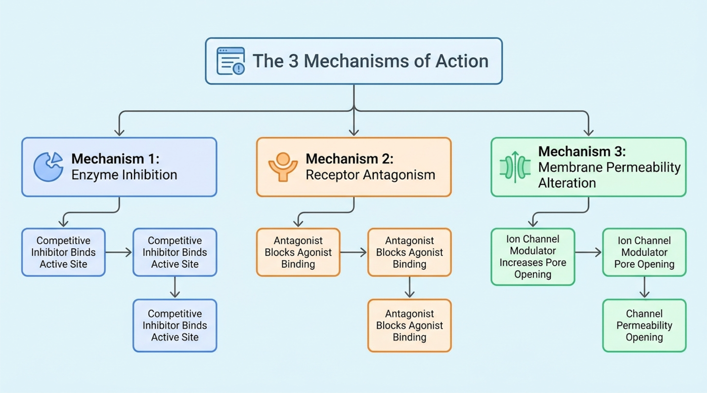
Figure 3: The paracrine effect dominates the healing process, shifting the joint microenvironment from destructive to reparative.
While paracrine signaling drives the majority of the clinical improvement, direct cellular differentiation plays a supplementary role in structural repair.
Let’s address the elephant in the room: stem cell cartilage regeneration. Can these cells actually become new cartilage?
Yes, but with heavy caveats. In controlled in vitro (laboratory petri dish) environments, we can absolutely force MSCs to differentiate directly into mature chondrocytes. However, when injected in vivo (into a living human hip), the actual engraftment rates—the percentage of injected cells that physically embed themselves and become permanent cartilage—are much lower than historically believed.
The primary mechanism for tissue preservation is trophic support.
Trophic support means the UC-MSCs optimize the environment to prevent further chondrocyte apoptosis (cell death) while stimulating the host tissue to repair itself. They lay down a supportive
We must manage clinical expectations here. The goal of cellular therapy is not to build a brand-new, 18-year-old hip joint out of stem cells. The goal is to restore a functional, pain-free tissue matrix that allows a 60-year-old to hike, golf, and sleep through the night without surgical intervention.
Understanding these biological realities is paramount when patients begin comparing this regenerative approach to traditional orthopedic interventions.
Traditional treatment for severe hip osteoarthritis culminates in total joint replacement surgery, involving the removal of damaged bone and cartilage (Mayo Clinic, 2023). However, for patients seeking alternatives to hip surgery, UC-MSC therapy presents a non-surgical intervention that targets biological healing rather than mechanical replacement, dramatically altering the risk profile and recovery timeline.
Total Hip Arthroplasty (THA) the surgical replacement of the hip joint is an engineering marvel. It is also a massive, traumatic surgical event. The surgeon must dislocate the femoral head, saw it off entirely, ream out the acetabulum (the pelvic socket), and hammer in titanium, ceramic, or cross-linked polyethylene prosthetic components.
THA carries the inherent risks of major open surgery: deep vein thrombosis (blood clots), periprosthetic joint infection, nerve damage, and prosthesis dislocation. A standard THA requires a grueling 3-to-6 month physical therapy protocol before the patient regains natural gait mechanics.
Compare that procedural reality to UC-MSC therapy. Cellular therapy is a same-day, outpatient intra-articular injection. It requires no general anesthesia, no hospital stay, and zero bone removal. The downtime is measured in days, not months.
But we must establish an explicit boundary for clinical trust. If a patient presents with severe Stage 4 osteoarthritis, absolute joint space collapse, severe mechanical deformity, or advanced avascular necrosis (bone death), stem cells will fail. Period. I have seen clinics promise miracles to bone-on-bone patients, and it is medical malpractice. In those specific scenarios, surgery is the only viable option. Biological solutions cannot override the physics of severely deformed, necrotic bone.
While avoiding surgery is a primary goal for many, patients must also weigh cellular therapy against more common conservative injection options.
When patients look for non-surgical hip arthritis treatment, they are almost universally funneled into the corticosteroid trap.
Corticosteroids are synthetic drugs used to temporarily reduce inflammation. They work beautifully—for about four to eight weeks. They forcefully shut down the inflammatory response, providing rapid, potent pain relief. But the biological cost is severe.
A growing body of orthopedic literature, including recent peer-reviewed evaluations on PubMed, demonstrates that repeated intra-articular cortisone injections are highly chondrotoxic. They literally poison the cartilage. They accelerate cartilage degradation, increase the risk of subchondral bone collapse, and hasten the need for a total hip replacement. You are trading a few weeks of pain relief for the permanent destruction of your joint matrix.
Hyaluronic acid (viscosupplementation) is the other common alternative. It acts as a temporary biological lubricant. Think of it like a heavy oil change for a squeaky hinge. It’s much safer than cortisone, but it does not alter the underlying disease progression. It doesn’t stop the MMPs from eating the cartilage, and it doesn’t resolve synovial inflammation long-term.
This highlights why UC-MSCs represent a regenerative paradigm shift. We are moving away from merely masking pain or applying temporary grease. Cortisone vs stem cells is a debate of palliation versus regeneration. Stem cell therapy aims to modify the disease environment, halt the degradation cycle, and restore structural homeostasis.
Shifting from palliative masking to biological regeneration inherently alters the treatment timeline, requiring patients to understand the procedural protocols and realistic clinical outcomes.
Clinical trials demonstrate that human umbilical cord mesenchymal stem cells have strong immunomodulatory capabilities, resulting in significant pain reduction in osteoarthritic joints (National Library of Medicine, 2021). Entering UC-MSC treatment for hip osteoarthritis requires an understanding that biological repair is a gradual process, necessitating precise procedural execution and managed clinical expectations to achieve optimal success rates.
What actually happens on the day of the procedure? For patients riddled with anxiety about a hip stem cell therapy, the reality is surprisingly anti-climactic.
First, let’s address precision. The hip is a deep, complex joint encapsulated by dense ligaments and thick gluteal musculature. You cannot simply guess where the joint capsule is. Therefore, an intra-articular hip injection MUST be performed under ultrasound-guided or fluoroscopic imaging. Precision imaging techniques ensure the needle bypasses the surrounding neurovascular bundles and delivers the UC-MSCs precisely into the intra-articular space. If a provider offers to inject your hip blind (without imaging), walk out immediately.
The entire procedural flow occurs in an outpatient setting.
The actual injection takes less than a few minutes. From walking into the clinic to walking out, the process typically takes under an hour. Because the cells are allogeneic, you bypass the grueling hours of bone marrow aspiration completely.
Following successful delivery into the joint space, the biological clock begins ticking on cellular repair and symptom relief.
We have to manage timeline expectations immediately. A stem cell injection for hip pain does not work like a cortisone shot. You will not wake up the next morning feeling 100% cured.
Because we are relying on biological processes immunomodulation and trophic support—the timeline requires patience.
When discussing the stem cell therapy success rate hip, clinical literature generally reports that 70% to 80% of properly selected candidates experience clinically meaningful improvements. These improvements are tracked via WOMAC scores (a standardized metric for arthritis pain and stiffness). A successful outcome means the patient experiences a massive reduction in daily pain, improved range of motion, and a return to functional activities without reliance on NSAIDs.
Let’s discuss the elephant in the room: hip stem cell therapy cost.
Because regenerative joint injections are largely classified by regulatory bodies as investigational, they are almost universally NOT covered by standard health insurance, Medicare, or Medicaid. Patients must view this as an out-of-pocket, cash-pay investment in joint preservation.
The Arthritis Foundation clearly notes that expected out-of-pocket costs for cellular therapies can range from several thousand dollars upward, depending on the cell count required and the geographic location of the clinic.
However, patients need to run a broader financial calculus. You must evaluate the upfront cost of stem cell therapy against the massive hidden costs of a surgical alternative. Total hip arthroplasty often incurs high insurance deductibles, copays for months of mandatory physical therapy, potential costs for home care assistance, and crucially, lost wages during the 3-to-6 month surgical downtime. When framed against the comprehensive financial burden of joint replacement, the cost of a same-day cellular injection often becomes highly competitive.
| 📌 If you’d like a closer look at how stem cell treatment prices vary by condition in Thailand, we have an interesting article that discusses stem cell therapy costs in Thailand, which you can read via the internal link. |
The lack of insurance coverage is directly tied to the regulatory status of cellular medicine, making patient safety and provider vetting paramount.
Stem cells work in hip osteoarthritis by releasing bioactive molecules that halt joint inflammation and stimulate tissue repair. This process, known as paracrine signaling, fundamentally alters the toxic microenvironment inside the joint. Rather than simply turning into new cartilage, the cells instruct the patient’s existing cells to synthesize new extracellular matrix proteins. Patients typically experience a reduction in pain and stiffness as the inflammatory cascade is neutralized. Results are most optimal when applied before severe structural deformity occurs.
Stem cells have the biological potential to promote the synthesis of new cartilage matrix, but they cannot completely regrow a severely destroyed hip joint. Through trophic support, UC-
Stem cell therapy is biologically superior to cortisone for long-term joint preservation, whereas cortisone provides faster but strictly temporary pain relief. Corticosteroids forcefully mask inflammation for a few months but have been clinically shown to accelerate cartilage toxicity and degradation with repeated use. Conversely, UC-MSCs actively modulate the immune environment to promote cellular healing and slow disease progression over several months. Patients prioritizing immediate, short-term relief may choose cortisone, while those seeking disease modification generally pursue regenerative cellular therapy.
Umbilical cord stem cells are favored because they are highly potent, ethically sourced, and eliminate the need for a painful bone marrow extraction surgery. UC-MSCs from Wharton’s jelly are “Day 0” cells, meaning they lack the cellular aging and environmental damage present in a patient’s own bone marrow cells. They also boast a higher proliferation rate and secrete a denser concentration of anti-inflammatory cytokines. This makes the outpatient procedure vastly more comfortable while delivering a biologically superior cellular product.
Patients typically experience initial pain relief from stem cell therapy in the hip within 4 to 8 weeks as joint inflammation subsides. Because cellular repair is a biological process, structural tissue remodeling continues to progress over the following 3 to 6 months. Some patients report continued functional improvements up to a year post-injection as the extracellular matrix strengthens. Individual recovery timelines vary significantly based on the patient’s baseline joint health, age, and adherence to post-procedure physical rehabilitation protocols.
For patients navigating chronic joint degeneration, stem cell therapy for hip osteoarthritis offers a scientifically viable alternative to surgery by utilizing UC-MSCs to suppress inflammation and stimulate matrix repair. Clinical data from the National Institutes of Health and ongoing orthopedic trials confirm that these cells act as powerful immunomodulators, fundamentally altering the disease environment rather than temporarily masking symptoms.
The key to clinical success lies entirely within the Regenerative Viability Window. By intervening during Stages 2 or 3 of osteoarthritis, patients leverage the paracrine effect to preserve their native joint architecture before mechanical bone-on-bone friction necessitates an invasive total hip arthroplasty.
Patients evaluating regenerative medicine should gather their most recent MRI imaging and orthopedic records to facilitate a comprehensive clinical review. Schedule a consultation with a board-certified regenerative medicine physician to determine if your specific joint pathology qualifies for advanced cellular intervention.