Our Services
Others
- DFPP
- Shockwave
- IV Drip


“This comprehensive literature review delineates the biophysical and molecular mechanisms governing the collapse of the endocochlear potential, detailing how the structural and functional breakdown of strial potassium channels, active transporters, and tight junctions deprives the cochlea of its vital bioelectric driving force and triggers progressive sensorineural hearing loss.”
The mammalian cochlea relies on an extraordinary electrochemical driving force known as the endocochlear potential (EP), a resting potential of approximately +80 to +100 mV maintained within the endolymph of the scala media. Operating as a biological battery, the stria vascularis—a non-sensory, stratified vascular epithelium residing in the lateral cochlear wall—generates and sustains this voltage through continuous, highly coordinated potassium ion (K+) recycling. Sensory hair cell transduction and the active mechanical amplification provided by outer hair cell electromotility fundamentally depend on this bioelectric voltage. Disruption of strial physiology causes the EP to collapse, which modern clinical and translational evidence identifies as a primary driver of metabolic presbycusis and hereditary sensorineural deafness
This literature review examines the molecular, cellular, and biophysical mechanisms responsible for EP collapse. Special emphasis is placed on the failure of critical potassium transport apparatuses, including the intermediate cell inwardly rectifying channel Kir4.1 (KCNJ10) and the apical marginal cell voltage-gated complex KCNQ1/KCNE1, alongside the active pumps Na+/K+-ATPase and NKCC1. We explore the dual-diffusion potential model of the stria vascularis, demonstrate how breakdown of tight-junctional compartmentalization and microvascular regression exacerbate ion transport failure, and chart the progression from early metabolic fatigue to irreversible strial atrophy. Finally, we discuss current diagnostic hurdles, emerging gene and redox therapies, and key avenues for inner ear regenerative medicine.
The mammalian auditory system displays sensitivity and frequency discrimination that border on physical limits.(Vlajkovic, Suzuki-Kerr, & Nayagam, 2026) The cochlea is the star of this sensory show. It translates mechanical pressure waves into electrochemical signals the central nervous system can interpret.(Thulasiram, Ogier, & Dabdoub, 2022) The primary sensory receptors, the inner hair cells (IHCs) and the outer hair cells (OHCs) do not, however, generate their own primary driving force.(Bovee, Klump, Köppl, & Pyott, 2024; Keithley, 2020) Rather, sensory transduction occurs only in the special chemical and electrical environment provided by the endolymph, an extracellular fluid that bathes the scala media. (Hiroshi Hibino & Kurachi, 2006). The perilymph is a typical extracellular fluid with low K+ (~4–5 mM) and high Na+ (~140–150 mM) whereas the endolymph is maintained at a very high K+ (~150–157 mM) and essentially no Na+ (~1.3 mM).(Kirwin, Lewis, & Steel, 2026) n addition to this chemical gradient, the endolymph has a resting direct current (DC) potential of +80 to +100 mV relative to perilymph, termed the endocochlear potential (EP).(Ma, Wise, Shepherd, & Richardson, 2019)
Hair cells have an intracellular potential of ~–40 to –70 mV, resulting in a huge total electrochemical driving force of ~140 to 170 mV for K+ entry into stereociliary mechanoelectrical transduction (MET) channels.(BÉKÉSy, 1952). Upon acoustic deflection of the hair bundles, K+ rushes down this combined electrical and chemical gradient, depolarizing the hair cell. In inner hair cells, this depolarization leads to opening of basolateral voltage-gated calcium channels, with vesicular glutamate release to auditory nerve fibers. Influx of charge in outer hair cells induces prestin-dependent somatic electromotility. This electromotility acts as a mechanical amplifier providing 50 to 70 dB gain in the cochlear base and about 20 dB at the apex, thus permitting detection of quiet sounds and fine frequency tuning.(Wangemann, 2006)
The EP fades The cascade of senses collapses.(Wilson, Tucci, O’Donoghue, Merson, & Frankish, 2019) A reduction in EP decreases OHC electromotility which reduces mechanical amplification and increases auditory thresholds dramatically as a function of frequency.(Keithley, 2020) Though traditional models of age-related hearing loss (presbycusis) and acoustic trauma have often focused on the death of primary sensory cells, early histopathologist Harold Schuknecht identified a major and separate type of inner ear disease in which the EP-producing lateral wall tissue atrophies – a condition he named metabolic presbycusis.(Keithley, 2020) Recent audiological and translational studies support strial degeneration and loss of voltage, not just hair cell loss, as a basis for significant threshold elevations in quiet-aged animals and aging humans.(Dubno, Eckert, Lee, Matthews, & Schmiedt, 2013) Thus, understanding the cellular machinery that generates this voltage, and the particular failure points that cause its collapse, is critical to maintaining auditory function.
2. Architectural Blueprint of the Stria Vascularis: Two Diffusion Potentials and an Electrical Barrier
The generator of the EP is the stria vascularis (SV), a specialized stratified, vascularized epithelial ribbon residing on the inner surface of the spiral ligament along the cochlear lateral wall.(Hiroshi Hibino & Kurachi, 2006) Unlike almost all other transporting epithelia in mammalian biology, the stria vascularis contains an embedded capillary bed within its cellular strata and lacks direct contact with an underlying basement membrane across its whole depth.(Thulasiram et al., 2022) It consists of three distinct embryonic lineages of morphological layers: an inner monolayer of marginal cells (from the otic epithelium), a middle layer of intermediate cells intercalated with pericytes and capillaries, and an outer layer of basal cells (from the otic mesenchyme) adjacent to the spiral ligament. (Thulasiram et al., 2022) The pigment bearing melanocytes that arise from migratory neural crest cells are called intermediate cells and have long cellular processes that interdigitate with the basolateral invaginations of the marginal cells.(S. I. Kitajiri et al., 2004) The anatomical structure of the stria vascularis, as shown in Figure 1., generates a compartmentalized, low-K+ extracellular space, the intrastrial space (IS) , physically sandwiched between the basolateral membranes of marginal cells and the apical faces of intermediate cells.(Salt, Melichar, & Thalmann, 1987) The cochlear bioelectric battery is contained in this tight extracellular cleft that is only 15 to 20 nanometers wide. (Kikuchi, Kimura, Paul, & Adams, 1995; Takeuchi & Ando, 1998)
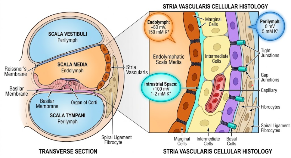
The biophysical origin of the EP was debated for decades until seminal microelectrode recordings elucidated the “two-diffusion-potential” mechanism.(Nin et al., 2008) The basal cells, intermediate cells, and fibrocytes of the spiral ligament are coupled by extensive connexin gap junctions (chiefly Connexin 26 and Connexin 30), effectively forming a continuous electrical syncytium.(Wangemann, 2002) Because this syncytium is connected to perilymph and possesses a massive aggregate surface area and capacitance, its resting membrane potential is clamped near 0 to –5 mV.(Salt et al., 1987)
Within the intrastrial space, the concentration of potassium is kept exceptionally low—ranging between 1 and 2 mM.(Nin et al., 2008) In contrast, the cytoplasm of intermediate cells within the syncytium maintains a high potassium concentration of approximately 100 to 140 mM.(Hiroshi Hibino & Kurachi, 2006) Intermediate cells express an extraordinarily high density of inwardly rectifying potassium channels, Kir4.1, on their apical membranes facing the intrastrial space.(H. Hibino et al., 1997) Driven by the steep chemical concentration gradient between the intermediate cell cytoplasm and the intrastrial fluid, K+ diffuses out through Kir4.1 channels into the intrastrial space.(Salt et al., 1987)
Because the basal cell layer forms a high-resistance electrical barrier sealed by Claudin-11 tight junctions, positive charge cannot leak laterally into the spiral ligament.(Gow et al., 2004; S. Kitajiri et al., 2004) Consequently, this K+ efflux produces an enormous positive diffusion potential across the intermediate cell membrane, driving the intrastrial space potential (ISP) up to +90 to +100 mV relative to perilymph. (Nin et al., 2008)
The second diffusion potential occurs across the apical membrane of strial marginal cells. Marginal cells actively clear K+ from the intrastrial space using basolateral Na+/K+-ATPase pumps and Na+-K+-2Cl- cotransporters (NKCC1), concentrating K+ within their cytoplasm up to ~70–80 mM while keeping intrastrial K+ at baseline trace levels.(Salt et al., 1987) At the apical membrane facing the scala media, voltage-gated KCNQ1/KCNE1 potassium channels allow for the diffusion of K+ into the endolymph.(Takeuchi, Ando, & Kakigi, 2000) Therefore, the total EP recorded in the endolymph (+80 mV) is the algebraic sum of the intrastrial potential, the electrical resistance of the marginal cell epithelium, and the trans-apical marginal cell diffusion potential.(Nin et al., 2008)
The integrity of the endocochlear potential is fundamentally governed by the biophysical fidelity of its principal potassium channels. Selective deletion or loss of function of either Kir4.1 or KCNQ1/KCNE1 results in rapid or progressive collapse of EP and total loss of sensory transduction.(Estévez et al., 2001)
Kir4.1, encoded by KCNJ10, is an ATP-sensitive, inward rectifying potassium channel that is selectively expressed on the apical digitations of intermediate cells.(H. Hibino et al., 1997) Under physiological conditions, its open probability ensures high resting K+ conductance. The crucial role of Kir4.1 was definitively established in Kcnj10-null mice: targeted ablation of this channel abolishes the EP completely, dropping it from +80–100 mV to 0 mV.(Estévez et al., 2001) Remarkably, while the EP is obliterated in Kcnj10-null mice, the endolymphatic K+ concentration in the cochlea decreases by only ~50%, and the vestibular endolymphatic potential and vestibular K+ levels remain unaffected. This demonstrates that Kir4.1 is not required merely for bulk potassium transit, but is the non-redundant generator of the positive trans-intermediate potential.(H. Hibino et al., 1997)
Pharmacological studies with barium (Ba2+), a potent blocker of Kir channels, applied through the vasculature reproduce the knockout phenotype with an immediate reduction of ISP and EP without an immediate effect on bulk fluid volumes.(Keithley, 2020) In human pathology, loss-of-function mutations in KCNJ10 cause EAST/SeSAME syndrome (epilepsy, ataxia, sensorineural deafness, and tubulopathy), directly tying the human channelopathy to strial bioelectric failure.
Moreover, Kir4.1 expression is tightly coupled to intermediate cell physiology. In mouse models of Pendred syndrome (Slc26a4 deficiency), the primary strial defect is a progressive down-regulation and ultimate disappearance of Kir4.1 from intermediate cells, which leads to total loss of the EP, severe strial edema, and irreversible neuroepithelial damage.(Ito, Nishio, Wangemann, & Griffith, 2015)
The slow voltage-gated potassium channel complex composed of the pore-forming α-subunit KCNQ1 (KvLQT1) and the single transmembrane β-regulatory subunit KCNE1 (IsK or minK) is expressed at the apical pole of marginal cells at the epithelia of the stria vascularis.(Vetter et al., 1996) This heteromeric channel has unusual gating kinetics, it opens slowly upon depolarization and stays open to allow massive, sustained K+ excretion against a standing positive voltage into the scala media.(Rivas & Francis, 2005)
Extending findings discussed in our prior review which showed mitochondrial DNA lesions and reactive oxygen species (ROS) cascades that disrupted structural outer hair cell proteins and baseline bioenergetics.(Keithley, 2020), similar metabolic and oxidative stresses wreak havoc on strial marginal cell ion channels. One of the highest mitochondrial densities in the whole organism is found in marginal cells.(Hiroshi Hibino & Kurachi, 2006) Oxidative stress or genetic insults impair marginal cell viability and dissociate KCNQ1/KCNE1 channel clustering and surface trafficking toward the apical microvilli.(Keithley, 2020)
Mutations in human KCNQ1 or KCNE1 cause Jervell and Lange-Nielsen syndrome (JLNS) characterized by severe bilateral sensorineural deafness and cardiac arrhythmias (long QT interval). Marginal cells in Kcnq1- and Kcne1-knockout mice are unable to secrete K+ into the endolymph, thereby precluding normal EP formation, leading to complete dehydration and architectural collapse of the scala media (endolymphatic collapse).(Tranebjaerg, Bathen, Tyson, & Bitner-Glindzicz, 1999) shown in Figure 2., the spatial distribution and functional interaction of Kir4.1 and KCNQ1/KCNE1 show how isolated channelopathy can disturb the entire strial circuit, resulting in a sharp decline of endolymphatic potential.
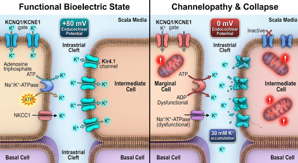
Figure 2. Molecular Failure of Strial Potassium Channels and the Breakdown of the Intrastrial Bioelectric Circuit.
4. Upstream Transport Collapse: The Metabolic Fragility of Na+/K+-ATPase, NKCC1, and Syncytial Coupling
While Kir4.1 produces the ISP, its ability to do so depends entirely on the upstream maintenance of an ultra-low potassium concentration in the intrastrial space. If K+ accumulates in this microscopic cleft, the chemical gradient across the intermediate cell apical membrane is erased, collapsing the equilibrium potential (EK) to 0.(Hiroshi Hibino & Kurachi, 2006)
The low-K+ microenvironment of the intrastrial space is cleared primarily by two transport systems situated in the heavily infolded basolateral membranes of marginal cells: the Na+/K+-ATPase catalytic pump and the electroneutral Na+-K+-2Cl- cotransporter 1 (NKCC1, encoded by SLC12A2).(S. I. Kitajiri et al., 2004)
The physiological interdependence of these transporters is absolute. Perfusion with ouabain (a specific inhibitor of Na+/K+-ATPase) or bumetanide/furosemide (inhibitors of NKCC1) causes intrastrial K+ to surge from ~2 mM to over 25–30 mM.(Kakigi et al., 2002) Consequently, the ISP drops from +75 mV to less than +20 mV, and the EP drops precipitously.
Furthermore, because Na+/K+-ATPase is an enzymatic motor that consumes immense metabolic energy, it represents the primary point of vulnerability during cochlear hypoxia, ischemia, and aging.(Keithley, 2020) Experimental anoxia suppresses Na+/K+-ATPase activity within minutes, triggering rapid intrastrial K+ accumulation and an immediate plunge in EP from +80 mV into negative territory (–30 to –40 mV), a negative potential driven by the organ of Corti’s remaining hair cell conductance.(Nin et al., 2008)
Potassium exiting hair cells during auditory reception does not simply dissipate into the general circulation; it is taken up by Deiters’ supporting cells and shuttled back to the cochlear lateral wall via a complex recycling network.(Hiroshi Hibino & Kurachi, 2006) Types II, IV, and V fibrocytes in the spiral ligament express high levels of Na+/K+-ATPase (α1 and α2 isoforms) and NKCC1, actively absorbing K+ from the perilymph.These fibrocytes also express the inwardly rectifying channel Kir5.1, which fine-tunes local K+ buffering.(Takumi et al., 1995)
Once inside the fibrocytes, K+ travels through the connective-tissue gap junction syncytium directly into strial basal and intermediate cells. Mutations in the GJB2 (Connexin 26) and GJB6 (Connexin 30) genes, which cause the most common form of congenital nonsyndromic hereditary deafness (DFNB1 and DFNA3), affect this intercellular syncytial bridge. In the absence of functional gap junction hemichannels, intermediate cells fail to efficiently replenish K+, thereby starving the Kir4.1 channel of its ionic substrate and destabilizing lateral wall bioenergetics.(Kikuchi, Adams, Miyabe, So, & Kobayashi, 2000)
The hallmark of metabolic presbycusis, acoustic trauma recovery failures, and hereditary inner ear dysplasias is the degeneration of the stria vascularis.(Bovee et al., 2024) The failure of strial function is not sudden but occurs in discrete histopathological stages, from acute metabolic exhaustion and edema to permanent architectural atrophy.(Ito et al., 2015)
Age-related loss of strial Na+/K+-ATPase has been demonstrated in quiet-aged mammalian models such as the aging Mongolian gerbil (Meriones unguiculatus) and CBA/CaJ mice through structural and quantitative immunohistochemical analyzes.(Keithley, 2020) In CBA/CaJ mice strial Na+/K+-ATPase expression is reduced by up to 80% with advanced age even when total strial thickness is reduced by only ~20%.(Bovee et al., 2024) This critical mismatch indicates that molecular transport failure precedes gross anatomical tissue loss: the strial battery loses its power long before its cellular components vanish entirely.(Thulasiram et al., 2022)
This enzymatic decrease is associated with local vascular regression. High-resolution corrosion casts and transmission electron microscopy reveal considerably reduced capillary diameters, thickening of the vascular basement membrane, and loss of strial pericytes, especially in the extreme basal and apical turns of the cochlea. (Keithley, 2020) Because basal turns demand the greatest rate of active K+ throughput to sustain high-frequency auditory amplification, vascular rarefaction and hypoperfusion hit the basal stria hardest, explaining the pronounced high-frequency hearing loss observed in early presbycusis. (Keithley, 2020)
At the subcellular level, aging strial marginal cells show pronounced regression of their secondary and primary basolateral interdigitations. Ultrastructural evidence reveals that local mitochondrial self-damage in marginal cell foot-processes diminishes ATP production, starving the membrane-bound Na+/K+-ATPase and causing structural retraction of the interdigitating membranes. Over time, this degradation flattens the complex strial architecture into a thin, disorganized, non-functional squamous monolayer. (Keithley, 2020)
The response of the stria vascularis to metabolic stress is biphasic, as shown in mouse models of fluctuating and progressive deafness such as Slc26a4-insufficient (DE17.5) mice. In the early stages or in the milder forms of insult the stria vascularis is markedly thickened and edematous.(Ito et al., 2015) This strial edema is due to osmotic imbalance in the intrastrial space. If Na+/K+-ATPase or NKCC1 cannot remove K+ and accompanying ions fast enough from the intrastrial space, water will enter the intrastrial cleft and swell the tissue.(Salt et al., 1987) At this stage of edema, loss of EP and elevations in auditory thresholds are still largely reversible if metabolic homeostasis can be reestablished. (Ito et al., 2015)
Conversely, chronic unrelieved metabolic stress transforms reversible edema into irreversible atrophy. In mice with profound hearing loss there is extensive degeneration of intermediate and marginal cells.(Ito et al., 2015) Immunohistochemistry shows complete loss of Kir4.1 in intermediate cells, severe disorganization of the apical geometry of marginal cells, downregulation of KCNQ1, and heavy accumulation of pigment granules. Furthermore, tissue-resident macrophages are recruited and activated, promoting phagocytic clearance of collapsed strial elements and permanently consolidating the loss of the endocochlear potential.(Ito et al., 2015) As shown in Figure 3., this pathological progression traces the clear morphologic transition from a healthy, interdigitated strial epithelium into edematous swelling, followed by terminal atrophic thinning and voltage collapse.
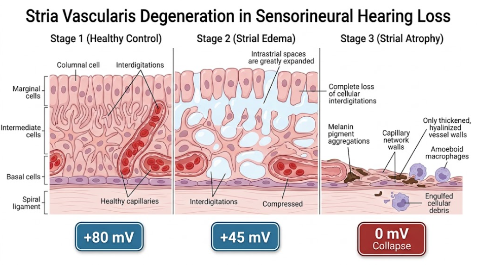
Figure 3. Pathological Spectrum of Strial Degeneration from Metabolic Edema to Irreversible Atrophy.
The finding that stria vascularis degeneration can be a primary and independent cause of sensorineural hearing loss, implies a need to re-evaluate current diagnostic and therapeutic approaches to the inner ear. Pure tone audiometry has traditionally been unable to differentiate between sensory presbycusis (death of hair cells) and metabolic presbycusis (atrophy of the stria).(Keithley, 2020) However, physiological and biophysical modeling confirms that metabolic presbycusis typically produces a slowly progressive, flatter audiometric threshold shift that impacts low-to-mid frequencies earlier than classical high-frequency sensory presbycusis. Recent algorithmic decompositions of human audiometric databases, validated by post-mortem histopathology, confirm that loss of the endocochlear potential is the principal contributor to hearing threshold shifts across large cohorts of aging adults.(Keithley, 2020)
Nevertheless, non-invasive clinical evaluation of the stria vascularis in living human patients remains a formidable challenge. Direct electrophysiological recording of the EP requires an invasive technique of advancing a microelectrode through the round window into the scala media, which is limited to animal models.(Thulasiram et al., 2022) Consequently, clinical diagnostics must depend on indirect surrogates such as electrocochleography (measuring the summating potential and compound action potential) and ultra-high-resolution magnetic resonance imaging of inner ear fluid dynamics and the integrity of the cochlear blood-labyrinth barrier.(Vlajkovic et al., 2026)
One of the major pathologies identified on the therapeutic front, potassium channelopathies and strial transport failure, opens promising translational avenues:
The endocochlear potential is the fundamental bioelectric driver of mammalian audition, powering the sensory hair cell transduction channels and outer hair cell cochlear amplifier. This voltage is not an intrinsic property of sensory hair cells, but the product of a biophysical battery within the stria vascularis. As detailed in this review, EP generation hinges upon a dual-diffusion potential system governed by the precise spatial arrangement and activity of intermediate cell Kir4.1 channels, apical marginal cell KCNQ1/KCNE1 complexes, and basolateral Na+/K+-ATPase and NKCC1 transporters, all insulated by Claudin-11 tight junctions.
When age-related vascular regression, chronic oxidative damage, or inherited genetic variants degrade these potassium channels and active pumps, this homeostatic engine collapses. The progression from early bioenergetic insufficiency and strial edema to permanent, irreversible strial atrophy explains a substantial portion of age-related and progressive hearing loss. Future therapeutic advances will necessitate a transition from conventional sensory prostheses to precision, mechanism-based interventions—using targeted gene delivery, bioenergetic stabilization, and vascular preservation to restore the inner ear’s battery before irreversible structural atrophy seals its silence.
BÉKÉSy, G. V. (1952). Resting Potentials Inside the Cochlear Partition of the Guinea Pig. Nature, 169(4293), 241-242. doi:10.1038/169241a0
Bovee, S., Klump, G. M., Köppl, C., & Pyott, S. J. (2024). The Stria Vascularis: Renewed Attention on a Key Player in Age-Related Hearing Loss. Int J Mol Sci, 25(10). doi:10.3390/ijms25105391
Dubno, J. R., Eckert, M. A., Lee, F. S., Matthews, L. J., & Schmiedt, R. A. (2013). Classifying human audiometric phenotypes of age-related hearing loss from animal models. J Assoc Res Otolaryngol, 14(5), 687-701. doi:10.1007/s10162-013-0396-x
Esteves, F., & Caria, H. (2026). Genetic and Environmental Factors Shaping Hearing Loss: Xenobiotics, Mechanisms and Translational Perspectives. Journal of Xenobiotics, 16(1), 27.
Estévez, R., Boettger, T., Stein, V., Birkenhäger, R., Otto, E., Hildebrandt, F., & Jentsch, T. J. (2001). Barttin is a Cl- channel beta-subunit crucial for renal Cl- reabsorption and inner ear K+ secretion. Nature, 414(6863), 558-561. doi:10.1038/35107099
Gow, A., Davies, C., Southwood, C. M., Frolenkov, G., Chrustowski, M., Ng, L., . . . Kachar, B. (2004). Deafness in Claudin 11-null mice reveals the critical contribution of basal cell tight junctions to stria vascularis function. J Neurosci, 24(32), 7051-7062. doi:10.1523/jneurosci.1640-04.2004
Hibino, H., Horio, Y., Inanobe, A., Doi, K., Ito, M., Yamada, M., . . . Kurachi, Y. (1997). An ATP-dependent inwardly rectifying potassium channel, KAB-2 (Kir4. 1), in cochlear stria vascularis of inner ear: its specific subcellular localization and correlation with the formation of endocochlear potential. J Neurosci, 17(12), 4711-4721. doi:10.1523/jneurosci.17-12-04711.1997
Hibino, H., & Kurachi, Y. (2006). Molecular and Physiological Bases of the K+ Circulation in the Mammalian Inner Ear. Physiology, 21(5), 336-345. doi:10.1152/physiol.00023.2006
Ito, T., Nishio, A., Wangemann, P., & Griffith, A. J. (2015). Progressive irreversible hearing loss is caused by stria vascularis degeneration in an Slc26a4-insufficient mouse model of large vestibular aqueduct syndrome. Neuroscience, 310, 188-197. doi:10.1016/j.neuroscience.2015.09.016
Kakigi, A., Takeuchi, S., Ando, M., Higashiyama, K., Azuma, H., Sato, T., & Takeda, T. (2002). Reduction in the endocochlear potential caused by Cs(+) in the perilymph can be explained by the five-compartment model of the stria vascularis. Hear Res, 166(1-2), 54-61. doi:10.1016/s0378-5955(01)00412-9
Keithley, E. M. (2020). Pathology and mechanisms of cochlear aging. J Neurosci Res, 98(9), 1674-1684. doi:10.1002/jnr.24439
Kikuchi, T., Adams, J. C., Miyabe, Y., So, E., & Kobayashi, T. (2000). Potassium ion recycling pathway via gap junction systems in the mammalian cochlea and its interruption in hereditary nonsyndromic deafness. Med Electron Microsc, 33(2), 51-56. doi:10.1007/s007950070001
Kikuchi, T., Kimura, R. S., Paul, D. L., & Adams, J. C. (1995). Gap junctions in the rat cochlea: immunohistochemical and ultrastructural analysis. Anat Embryol (Berl), 191(2), 101-118. doi:10.1007/bf00186783
Kirwin, D. A., Lewis, M. A., & Steel, K. P. (2026). A Systematic Review of Genes Affecting Endocochlear Potential. J Assoc Res Otolaryngol, 27(3), 361-378. doi:10.1007/s10162-026-01046-y
Kitajiri, S., Miyamoto, T., Mineharu, A., Sonoda, N., Furuse, K., Hata, M., . . . Tsukita, S. (2004). Compartmentalization established by claudin-11-based tight junctions in stria vascularis is required for hearing through generation of endocochlear potential. J Cell Sci, 117(Pt 21), 5087-5096. doi:10.1242/jcs.01393
Kitajiri, S. I., Furuse, M., Morita, K., Saishin-Kiuchi, Y., Kido, H., Ito, J., & Tsukita, S. (2004). Expression patterns of claudins, tight junction adhesion molecules, in the inner ear. Hear Res, 187(1-2), 25-34. doi:10.1016/s0378-5955(03)00338-1
Ma, Y., Wise, A. K., Shepherd, R. K., & Richardson, R. T. (2019). New molecular therapies for the treatment of hearing loss. Pharmacology & Therapeutics, 200, 190-209. doi:https://doi.org/10.1016/j.pharmthera.2019.05.003
Nin, F., Hibino, H., Doi, K., Suzuki, T., Hisa, Y., & Kurachi, Y. (2008). The endocochlear potential depends on two K+ diffusion potentials and an electrical barrier in the stria vascularis of the inner ear. Proc Natl Acad Sci U S A, 105(5), 1751-1756. doi:10.1073/pnas.0711463105
Rivas, A., & Francis, H. (2005). Inner Ear Abnormalities in a Kcnq1 (Kvlqt1) Knockout Mouse: A Model of Jervell and Lange-Nielsen Syndrome. Otology & Neurotology, 26(3), 415-424. doi:10.1097/01.mao.0000169764.00798.84
Salt, A. N., Melichar, I., & Thalmann, R. (1987). Mechanisms of endocochlear potential generation by stria vascularis. Laryngoscope, 97(8 Pt 1), 984-991.
Takeuchi, S., & Ando, M. (1998). Dye-coupling of melanocytes with endothelial cells and pericytes in the cochlea of gerbils. Cell Tissue Res, 293(2), 271-275. doi:10.1007/s004410051118
Takeuchi, S., Ando, M., & Kakigi, A. (2000). Mechanism generating endocochlear potential: role played by intermediate cells in stria vascularis. Biophys J, 79(5), 2572-2582. doi:10.1016/s0006-3495(00)76497-6
Takumi, T., Ishii, T., Horio, Y., Morishige, K., Takahashi, N., Yamada, M., . . . et al. (1995). A novel ATP-dependent inward rectifier potassium channel expressed predominantly in glial cells. J Biol Chem, 270(27), 16339-16346. doi:10.1074/jbc.270.27.16339
Thulasiram, M. R., Ogier, J. M., & Dabdoub, A. (2022). Hearing Function, Degeneration, and Disease: Spotlight on the Stria Vascularis. Front Cell Dev Biol, 10, 841708. doi:10.3389/fcell.2022.841708
Tranebjaerg, L., Bathen, J., Tyson, J., & Bitner-Glindzicz, M. (1999). Jervell and Lange-Nielsen syndrome: a Norwegian perspective. Am J Med Genet, 89(3), 137-146.
Vetter, D. E., Mann, J. R., Wangemann, P., Liu, J., McLaughlin, K. J., Lesage, F., . . . Barhanin, J. (1996). Inner ear defects induced by null mutation of the isk gene. Neuron, 17(6), 1251-1264. doi:10.1016/s0896-6273(00)80255-x
Vlajkovic, S. M., Suzuki-Kerr, H., & Nayagam, B. A. (2026). Cochlear Homeostasis in Sensorineural Hearing Loss: Mechanisms, Implications, and Therapeutic Prospects. International Journal of Molecular Sciences, 27(1), 102.
Wangemann, P. (2002). K+ cycling and the endocochlear potential. Hear Res, 165(1-2), 1-9. doi:10.1016/s0378-5955(02)00279-4
Wangemann, P. (2006). Supporting sensory transduction: cochlear fluid homeostasis and the endocochlear potential. J Physiol, 576(Pt 1), 11-21. doi:10.1113/jphysiol.2006.112888
Wilson, B. S., Tucci, D. L., O’Donoghue, G. M., Merson, M. H., & Frankish, H. (2019). A Lancet Commission to address the global burden of hearing loss. The Lancet, 393(10186), 2106-2108. doi:10.1016/S0140-6736(19)30484-2