Our Services
Others
- DFPP
- Shockwave
- IV Drip

Patient Profile: Adult male | History of Previous Contralateral Knee Surgery & High-Impact Activity
The medical team evaluated an adult male patient presenting with recurrent left knee swelling and mechanical symptoms. The patient has a demanding occupational history requiring prolonged standing and is an active surfer. While initial conservative management with physiotherapy yielded temporary symptom relief, a recent return to surfing precipitated a significant exacerbation of joint effusion and pain.
The patient’s clinical history is further complicated by a prior meniscal tear in the contralateral (right) knee during his youth, which was managed surgically but continues to present minor residual issues. This bilateral history suggests altered lower-extremity biomechanics and compensatory loading, which the medical team must account for in his long-term recovery strategy.
To determine the precise structural pathology driving the recurrent swelling, the medical team reviewed the patient’s non-contrast 1.5T MRI of the left knee (referencing the medical report “Francesco Angeloni.pdf”). The imaging demonstrated several significant findings:
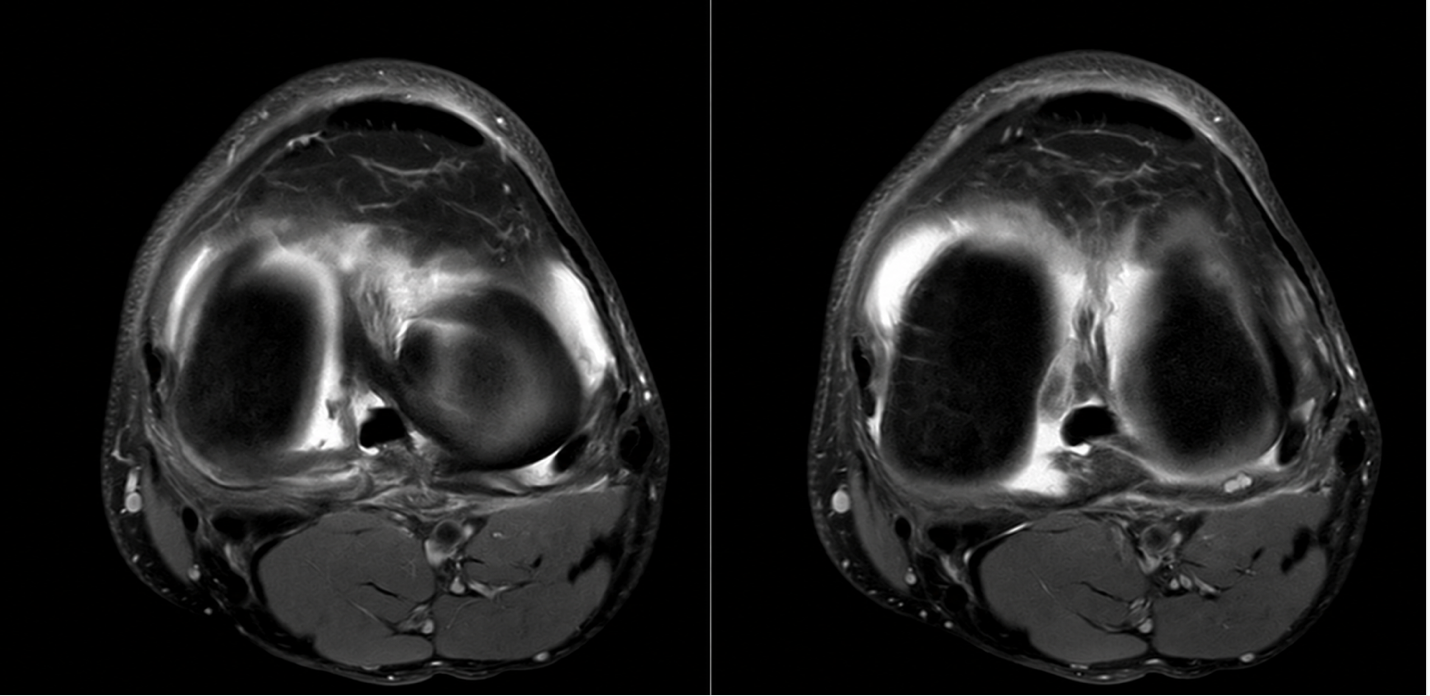
Figure 1. Axial MRI Images of the Knee Joint Demonstrating Joint Effusion.
In this case, the medical team recommends distinguishing clearly between the biological inflammatory symptoms and the structural mechanical defects. Because the patient has a complex meniscal root tear and a full-thickness cartilage defect, conservative therapy and biological injections alone cannot physically repair the torn tissue or restore the mechanical stability of the joint.
The primary recommended intervention is an arthroscopic surgical approach. This allows the surgeon to directly address the meniscal pathology through meniscal repair or partial meniscectomy with reshaping, depending on the intraoperative tissue quality. Concurrently, a microfracture procedure can be performed to address the focal full-thickness cartilage defects. Treatment of the ACL will be determined based on clinical instability assessments and intraoperative findings.
Following the structural correction of the knee, the medical team positions targeted cell therapy as an adjunctive biological support option to enhance the postoperative healing environment.
Rather than using cell therapy as a substitute for surgery, the proposed protocol integrates an intra-articular administration of supportive cell therapy at approximately 3 months postoperatively, contingent on the patient’s recovery trajectory. The goals of this biological support are to:
It is clearly communicated that cell therapy is designed to optimize the biological conditions for recovery, but cannot physically suture a meniscus or reconstruct an ACL.
The medical team’s recommendation is to manage this case through a comprehensive, staged approach combining structural, biological, and rehabilitative strategies:
This integrative pathway provides the patient with a medically responsible, structurally sound treatment plan while utilizing advanced cellular therapies safely within their evidence-based, supportive capabilities.