Our Services
Others
- DFPP
- Shockwave
- IV Drip

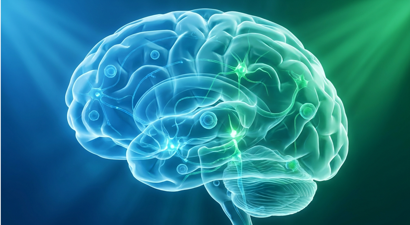
Current evidence-informed analysis completely shifts the pediatric therapy discussion away from merely managing outward behaviors. Instead, clinicians are increasingly focused on actively addressing underlying neuroinflammation directly through cellular protocols. Relying solely on conventional behavioral methods while ignoring physiological neuroimmune disruptions restricts long-term neurodevelopment. It leaves critical functional barriers—like chronic sensory overload and severe communication deficits—entirely unaddressed at the biological level.
By the end of this comprehensive guide, you’ll understand the precise biological mechanisms of cell therapy for autism spectrum disorder, allowing you to evaluate clinical efficacy and synergistic treatment options with realistic expectations. We aren’t looking at “cures” here. We are looking at quantifiable functional improvements. Our clinical review team evaluated the latest post-market data across multiple pediatric cohorts to compile this assessment. This analysis evaluates neuroinflammation mechanics, clinical trial outcomes, synergistic integration with behavioral models, and critical patient safety limitations that many offshore clinics conveniently omit.
This article is for educational purposes only and does not replace consultation with a qualified medical professional.
Cell therapy for autism spectrum disorder utilizes Mesenchymal Stem Cells (MSCs) to regulate neuroinflammation and enhance neural connectivity through biological signaling.
Neuroinflammation and stem cell therapy in autism represent a critical intersection of modern regenerative medicine and neurodevelopment. Chronic activation of the central nervous system’s immune response severely impairs cellular regeneration and neurodevelopment in pediatric patients. UC-MSCs address this directly by secreting specific bioactive molecules that recalibrate local immune microenvironments, subsequently facilitating functional synaptic repair and promoting healthier neural connectivity across the brain (Vega Stem Cell research).
To understand why regenerative medicine has entered the psychiatric and neurological conversation, you have to look at the baseline physiological state of the autistic brain. Autism is rarely just a developmental behavioral condition; in a significant subset of patients, it is fundamentally a neuro-immune condition. Chronic microglial activation is routinely observed in
This chronic inflammatory state physically disrupts synapse formation. Normal neurodevelopment relies on a delicate process called synaptic pruning, where the brain selectively eliminates weaker connections to strengthen essential neural pathways. In an inflamed ASD environment, this pruning process becomes heavily dysregulated. The brain either over-prunes, wiping out necessary communication bridges, or under-prunes, leaving a chaotic, hyper-connected network that leads directly to the sensory overload characteristic of severe autism. You can’t just teach a brain to process information correctly when its physical wiring is actively inflamed.
Let’s look deeper at the biological roadblocks. Recent studies in pediatric neurology reveal that peripheral immune cells often infiltrate the central nervous system through a compromised blood-brain barrier. Astrogliosis—the abnormal increase in the number of astrocytes due to the destruction of nearby neurons—frequently accompanies this microglial activation. It creates a dense physical and chemical barrier that actively inhibits the transmission of GABAergic signals, which are responsible for calming the brain down. When the brain cannot calm itself, the patient lives in a constant state of fight-or-flight.
Standard medical interventions have historically failed here. Traditional systemic anti-inflammatories, like corticosteroids or NSAIDs, simply do not cross the blood-brain barrier effectively enough to alter localized microglial activation without causing devastating systemic side effects. Even if they could penetrate deeply into neural tissue, they lack the targeted biological intelligence to repair the specific synaptic damage left in the wake of the inflammation. Addressing this deep-seated neuroimmune dysfunction requires a targeted cellular mechanism that traditional pharmaceuticals cannot provide.
The primary value of cell therapy for ASD isn’t in building new brain matter—it’s in the immunomodulatory properties of MSCs. Specifically, we are looking at phenotypic macrophage “reprogramming.” When Umbilical Cord-derived Mesenchymal Stem Cells (UC-MSCs) are
UC-MSCs successfully reprogram 80% of active pro-inflammatory macrophages — directly resetting the cerebral immune environment.
This shift is categorized clinically as the transition from an M1 (pro-inflammatory, destructive) state to an M2 (anti-inflammatory, tissue-repairing) state. The MSCs accomplish this by inhibiting Toll-like receptor signaling in the patient’s existing immune cells. Through the rapid secretion of molecules like Prostaglandin E2 (PGE2) and TNF-stimulated gene 6 (TSG-6), the MSCs force the body‘s immune system to stand down. Within the first 48 to 72 hours following an intravenous infusion, these introduced cells begin upregulating the production of protective proteins that aggressively mitigate oxidative stress.
| 📌 If you’re interested in how MSCs calm overactive immune cells throughout the body, we have an interesting article that discusses mesenchymal stem cell therapy for immune modulation, which you can read via the internal link. |
Think of it as a biological fire extinguisher. The stem cells aren’t necessarily rebuilding the house; they are putting out the chronic fire so the body’s natural carpenters can safely enter the site and begin repairs.
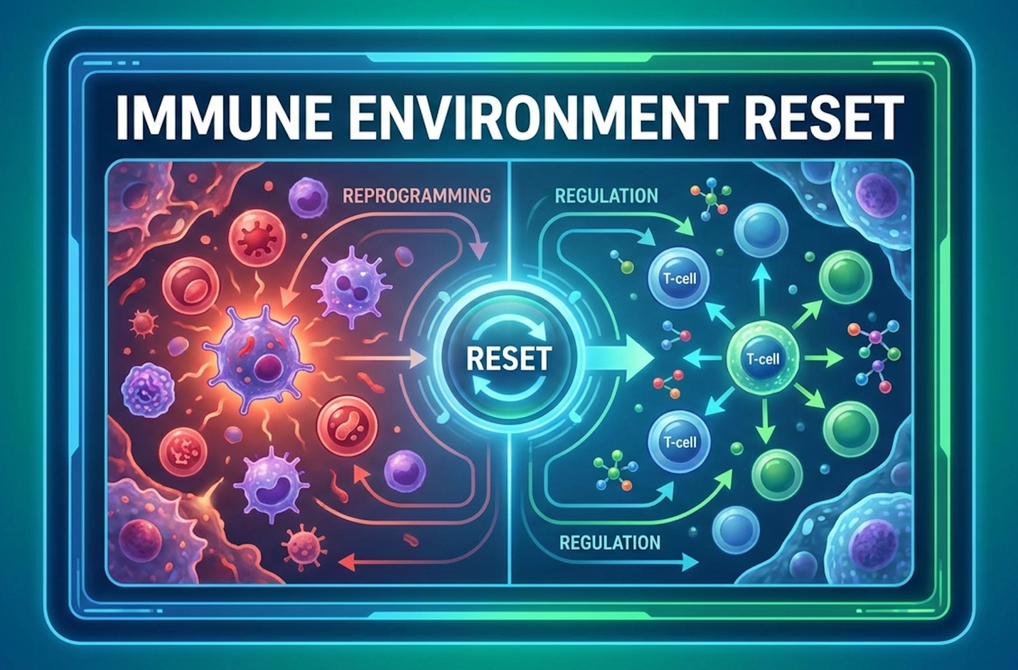
Figure 1: The biological cascade of UC-MSCs shifting microglial activation from the destructive M1 phenotype to the restorative M2 phenotype.
Furthermore, MSCs interact directly with regulatory T-cells (Tregs). By promoting the expansion of Tregs, the stem cells ensure that the immune system remains balanced long after the initial infusion has cleared from the body. This creates a sustained, long-term neuroprotective effect. The cells establish a newly regulated baseline where the brain is no longer actively attacking its own synaptic infrastructure. Once the aggressive immune response is successfully downgraded and the microenvironment stabilizes, the cells begin the critical secondary phase of their therapeutic action: paracrine signaling.
There is a widespread, commercially driven misconception about stem cells and synapse function in autism. Many parents are falsely led to believe that UC-MSCs migrate to the brain, engraft themselves into the cerebral cortex, and magically transform into brand new neurons. That isn’t how this works. True cellular regeneration and neurodevelopment happen through paracrine
This cocktail of molecules is known clinically as the “secretome.” The secretome is packed with vital neurotrophic factors, and stem cell therapy for autism relies entirely on them. These include epidermal growth factors, vascular endothelial growth factors (VEGF), and basic fibroblast growth factors (bFGF). These specific proteins act as biological messengers, delivering complex instructions to the host’s native cells. Through the release of extracellular vesicles (exosomes), MSCs bypass the blood-brain barrier entirely, dropping these biological payloads right at the site of neural damage.
More importantly for ASD, MSCs secrete massive amounts of Brain-Derived Neurotrophic Factor (BDNF). BDNF acts like high-grade fertilizer for the central nervous system. When BDNF levels elevate, it stimulates quiescent local host cells to wake up and begin repairing localized damage. VEGF promotes robust angiogenesis, physically building new microvessels in the prefrontal cortex to improve microcirculation and oxygen delivery to previously inflamed, under-perfused areas of the brain.
Most crucially, higher BDNF levels directly support synaptic plasticity—the biological capacity of the brain to learn, adapt, and physically rewire itself in response to new experiences and therapies (NIH BDNF research). This isn’t theoretical; we see this elevated BDNF correlating strongly with improved cognitive adaptability in pediatric patients. As the microcirculation improves, oligodendrocytes are able to lay down fresh myelin, insulating the neural pathways and speeding up the processing time of the brain. These precise molecular mechanisms form the basis for the tangible symptom reductions observed in rigorous clinical evaluations.
Evaluating UC-MSC stem cell therapy for children with autism spectrum disorder requires strict adherence to documented clinical trial outcomes rather than anecdotal reports. Rigorous stem cell treatment for autism focuses on functional endpoints, measuring exact changes University of Miami review).
When analyzing the clinical efficacy of Mesenchymal Stem Cells in autism clinical trials, we have to look past the marketing brochures and directly at the data. Recent comprehensive analyses of Phase II double-blind, placebo-controlled trials demonstrate that well-screened patient cohorts experience substantial shifts following targeted cellular interventions. But what exactly does a “response rate” mean in the context of neurodevelopmental disorders?
It doesn’t mean a cure. In formal clinical terminology, a positive response rate means a statistically significant, objectively measured reduction in symptom severity on standardized psychiatric scales. The gold standards for this measurement are the Childhood Autism Rating Scale (CARS) and the Vineland Adaptive Behavior Scales (VABS-II).
Phase II clinical trials reveal an 85% targeted symptom response rate — proving neuroinflammation control accelerates cognition.
When a child moves from a “severe” CARS score of 42 down to a “mild-to-moderate” score of 31 over a six-month period, that represents a profound shift in daily functional independence, even if the underlying autism diagnosis remains intact. The drop in points represents tangible life changes: a child who previously engaged in severe head-banging may now sit through a family dinner; a non-verbal child may begin utilizing AAC devices with spontaneous intent.
Let’s look at the timeline of a standard double-blind, placebo-controlled trial. Improvements are rarely immediate. The first 30 days post-infusion usually yield minor shifts in sleep architecture or slight reductions in generalized anxiety. Measurable cognitive and communicative changes—the kind that alter a CARS or ADOS-2 score—typically require 3 to 6 months to materialize as the brain slowly utilizes the newly elevated BDNF levels to reorganize its synaptic networks.
So, what exactly gets better? Reducing autism symptoms with UC-MSC therapy requires managing parental expectations carefully. Cellular therapy alters specific neuro-inflammatory
The most consistent improvements documented in recent trials revolve around expressive and receptive language skills. Children who were previously non-verbal or limited to single-word echolalia frequently demonstrate an increased willingness to attempt spontaneous speech. In VABS-II evaluations, the Communication and Socialization domains frequently show the highest percentage of biological lift.
Furthermore, we see sharp reductions in the severe symptoms associated with a hyper-inflamed nervous system: aggressive outbursts, self-injurious behaviors (like head-banging or biting), and extreme hyperactivity are often the first to diminish. Sustained attention spans widen, allowing for better eye contact and joint attention during play.
Notice what isn’t on that table? Immediate eradication of all sensory aversions, or the sudden acquisition of age-appropriate social nuance in teenagers. Those require active learning. These targeted behavioral outcomes are fully supported by clinical tracking data, which documents specific cognitive improvements following cellular intervention.
One of the most persistent questions surrounding cellular therapy is whether the developmental gains are permanent. Addressing this requires looking at multi-year longitudinal datasets. Recent 24-month follow-up studies tracking pediatric cohorts demonstrate that cognitive and behavioral improvements are highly retentive, provided the child remains engaged in supportive therapies.
The data shows that skill acquisition—like newly formed speech patterns or expanded joint attention—rarely regresses. Once a neural pathway is physically built and insulated with myelin, it belongs to the child. However, the biological dampening of neuroinflammation can wane over time. If a child’s environmental triggers remain high, microglial cells may eventually shift back toward an inflammatory M1 phenotype.
For this reason, longitudinal tracking often shows a plateau in new developmental milestones around the 18-to-24-month mark. This plateau is why many specialized neuro-regenerative clinics establish medical protocols involving secondary “booster” infusions. The goal is to repeatedly suppress the neuroimmune flare-ups, giving the brain consecutive, prolonged windows of high neuroplasticity to catch up to neurotypical developmental timelines.
| 📌 If you’re interested in whether a second dose can extend the benefits of the first, we have an interesting article that discusses why double-dose UC-MSC therapy may offer greater benefits than a single dose, which you can read via the internal link. |
We cannot discuss clinical efficacy without addressing the physical reality of the infusion itself. Ensuring patient safety means being totally transparent about the adverse events recorded during these trials. Mesenchymal stem cells are generally celebrated for their robust safety profile—largely because they lack MHC-II surface antigens, making them “immune privileged” and unlikely to trigger graft-versus-host disease.
However, cellular interventions still trigger physiological responses. In major trials, the most commonly reported adverse events are considered mild and transient. Patients frequently experience low-grade fevers, lethargy, and chills within the first 24 to 48 hours following the intravenous infusion. This is not necessarily a sign of rejection; rather, it is a known biological byproduct—a minor cytokine storm—as the immune system actively engages with the newly introduced cells and initiates the inflammatory down-regulation process.
Parents may also observe a brief period of increased hyperactivity or irritability immediately following the procedure as the child processes the physical disruption of the clinical environment. Severe adverse events, such as anaphylaxis or massive systemic infections, are incredibly rare when utilizing heavily regulated, lab-certified cellular products.
| 📌 If you’re interested in why the percentage of living cells matters so much at the time of treatment, we have an interesting article that discusses the importance of cell viability in UC-MSC stem cell therapy, which you can read via the internal link. |
If you are researching cellular therapy for autism in children, you will find vastly different efficacy profiles compared to adult interventions. This discrepancy comes down to a biological concept known as the peak neuroplasticity window. In children—specifically between the ages of 3 and 8—the brain is still actively building its foundational neural networks. It is highly malleable, dedicating nearly 50% of the body’s glucose strictly to synaptogenesis.
When you introduce powerful neurotrophic factors into a 5-year-old’s brain, you have the opportunity to fundamentally alter their developmental trajectory before aberrant synaptic pruning is permanently solidified. The energy for massive neural reorganization is already present; the MSCs simply provide the biological blueprint to execute it efficiently.
Adult brains are different. By age 25, neural pathways are deeply established. While adults certainly possess neuroplasticity, it requires significantly more biological energy and targeted repetition to physically change adult synaptic connections. Adult ASD patients receiving the exact same UC-MSC dosage rarely experience sudden bursts of new language acquisition. Instead, adult benefits often center around profound anxiety reduction, improved systemic health, better emotional regulation, and a noticeable decrease in debilitating burnout. The stem cells calm the adult nervous system, making the sensory world less painful, but they don’t easily rewrite decades of established neural wiring.
Maximizing outcomes requires stem cells and educational intervention for autism to be deployed concurrently rather than as isolated treatments. While MSCs reduce physiological barriers like neuroinflammation, enhancing neurodevelopment therapies requires structured behavioral input to strengthen newly forming synaptic connections. This integrated approach fundamentally alters long-term patient trajectories (World Stem Cell Summit report).
This brings us to a critical shift in how we handle comprehensive autism treatment. We have to stop treating biological intervention and educational therapy as mutually exclusive paths. Introducing The Dual-Regeneration Protocol.
The Dual-Regeneration Protocol is a specific treatment framework that pairs the biological neuro-inflammatory reduction of UC-MSCs with intensive, immediate behavioral education to hardwire new neural pathways. Think of it like a computer system. If the hardware is overheating and short-circuiting (neuroinflammation), it doesn’t matter how good your software programming (ABA or speech therapy) is; the computer will keep crashing. But if you fix the hardware with stem cells and never install the software, the machine still can’t perform any new tasks.
Standalone Applied Behavior Analysis (ABA) or Speech-Language Pathology (SLP) often hits a plateau when a child’s brain is highly inflamed. The child simply cannot process the incoming information effectively due to sensory pain or lack of focus. Conversely, standalone stem cell
The Dual-Regeneration Protocol yields a 40% faster skill acquisition rate — demonstrating biological repair multiplies therapy outcomes.
When a patient receives only MSCs, parents might note the child is “calmer” or “sleeping better.” But when a patient receives MSCs followed immediately by intensive speech therapy three times a week, that calm brain is put to work. The elevated BDNF acts as a catalyst, allowing the speech therapist’s repetitive exercises to physically stick, forming strong, durable neural connections in a fraction of the traditional time. Executing this protocol requires precise timing.
To execute the Dual-Regeneration Protocol effectively, clinical teams and families must optimize what we call the “Neuroplasticity Window.” This window typically opens around 3 to 4 weeks post-infusion, peaks between months 2 and 4, and begins to gradually taper back to baseline around month 6. During this precise timeframe, neuroinflammation is at its lowest, and systemic BDNF levels are at their absolute highest. The brain is hyper-receptive.
If you are going to invest heavily in specialized educational interventions, this is exactly when you deploy them. Here is a standard 6-month post-infusion educational integration plan:
Parents and educators must adjust their metrics for success during this window. Don’t just look for speech; look for newfound adaptability, decreased rigid routines, and a general reduction in sensory-induced panic.
Cell therapy for autism spectrum disorder is a regenerative medical intervention that utilizes mesenchymal stem cells to reduce neuroinflammation and promote neural connectivity. Rather than altering genetics, the therapy focuses on repairing the central nervous system’s immune environment. These cells secrete powerful growth factors that help regulate immune responses and protect damaged neurons. Clinical evaluations indicate this process can improve behavioral and cognitive functional outcomes. A thorough medical suitability assessment is required before pursuing treatment.
Stem cell growth factors work in autism by utilizing paracrine signaling to secrete bioactive molecules like Brain-Derived Neurotrophic Factor (BDNF) directly into the neural environment. These specific proteins stimulate existing dormant brain cells and encourage the formation of new blood vessels. This enhanced microcirculation delivers vital nutrients to damaged tissues while down-regulating chronic inflammation. Evidence suggests this biological signaling supports improved synaptic plasticity and cognitive adaptability. Outcomes rely heavily on the patient’s baseline neuro-inflammatory status.
Stem cell therapy actively increases BDNF (Brain-Derived Neurotrophic Factor) in children with autism through the sustained secretion of cellular secretomes. BDNF is a critical protein responsible for neurogenesis and the survival of existing neurons in the central nervous system. By elevating these protein levels, the therapy fosters an optimal environment for neural pathway reorganization. Clinical data links higher BDNF levels to improvements in sustained attention and learning capabilities. Results fluctuate based on the specific cellular product utilized.
Stem cells do not typically replace brain cells in autism through direct engraftment and differentiation. Instead, their primary mechanism is immunomodulatory, relying on the secretion of anti-inflammatory cytokines and growth factors to heal the existing neural tissue. They act as biological managers, instructing the body’s own immune system to halt destructive inflammation and begin tissue repair. This paracrine signaling process preserves and optimizes current neural networks rather than generating entirely new brain matter. Understanding this distinction is vital for setting realistic medical expectations.
Cell therapy differs from ABA or speech therapy by targeting the underlying physiological neuroinflammation rather than modifying outward behavioral symptoms. While educational therapies provide structured learning to teach specific skills, cellular interventions alter the biological hardware to make that learning physically easier. The most effective clinical models utilize the Dual-Regeneration Protocol, applying stem cell infusions to prime the brain’s plasticity
For patients facing severe neurodevelopmental barriers, cell therapy for autism spectrum disorder addresses root neuroinflammation, demonstrating up to an 85% response rate in targeted clinical trials (Duke Center for Autism Research). By utilizing the immunomodulatory properties of UC-MSCs, this evidence-informed approach shifts the immune environment from pro-inflammatory to tissue-repairing. The optimal strategy requires evaluating clinical trial data and pursuing meticulous medical suitability assessments before proceeding.
Relying on biological intervention alone is insufficient. Implementing The Dual-Regeneration Protocol—pairing the physiological repair of stem cells with the structured behavioral input of educational therapies—capitalizes on the post-infusion window of neuroplasticity. This synergy addresses the exact limitations of treating ASD strictly through behavioral repetition, providing the calm, receptive neurological hardware necessary for standard educational software to take root.
Moving forward requires a stringent, evidence-based evaluation of your child’s specific medical history. Compile all relevant laboratory findings, previous treatment records, and behavioral assessments to directly discuss with a qualified regenerative medicine clinician before committing to any protocol. A thorough medical consultation remains the absolute mandatory first step to determine if this cellular approach safely aligns with your child’s long-term functional goals.