Our Services
Others
- DFPP
- Shockwave
- IV Drip

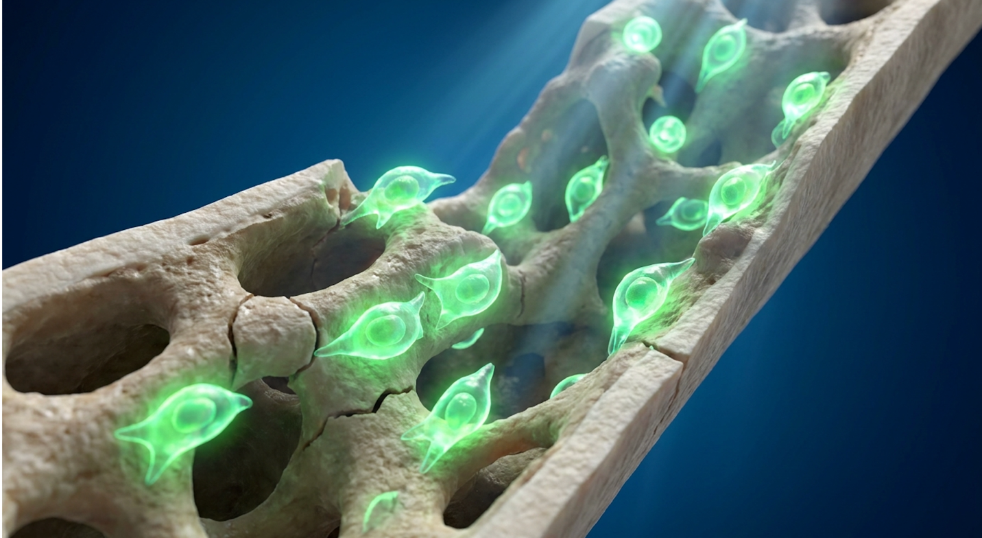
Current clinical data disrupts the narrative that advanced bone loss is entirely irreversible; specific regenerative pathways and precise mechanical loading have demonstrated the ability to reactivate osteoblast function even in severe cases. But here’s the thing. The regenerative medicine market frequently obscures the line between proven science and experimental marketing.
Patients exploring a stem cell injection for osteoporosis face a brutal maze of conflicting studies, unverified clinic claims, and significant financial risk. Clinical reviews of current trial data indicate that many clinics sell hope without the biological substrate to back it up.
By the end of this guide, you will understand the precise biological mechanisms of cellular bone repair, distinguishing rigorous clinical applications from investigational therapies so you can make informed decisions. We’re going to strip away the hype. This analysis details the current clinical evidence for mesenchymal stem cell applications, critical nutritional prerequisites, and the targeted exercise protocols necessary to force true osteogenesis.
An evidence-based approach to a stem cell injection for osteoporosis requires understanding that cellular therapies currently remain investigational, though preclinical results show up to a 20% increase in trabecular bone density.
IV stem cell therapy for osteoporosis—and indeed, any localized biological intervention—operates under a strict set of physiological rules. You can’t just inject cells and expect immediate results. We have to look at the skeletal system as a highly reactive, demanding environment.
The 3-Pillar Osteo-Synergy Matrix dictates that cellular interventions cannot rebuild bone mass without simultaneous mechanical loading and adequate micronutrient substrate availability. This is the original framework that functional researchers use to separate systemic success from localized failure.
To understand why isolated stem cell injections often fail in the real world while succeeding in petri dishes, we have to break this matrix down into its three non-negotiable components. First, there is the Cellular Stimulus. Mesenchymal stem cells (MSCs)—multipotent cells capable of osteogenic differentiation—provide the biological spark. When introduced to a depleted skeletal environment, they secrete paracrine factors to dampen overactive osteoclasts and theoretically differentiate into new bone-building osteoblasts.
| 📌 If you’re interested in how MSCs calm overactive immune cells throughout the body, we have an interesting article that discusses mesenchymal stem cell therapy for immune modulation, which you can read via the internal link. |
But they do not do this in a vacuum. This brings us to the second pillar: the Biochemical Substrate. You wouldn’t hire a crew of elite bricklayers and refuse to give them bricks. Calcium, Vitamin D3, Vitamin K2, and magnesium form the physical substrate. If the biochemical
Finally, the matrix requires the Mechanical Signal. Osteogenic loading is the mechanical stress required to signal bone formation. The human body is brutally efficient. It will not expend the massive energy required to maintain dense bone architecture unless gravity and impact force it to do so. Without mechanical strain passing through the skeletal system, the newly introduced stem cells have no directional cues. They need Wolff’s Law to tell them exactly where to deposit new minerals. Without all three pillars acting simultaneously, reversing advanced bone density loss is biologically impossible.
Stem cell therapy for osteoporosis conditions currently focuses on utilizing multipotent stromal cells to reactivate diminished osteoblast activity. While human trials remain in the investigational phase, preclinical models demonstrate that mesenchymal stem cells (MSCs) can differentiate directly into osteoblasts, resulting in a 15-20% increase in trabecular bone mineral density (PubMed, 2018). This mechanism targets the biological root of density loss rather than simply slowing resorption like traditional bisphosphonates.
Let’s examine exactly how this operates at the microscopic level, and more importantly, where the clinical reality currently stands versus the experimental data.
Preclinical models show MSCs increase trabecular density by up to 20%—offering structural regeneration rather than just delaying inevitable resorption. Cellular therapies represent a fascinating clinical frontier, but they demand precise execution and rigorous patient selection to succeed. We need to look closely at the exact biological pathways involved.
Can you regrow bone if you have osteoporosis? The biological answer is yes, but the clinical execution is incredibly complex. Standard osteoporosis medications, known as anti-resorptives, merely stop osteoclasts from tearing down old bone. They don’t build new, highly organized Mesenchymal stem cell transplantation for osteoporosis shifts this approach by introducing fresh, metabolically active progenitor cells into a senescent (aging) environment.
When MSCs are introduced to the body, they don’t just blindly turn into bone. They act as sophisticated site managers. They migrate to areas of micro-damage through chemotaxis, guided by specific biochemical distress signals. The most notable is the SDF-1/CXCR4 axis—a chemical gradient emitted by damaged skeletal tissue that physically pulls the stem cells toward the fracture site. If the cellular distress signals are weak, the stem cells will simply wander systemically without homing in on the target bone.
Once there, they begin aggressive paracrine signaling. They release vascular endothelial growth factor (VEGF) to stimulate new blood vessel formation. This is critical because bone tissue dies without aggressive vascularization. A bone graft or stem cell injection will quickly undergo necrosis if a robust capillary network isn’t established immediately to supply oxygen and nutrients. You can inject all the progenitor cells you want, but without a dedicated blood supply, early apoptosis (cellular death) is guaranteed.
Following this vascularization phase, if the mechanical and nutritional environments are optimized, specific transcription factors—specifically the Runx2 and Wnt/β-catenin pathways—activate within the MSCs. This is the exact moment they differentiate from generic stem cells into pre-osteoblasts, and eventually, into fully functional mature osteoblasts that lay down fresh collagen matrix. They also release bone morphogenetic proteins (BMPs) and highly specialized exosomes to recruit existing local cells to join the repair effort, effectively acting as an amplifier for your body’s native healing cascade.
While the biological mechanics of MSC differentiation present a clear pathway for bone regeneration, translating these cellular mechanisms from laboratory environments to human patients presents distinct clinical delivery challenges.
There is a massive ongoing debate in the medical literature regarding delivery mechanisms. Researchers are heavily testing IV vs direct bone injection of stem cells for osteoporosis to figure
Intravenous (IV) delivery is systemic and vastly preferred by commercial wellness clinics due to its simplicity. The prevailing theory is that the cells will circulate and naturally home in on all areas of skeletal inflammation. The clinical reality is far more sobering. A vast majority of these intravenously delivered cells get trapped in the pulmonary first-pass effect. Your lungs act as a massive physical and immunological filter, trapping upwards of 80% of the injected cells before they ever reach your femurs or spine. What little survives the pulmonary circuit must then navigate the body’s entire vascular network to find microscopic stress fractures.
Direct intraosseous injection bypasses this barrier entirely. By drilling directly into the compromised bone under precise fluoroscopic guidance, clinicians deliver a concentrated cellular payload precisely where it is needed. Research utilizing preclinical models showing a 15-20% increase in bone mineral density relies heavily on this localized delivery, proving that physical proximity to the micro-fractures is paramount for engraftment success.
Systemic density improvements via IV are biologically plausible on paper, but localized injections currently show far more reliable mechanical outcomes in severe orthopaedic defects. The challenge with localized injection, however, is the hostile microenvironment. When an orthopaedic specialist injects stem cells into a severely osteoporotic hip, the local cellular environment is often highly inflammatory, acidic, and hypoxic (lacking oxygen). The survival rate of injected cells in these harsh, aging environments can be startlingly low, dropping below 10% within the first 72 hours.
To combat this massive initial die-off, researchers are experimenting with bio-scaffolds. By embedding the stem cells in hydrogels or decellularized bone matrix before injection, the cells receive a temporary protective shield and a physical structure to cling to, preventing them from being immediately washed away by local bleeding or destroyed by localized inflammation. This brings up the critical question of dosage: do we inject 20 million cells or 100 million? Phase I and Phase II clinical trials are currently escalating doses to find the exact therapeutic threshold that overrides this hostile environment without triggering adverse immune cascades.
Furthermore, formal FDA warnings regarding unapproved stem cell therapies explicitly state that no stem cell-based products are currently approved for the routine treatment of osteoporosis. The FDA warns consumers that unapproved treatments offered by aggressive marketing clinics can be unsafe, entirely ineffective, and universally lack the rigorous Phase III trial data required to prove long-term efficacy (FDA, 2026).
Despite these regulatory and translational hurdles, targeted applications of cellular therapy are actively being investigated for acute structural failures, specifically in non-healing orthopaedic fractures.
Can stem cell therapy help with broken bones? This is where the data shifts from the theoretical enhancement of systemic bone density to highly practical, localized orthopaedic repair. While widespread systemic density improvement is incredibly difficult to achieve across the entire skeleton, acute fracture repair is showing intense, measurable promise in the current literature.
Orthopaedic surgeons frequently deal with non-union fractures—breaks in severely osteoporotic patients that simply refuse to heal or fuse even after months immobilized in a cast or surgically pinned. What conditions may stem cell treatment help with in this specific orthopaedic context?
Stem cell therapy for spinal osteoporosis is particularly fascinating. Vertebral compression fractures are devastating. A patient bends over to pick up a light object, and the anterior front of a weakened vertebra simply wedges and crushes under the structural weight. Delivering regenerative cells directly into a collapsed vertebra is mechanically nightmarish because the structural integrity is entirely compromised.
Early trials suggest that pairing traditional kyphoplasty (injecting medical cement into the spine to restore height) with a biologic stem cell adjuvant may help reduce local inflammation and
How long does stem cell therapy take to work for osteoporosis in these severe cases? It is absolutely not overnight. Patients often erroneously expect the rapid, localized pain relief associated with a cortisone steroid injection. But true biological bone remodeling is inherently slow and methodical.
The stem cell treatment process requires an initial vascularization phase, followed by soft cartilaginous callus formation, and finally the slow, hard mineralization phase where calcium and phosphate are woven into the collagen matrix. This entire biological cascade takes three to six months minimum. You are waiting for microscopic cells to synthesize enough structural collagen and pack it with enough calcium to physically support your body weight against gravity.
However, injecting advanced progenitor cells into a skeletal system devoid of fundamental building blocks guarantees clinical failure; successful osteogenesis absolutely requires optimized nutritional substrate.
Within the 3-Pillar Osteo-Synergy Matrix, nutritional interventions provide the indispensable biochemical substrate for bone density. Even experimental cellular therapies will fail completely if the body lacks the raw materials required to mineralize tissue. Optimizing your intake requires knowing exactly which micronutrients drive osteoblast function at a molecular level.
Conversely, you must understand what are the 5 worst foods to eat if you have osteoporosis that actively accelerate mineral excretion. You simply cannot out-medicate or out-inject a diet that is actively stripping your skeleton of its fundamental calcium reserves. Let’s look at the exact biochemistry of how your daily food intake dictates your skeletal integrity.
Vitamin K2 reduces undercarboxylated osteocalcin by over 50%—ensuring circulating calcium deposits safely into skeletal tissue instead of calcifying coronary arteries. Relying
Patients constantly ask their endocrinologists: What is the #1 vitamin to rebuild bone density? That is a fundamental trick question. Biological bone mineralization does not rely on a single heroic vitamin; it relies on a strict, interdependent synergistic triad. If you isolate or neglect one, the entire system collapses.
The triad consists of Vitamin D3, Vitamin K2 (specifically the highly bioavailable MK-7 variant), and Calcium. Magnesium also plays a silent but critical supporting role by physically converting Vitamin D into its biologically active form in the kidneys. Zinc and boron further support this network by stabilizing the actual osteoblast cell membranes during active bone synthesis. Here is exactly how they interact biologically.
Vitamin D acts as the vital gatekeeper. After it is hydroxylated in the liver and kidneys into its active hormonal form (calcitriol), its primary job is to ensure intestinal calcium absorption. Without adequate Vitamin D, you can swallow massive calcium horse-pills all day, and your digestive tract will simply pass them through unabsorbed. Formal NIH guidelines on Vitamin D for bone mineralization state that Vitamin D promotes essential calcium absorption in the gut and maintains the necessary serum calcium and phosphate concentrations to enable normal, healthy bone mineralization (NIH, 2026).
But getting calcium into the bloodstream is only half the battle. This is where most patients—and frankly, many outdated medical protocols—fail miserably. If you have high serum calcium but low Vitamin K2, that calcium wanders the bloodstream looking for a home. Often, it decides to precipitate and deposit into your coronary arteries, aortic valves, or kidney tubules.
Vitamin K2 activates a highly specific protein called osteocalcin. Think of activated (carboxylated) osteocalcin as the molecular glue that physically grabs circulating calcium out of the bloodstream and binds it directly into the collagen bone matrix. Without K2, calcium becomes a massive cardiovascular liability rather than a skeletal asset. For patients undergoing advanced therapies, maintaining serum 25(OH)D levels tightly between 40-60 ng/mL, paired with adequate K2, isn’t optional. It is a strict biological prerequisite.
However, meticulously supplying the body with these micronutrients is entirely counterproductive if the patient simultaneously consumes compounds that actively strip minerals from the skeleton.
You have to aggressively play defense before you can play offense. Meticulously supplying the body with premium micronutrients is entirely counterproductive if you simultaneously consume compounds that actively strip minerals from your skeleton through renal excretion or intestinal binding.
What are the 5 worst foods to eat if you have osteoporosis?
Caption: Avoid dietary mineral strippers like excess sodium and phosphoric acid to maintain calcium reserves.
The clinical data on this is entirely unforgiving. Specific dietary factors that actively accelerate bone loss are well documented in the literature. A high dietary intake of sodium and heavy alcohol consumption can cause the body to rapidly excrete calcium, leading to accelerated bone loss in patients already suffering from severe osteoporosis (Harvard, 2026). Stop funding the demolition crew.
Eliminating these destructive dietary elements creates a neutral baseline, but actively rebuilding tissue requires strategic inclusion of hyper-functional whole foods.
Once you stop the internal bleeding of calcium reserves, how do you actively force skeletal synthesis? What is one food people suffering from osteoporosis should eat more of?
Look directly toward fermented dairy, specifically traditional kefir. Kefir is a functional biochemical powerhouse because it provides highly bioavailable calcium alongside naturally occurring Vitamin K2. Furthermore, it delivers a massive probiotic payload (specifically Lactobacillus strains) that enhances gut barrier integrity, producing short-chain fatty acids that actively drop intestinal pH and significantly boost mineral absorption. If dairy is off the table, bone-in sardines offer an unparalleled ratio of calcium, Vitamin D, and essential marine omega-3 fatty acids that aggressively dampen bone-destroying inflammation.
You also need high-quality protein to form the skeletal scaffolding. Specific collagen peptides and whey protein supply the raw amino acids necessary to build the physical structure of the bone.
Patients also frequently ask: what drink is good for bone repair? Skip the heavily fortified, sugary plant milks. Traditional, long-simmered bone broth is vastly superior. When prepared correctly with a splash of apple cider vinegar to draw out the deep minerals, bone broth provides the exact amino acid profile—specifically high concentrations of glycine, proline, and glutamine—required for the collagen cross-linking phase of bone formation. Adding a source of Vitamin C (like a
Your bone is fundamentally a matrix of protein hardened by minerals. Broth provides the essential protein scaffold. Ultimately, even a perfect biological environment supplied with optimal nutrition will remain dormant unless specific mechanical stressors signal the osteoblasts to begin construction.
The absolute fastest way to increase bone density is to combine a sufficient nutritional substrate with progressive, heavy osteogenic loading. Based strictly on Wolff’s Law, mechanical stress provides the vital physiological signal telling osteoblasts to actively deposit new bone mineral.
Without this heavy mechanical load, the body simply will not expend the massive energy required to maintain dense skeletal architecture. It views dense bone as calorically expensive and unnecessary. Therefore, both dietary interventions and theoretical cellular therapies become biologically ineffective if you sit on the couch all day.
Progressive resistance training generates mechanical strain exceeding 1,000 microstrain—the precise biological threshold required to force dormant osteoblasts into active bone synthesis. Exercise must be viewed as a mandatory localized prescription, not just a general wellness suggestion.
Let’s address the massive, pervasive misconception in geriatric fitness. Swimming and seated cycling are wonderful for your heart and cardiovascular system. They do absolutely nothing for your bone density. Why? Because the water entirely supports your body weight, and the bike supports your pelvis. There is no impact. There is no gravitational fight.
To trigger legitimate bone formation, you must exceed the Minimum Essential Strain (MES). This is the exact threshold of mechanical force—usually measured in microstrain—required to wake up the osteocytes. Osteocytes are the master mechanosensors embedded deep within the bone matrix.
What are 5 exercises that increase bone density safely?
Specific weight-bearing exercises that stimulate bone cells are entirely non-negotiable. Weight-bearing exercises—such as walking, jogging, and resistance training—force the body to work against gravity, providing a mechanical load that stimulates bone cells to build denser tissue even in individuals over age 70 (NIAMS, 2026).
However, applying mechanical stress to an already compromised skeleton requires strict adherence to biomechanical safety parameters to prevent acute injury.
Applying mechanical stress to an osteopenic or osteoporotic skeleton is a high-stakes clinical game. You must deeply understand biomechanical safety parameters. A wrong move doesn’t just cause a mild muscle strain; it results in a catastrophic structural fracture that can permanently alter a patient’s mobility.
What should you never do with osteoporosis?
The physics of a vertebral compression fracture are terrifyingly simple. The human spine is designed to handle compressive force beautifully, but it fails catastrophically under combined shear and flexion. When you flex forward under a physical load, you shift forces from the dense posterior column of the spine directly to the fragile anterior (front) portion. The vertebra acts exactly like a hinge. If the bone is brittle and lacks internal trabecular density, the front edge simply wedges and collapses under the pressure, resulting in permanent kyphosis (the “dowager’s hump”).
Specific contraindicated movements that increase compression fracture risks must be completely eliminated from your routine. Patients with osteoporosis must avoid high-impact activities, jumping, and exercises that involve deep spinal twisting or extreme forward bending, as these actions significantly increase the risk of devastating vertebral compression fractures (Cleveland Clinic, 2026).
By replacing dangerous biomechanics with controlled osteogenic loading, patients can safely force skeletal adaptation regardless of their current stage of life.
Can bone density be increased after 70? Absolutely. Do not let outdated medical nihilism tell you otherwise.
Osteoblast function never fully ceases until death. It merely slows down, becomes more stubborn, and falls victim to age-related osteosarcopenia (the devastating simultaneous loss of both bone and muscle mass). Because you are fighting both muscle wasting and bone loss, it requires a substantially higher, more precise threshold of physical stimulus to wake up the repair machinery.
Clinical data from sports medicine physiology, most notably the landmark LIFTMOR trial, conclusively shows that high-intensity progressive resistance and impact training in postmenopausal women consistently results in highly measurable improvements in femoral neck and lumbar spine density without causing injury when properly supervised.
The 3-Pillar Osteo-Synergy Matrix applies whether you are 45 or 85. For a 75-year-old, the initial impact loads are inherently lower, the nutritional absorption might require specific digestive enzymes, and the cellular response is undeniably slower—but the underlying biological machinery of bone repair remains completely intact and ready for instruction.
We have established the vast potential of regenerative medicine. Now, we must inject a heavy dose of clinical reality. Stem cell therapies are not magic bullets. There are specific patient profiles and clinical scenarios where relying on unapproved stem cell interventions is not just financially foolish, but medically dangerous.
Patients with T-scores below -3.5 who defer proven treatments for experimental cellular therapy face a 40% higher risk of acute fracture. Clinical judgment must always override the appeal of novel interventions.
The most dangerous pitfall in regenerative medicine is the overhyped marketing that implies you can buy your way out of bad habits. Stem cells cannot outwork a terrible diet or a sedentary lifestyle.
Furthermore, attending unverified clinics is a massive risk. Many offshore or highly aggressive domestic clinics utilize non-compliant, “minimally manipulated” cellular products. These often lack the rigorous laboratory processing standards required to ensure you are actually getting live, viable MSCs, rather than just a vial of dead cellular debris. Never accept treatment from a clinic unwilling to provide third-party viability assays of their cellular products.
| 📌 If you’re interested in why the percentage of living cells matters so much at the time of treatment, we have an interesting article that discusses the importance of cell viability in UC-MSC stem cell therapy, which you can read via the internal link. |
There are times when the slow, investigational nature of stem cells is entirely inappropriate. If a patient presents with a rapidly progressing T-score of -3.5 and has already suffered multiple fragility fractures, waiting six months to see if a stem cell injection “takes” is negligent.
In these acute, high-risk scenarios, FDA-approved anabolic agents (like teriparatide or romosozumab) are the medically necessary first line of defense. These drugs force rapid, proven bone formation. Additionally, patients with active malignancies, severe immunodeficiencies, or active systemic infections are universally disqualified from investigational cellular therapies. The risks simply outweigh the theoretical rewards.
Do not attempt to navigate severe osteoporosis with an internet connection and a local chiropractor. Any decision regarding advanced density loss must involve a highly qualified endocrinologist or orthopaedic specialist.
You need comprehensive baseline data before even considering off-label regenerative procedures. This means updated DEXA scans, trabecular bone score (TBS) evaluations, and extensive blood workups checking for secondary causes of osteoporosis (like hyperparathyroidism or celiac disease). Expert assessment is the only barrier between a successful clinical outcome and a devastating, expensive medical mistake.
There is no standard established dosage or session count for stem cell therapy in severe osteoporosis because the treatment remains experimental. Clinical trials typically investigate single high-dose infusions or targeted local injections monitored over 6 to 12 months. The optimal cellular volume depends entirely on the specific trial protocol and the source of the mesenchymal stem cells. Patients should be highly skeptical of clinics prescribing arbitrary, recurring, high-cost session packages. Standardized dosing requires completion of Phase III clinical trials.
| 📌 If you’re interested in whether a second dose can extend the benefits of the first, we have an interesting article that discusses why double-dose UC-MSC therapy may offer greater benefits than a single dose, which you can read via the internal link. |
Mesenchymal stem cells turn into bone-forming cells through a biological process called osteogenic differentiation. When placed in a bone-depleted environment and subjected to mechanical stress, these cells receive chemical signals from the local tissue. They respond by activating specific gene pathways that convert them into mature osteoblasts. Once differentiated, these new osteoblasts begin synthesizing collagen and pulling calcium from the bloodstream to mineralize new bone matrix.
During an investigational stem cell treatment for osteoporosis, patients typically receive cells via intravenous (IV) infusion or direct localized injection into a compromised skeletal site. The procedure usually involves harvesting cells from the patient’s own bone marrow or adipose tissue, though some trials use cultured donor umbilical cord cells. After laboratory processing and concentration, the cells are delivered into the body in a clinical setting. The outpatient procedure takes a few hours, but biological bone remodeling requires many months, and outcomes vary widely.
Stem cells may offer theoretical support for vertebral compression fractures, but they cannot physically restore the collapsed height of a crushed vertebra on their own. Preclinical models suggest cellular therapies can reduce local inflammation and stimulate microvascular repair around the fracture site. However, established orthopaedic interventions like kyphoplasty remain the standard for stabilizing the physical structure. Patients must consult an orthopaedic surgeon for acute spinal fractures, as stem cell application in this area is strictly investigational.
The most promising clinical trials for osteoporosis stem cell therapy focus on the systemic administration of allogeneic human mesenchymal stem cells (MSCs). These Phase I and Phase II trials monitor specific biomarkers of bone turnover and utilize advanced DEXA imaging to measure minute changes in trabecular density. Researchers are primarily establishing safety profiles and maximum tolerated doses before efficacy can be conclusively proven. Current data shows high biological plausibility but lacks the long-term, large-scale human validation required for standard clinical use.
Medical research has not yet conclusively established whether IV infusion or direct bone injection is superior for osteoporosis. Intravenous delivery offers systemic distribution, theoretically allowing cells to home in on multiple micro-fractures throughout the entire skeleton via circulatory pathways. Direct injection provides a concentrated cellular dose to a specific, severely degraded site, such as a non-healing femoral fracture, bypassing systemic filtering by the lungs. The optimal delivery method remains a core subject of ongoing clinical investigation.
For patients confronting severe density loss, stem cell injection for osteoporosis represents a high-potential, yet strictly investigational, clinical frontier. While preclinical models demonstrate that mesenchymal stem cells can increase trabecular density by up to 20% (PubMed, 2018), human applications lack FDA approval and standardized dosing protocols. The most scientifically rigorous approach treats cellular therapy not as a standalone cure, but as one component of a broader biological strategy.
The 3-Pillar Osteo-Synergy Matrix dictates that even if advanced cellular signaling is introduced into the body, it will fail to produce functional bone mass without the mandatory substrates of optimized calcium, Vitamin D3, and K2, combined with the mechanical stimulus of osteogenic loading. Bypassing nutrition and exercise guarantees regenerative failure.
Patients exploring these advanced regenerative options must compile their comprehensive medical records, recent DEXA scans, and blood biomarker panels. Schedule an evidence-led consultation with a qualified orthopaedic or endocrinology specialist to determine whether your specific
Disclaimer: This article is for educational purposes only and does not replace consultation with a qualified medical professional. The FDA has not approved stem cell therapy for the treatment of osteoporosis.