Our Services
Others
- DFPP
- Shockwave
- IV Drip

This case involves a male patient aged approximately 60–61 years with a long history of complex thoracic and lumbar spine problems. He had previously undergone several spinal surgeries, including fusion at T9–T11 and L5–S1, but continued to experience chronic back pain that significantly affected his sleep, work, mobility, and overall quality of life.
The patient enjoyed outdoor activities such as surfing, skiing, and snowboarding. He therefore wished to preserve his mobility and remain as physically active as possible. He contacted Vega Clinic to explore stem cell therapy as part of an integrated care plan for degenerative spinal disease, inflammation, and chronic pain.
His care was divided into three main stages:
The patient reported chronic spinal pain for many years. The severity of his symptoms affected his sleep, ability to work, daily activities, and personal life.
He had been using prescription pain medication for approximately 20 years, including OxyContin and Percocet. One of his goals was to reduce his long-term dependence on pain medication under the supervision of the prescribing physician.
His relevant medical history included:
Thoracolumbar X-rays showed previous spinal instrumentation and fusion. The hardware remained appropriately positioned, with no obvious evidence of complication.
However, the imaging also demonstrated multilevel degenerative disc disease, including moderate degeneration and mild rightward curvature of the lumbar spine.
A lumbar MRI performed on December 2024 identified abnormalities at several levels:
These findings indicated that the patient’s symptoms were not caused by a single abnormality. They were associated with a combination of disc degeneration, facet-joint disease, narrowing around the nerves, and changes adjacent to the previously fused spinal levels.
A thoracic MRI performed in January 2025 showed postoperative changes at T9–T11 and mild degenerative changes at several disc levels.
The thoracic spinal cord appeared normal, with no significant spinal-canal or neural-foraminal narrowing.
The patient later developed worsening neurological and structural problems, including:
He therefore underwent surgery on November 2025. The procedure included nerve decompression, spinal instrumentation, and fusion at L3–L5, together with additional procedures involving L3–L4, L4–L5, and L5–S1 as clinically appropriate.
During surgery, severe narrowing was found on the left side at L4–L5, with significant compression of the L5 nerve root.
A postoperative MRI performed in January 2026 showed spinal instrumentation and fusion at L3–L5, as well as the previous fusion at L5–S1.
No central spinal-canal stenosis was identified. However, some of the neural foramina could not be evaluated clearly because of artefact from the metallic implants.
The MRI also showed a postoperative subcutaneous haematoma above the fascia, This finding required assessment before any additional procedure was considered.
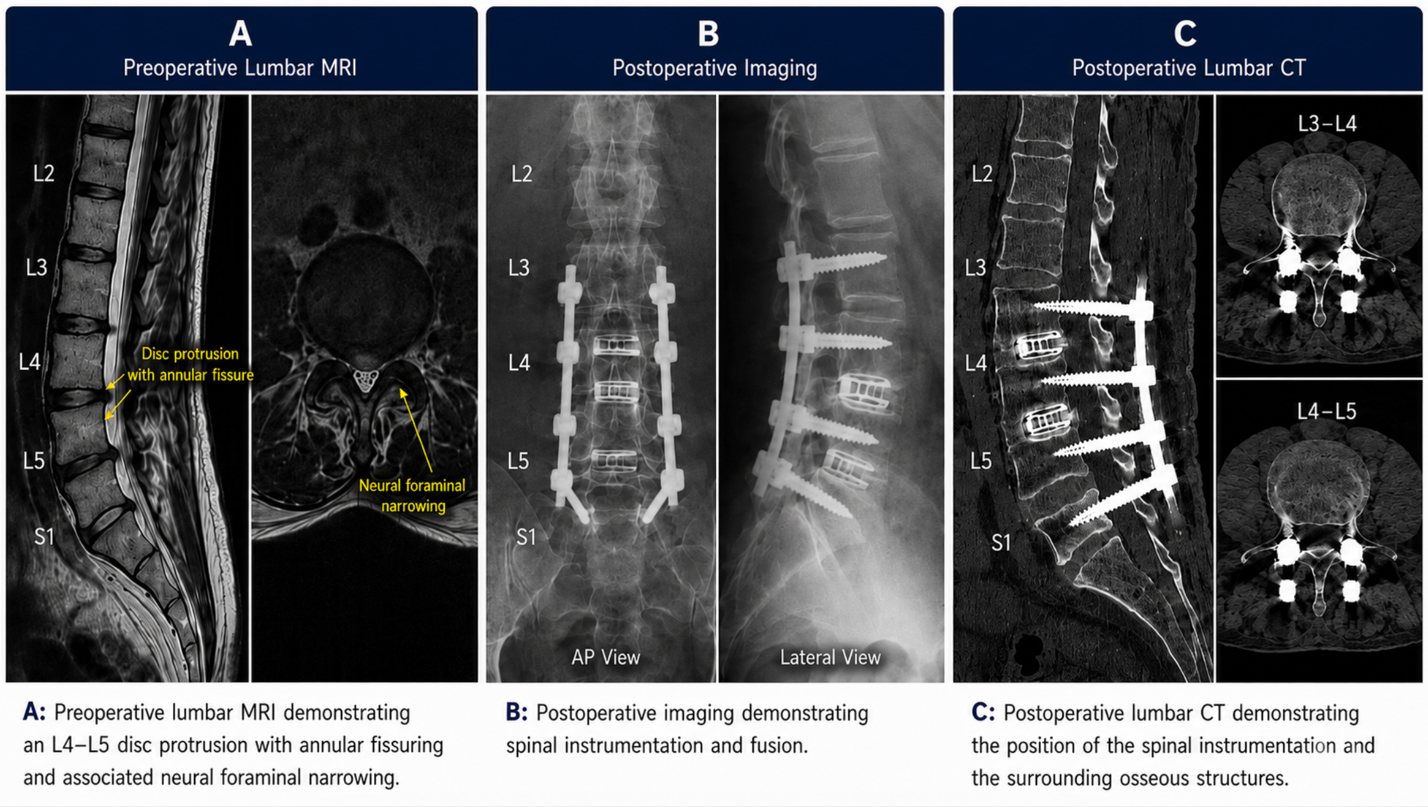
Figure 1: Composite lumbar imaging figure demonstrating the patient’s preoperative and postoperative findings. The preoperative lumbar MRI shows an L4–L5 disc protrusion with annular fissuring and associated neural foraminal narrowing. The postoperative images demonstrate spinal instrumentation and fusion, while the postoperative lumbar CT further shows the position of the spinal implants in relation to the surrounding bony structures.
Any identifying details, including the patient’s name, date of birth, medical-record number, and healthcare-provider information, should be removed before the images are published.
This was a complex degenerative spinal condition because the patient had disc degeneration, nerve-root compression, previous multilevel fusion surgery, and neurological symptoms.
Stem cell therapy cannot physically widen the spaces around compressed nerves, correct spinal deformity, reposition implanted hardware, or replace surgery when severe structural nerve compression is present.
Its potential role in selected cases is supportive and may include:
Stem cell therapy should not be described as rebuilding an entire disc, reversing spinal fusion, or guaranteeing the recovery of chronically damaged nerves.
The patient received care in several stages, combining UC-MSC therapy, surgery, and supportive treatment according to medical assessment.
In April 2025, the patient underwent a medical assessment and blood testing before starting the programme. He then received UC-MSCs through targeted injections around the lumbar and thoracic spine, as well as intravenous administration. Supportive treatments included NAD+ and intravenous vitamins.
The treatment schedule listed approximately 190 million cells in total. However, the final administration records should be reviewed because some entries in the schedule may have related to the patient’s companion.
As the patient’s nerve compression and neurological symptoms became more severe, he underwent decompression and fusion surgery in November 2025.
The purpose of surgery was to address the structural abnormalities and relieve nerve-root compression. These mechanical problems could not be corrected by stem cell therapy alone.
In February 2026, the patient returned for a postoperative recovery-support programme.
The programme included intravenous UC-MSC therapy, targeted injections around the facet joints and nerves with PRP, and other supportive therapies prescribed by the medical team.
Based on the entries clearly attributed to the patient, the February programme included approximately 150 million UC-MSCs.
The purpose of the postoperative programme was to support the tissues surrounding the spine and the overall recovery process. It was not intended to reverse the surgery, alter the position of the hardware, or guarantee neurological recovery.
| Period | Main treatment |
| April 2025 | Targeted and intravenous UC-MSC therapy with supportive treatment |
| November 2025 | L3–L5 decompression and spinal fusion surgery |
| February 2026 | Postoperative targeted and intravenous UC-MSC therapy with PRP |
The available records confirm that the patient completed stem cell programmes both before and after surgery. However, they do not yet include structured follow-up data concerning pain levels, mobility, neurological function, or changes in medication use.
It would therefore be premature to conclude that the treatment:
These outcomes should be included only after they have been confirmed through patient follow-up and medical evaluation.
This case demonstrates the difference between treatment intended to support the biological environment and treatment intended to correct structural spinal problems.
The stem cell treatment provided in April 2025 was intended to support the surrounding tissues and help regulate factors associated with inflammation. However, when the patient developed more severe nerve-root compression, foot drop, and progressive stenosis, surgery remained necessary.
The postoperative treatment in February 2026 therefore served as complementary support for recovery. It was not intended to correct structural abnormalities or replace the effects of surgery.
This case involves a patient with complex degenerative spinal disease, a history of multilevel fusion surgery, chronic pain, long-term pain-medication use, and nerve-root compression affecting mobility.
The patient first received UC-MSC therapy and supportive care in April 2025. When his structural and neurological problems became more severe, he underwent decompression and fusion surgery at L3–L5 in November 2025.
Following surgery, he completed another UC-MSC programme in February 2026. This programme combined targeted injections, intravenous administration, PRP, and other supportive therapies intended to support the surrounding tissues and the overall recovery process.
This case should be presented as an example of stem cell therapy being used as part of a multistage care plan. It should not be presented as a replacement for surgery or as a guarantee that damaged bone, discs, or nerves will regenerate.
Further follow-up information regarding pain, mobility, neurological function, medication use, and quality of life is needed to assess the longer-term outcome appropriately.