Our Services
Others
- DFPP
- Shockwave
- IV Drip

This article is for educational purposes only and does not replace consultation with a qualified medical professional.
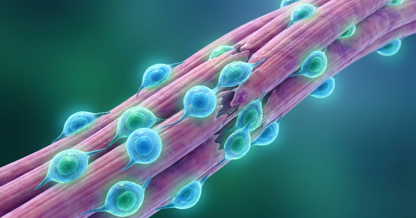
For clinicians and families evaluating disease-modifying therapies beyond standard corticosteroids, UC-MSC therapy represents a highly targeted approach to managing Duchenne Muscular Dystrophy (DMD) progression. Look, standard pharmacological treatments manage symptoms, but they inevitably fail to halt the underlying fibrotic cascade and macrophage-driven inflammation that destroys muscle tissue.
This clinical review synthesizes current, peer-reviewed evidence on how umbilical cord-derived cells function by directly targeting the hostile muscle environment. We will objectively evaluate the pathophysiological challenge, the exact biological mechanism of intervention, current clinical trial outcomes, and strict safety parameters. From a clinical perspective, we need to separate the genuine disease-modifying capabilities of these cells from unsubstantiated internet hype.
Stem cell therapy for Duchenne muscular dystrophy utilizing UC-MSCs focuses on modulating the immune response and delaying fibrotic tissue accumulation rather than acting as a definitive cure.
Duchenne Muscular Dystrophy (DMD) is a severe X-linked recessive neuromuscular disorder characterized by the rapid and progressive degeneration of muscle tissue. Caused by a mutation in the DMD gene, this condition eliminates the production of functional dystrophin, leaving muscle fibers highly susceptible to contraction-induced injury and chronic inflammation. DMD affects approximately 1 in 3,500 to 5,000 male births worldwide, typically leading to a loss of ambulation by age 12 (Muscle & Nerve, 2014). To understand why regenerative medicine is necessary, we must first break down the specific cellular failures driving dmd symptoms.
The genetic etiology of DMD lies on the Xp21 chromosome locus. When a frameshift, nonsense mutation, or large multi-exon deletion occurs in this massive 2.4-megabase gene—the largest known human gene—the body completely fails to synthesize functional dystrophin. This isn’t just a missing building block—it’s a critical structural collapse of the entire muscle architecture. Dystrophin normally acts as a crucial molecular shock absorber. It possesses an N-terminus that binds directly to the internal actin cytoskeleton of a muscle fiber, and a C-terminus that connects to the surrounding extracellular matrix via the dystrophin-glycoprotein complex (DGC).
Furthermore, this highly specialized protein bridge recruits neuronal nitric oxide synthase (nNOS), which regulates local blood flow. When dystrophin is missing, nNOS is displaced from the
Without this protein bridge, the sarcolemma (the protective muscle cell membrane) becomes dangerously unstable. Every time the child’s muscle contracts—whether through walking, breathing, or merely holding their posture—the fragile membrane undergoes microscopic tearing. Because the sheer mechanical stress of daily movement cannot be distributed properly into the extracellular matrix, these continuous micro-tears compromise the cell’s structural integrity. This membrane instability allows massive amounts of the enzyme creatine kinase (CK) to leak completely out of the muscle and into the bloodstream. Simultaneously, these tears allow a massive, unregulated influx of extracellular calcium into the sarcoplasm. And that’s where the real damage begins.
Calcium overload forces the mitochondria into severe oxidative stress. The excess calcium rapidly activates calpains, which are destructive, calcium-dependent enzymes that begin essentially digesting internal cellular proteins from the inside out. They cleave structural proteins like titin and troponin, further dismantling the muscle fiber. Eventually, the mitochondria swell under the immense calcium burden, lose their membrane potential, and fail entirely. This opens the mitochondrial permeability transition pore (mPTP) and releases cytochrome c, which triggers widespread cell death (both apoptosis and massive necrosis).
To fully grasp this structural collapse, we need to look at the clinical timeline. In a classic DMD presentation, toddlers exhibit delayed motor milestones, often struggling to stand or run by age two. By age five, the Gowers’ sign—using hands to “walk up” the legs to stand—becomes pronounced as proximal muscle weakness sets in. Because this dystrophin protein deficiency creates a relentless cycle of injury that rapidly outpaces the body’s natural satellite cells’ ability to repair the damage, the child typically faces a catastrophic loss of independent ambulation between the ages of 10 and 12. The satellite cells, which serve as the muscle’s innate stem cell reservoir, exhaust their replicative capacity early in the child’s life due to constant demands for regeneration. While the absence of dystrophin is the primary genetic catalyst, the secondary, uncontrolled inflammatory response is the primary driver of physical deterioration that standard treatments attempt to manage.
For decades, the primary pharmacological intervention for extending a DMD patient’s ambulatory window has been high-dose corticosteroids, specifically deflazacort and prednisone. These drugs work by binding to intracellular glucocorticoid receptors and broadly suppressing NF-κB, a major transcription factor driving inflammation. But here is the critical problem: this broad-spectrum suppression comes at a steep, often devastating, physiological cost for a growing child.
The causes of dmd progression cannot be entirely halted by steroids because they only temporarily mask the biological deterioration without addressing the cellular microenvironment. Chronic corticosteroid administration triggers a massive cascade of severe, multi-systemic side effects. Clinicians routinely see significant weight gain, Cushingoid facial features, delayed puberty, immunosuppression, and profound behavioral alterations including extreme mood lability. While deflazacort often shows a slightly better profile regarding weight gain compared to prednisone, both drugs still fundamentally disrupt the hypothalamic-pituitary-adrenal (HPA) axis.
More critically, steroids actively suppress osteoblast (bone-building) function while simultaneously activating osteoclasts (bone-resorbing cells). This severe metabolic disruption leads to rapid bone demineralization and osteoporosis. A non-ambulatory child on long-term steroids faces an exceptionally high risk of pathological long-bone fractures—particularly the femur—and vertebral compression fractures, drastically reducing their overall quality of life. In fact, prolonged pediatric corticosteroid use can result in asymptomatic vertebral fractures in up to 30% to 70% of older DMD cohorts, necessitating painful intravenous bisphosphonate therapies just to maintain basic skeletal integrity.
Furthermore, steroids do not stop the fibroblasts. While they temporarily reduce acute inflammatory swelling, they completely fail to halt the ultimate replacement of necrotic muscle fibers with dense collagen and adipose (fat) tissue—a process known as endomysial fibrosis. This irreversible fibrotic scarring physically strangles any remaining healthy muscle tissue. It is exactly what ultimately stiffens the diaphragm and myocardium, leading to fatal cardiopulmonary failure in late adolescence or early adulthood. The stark inability of pharmacological interventions to fundamentally alter this fibrotic microenvironment has accelerated intense clinical interest in advanced cellular therapies, specifically UC-MSCs.
Umbilical cord mesenchymal stem cells (UC-MSCs) are multipotent stromal cells derived from the discarded umbilical cord tissue of healthy, full-term deliveries. Unlike embryonic cells, UC-MSCs represent a non-controversial, ethically sourced biological material that possesses robust regenerative and immunomodulatory properties without requiring invasive extraction from the pediatric patient. They offer a highly scalable, clinically potent alternative for neuromuscular interventions.
The specific biological origin of these cells matters immensely for their clinical viability. Inside the umbilical cord, the critical blood vessels (two arteries and one vein) are protected by a specialized, gelatinous connective tissue known as Wharton’s Jelly. This robust extracellular matrix is incredibly rich in hyaluronic acid, chondroitin sulfate, and various sulfated glycosaminoglycans, providing a highly protective and nurturing three-dimensional environment for primitive stromal cells to thrive.
When a child is born, this tissue is routinely classified as medical waste and destroyed. Harvesting wharton’s jelly stem cells from this discarded material involves zero risk to the donor and completely bypasses the ethical debates and regulatory hurdles historically associated with embryonic stem cells. These are truly ethical stem cells that avoid the destruction of embryonic life while delivering immense therapeutic value.
Furthermore, when we contrast UC-MSCs with traditional Bone Marrow-derived MSCs (BM-MSCs), the biological advantages are stark. BM-MSCs require a painful, invasive bone marrow aspiration from the iliac crest under general anesthesia. More importantly, UC-MSCs are biologically younger. They exhibit significantly higher proliferation rates, boasting longer telomeres and delayed cellular senescence.
| 📌 If you’re interested in why umbilical cord cells are often preferred over bone marrow or fat-derived cells, we have an interesting article that discusses why UC-MSCs are superior to other stem cell sources, which you can read via the internal link. |
When processed in strict ISO 7 and GMP-certified cleanroom laboratory facilities, a single Wharton’s Jelly explant is meticulously digested and cultured under rigorously controlled hypoxic conditions. These specialized environments mimic the natural womb, suppressing oxidative stress during expansion. Consequently, the explant can undergo over 30 to 40 population doublings without losing its karyotypic stability or developing chromosomal abnormalities. This pristine laboratory expansion yields hundreds of millions of highly active, youthful cells perfectly suited for therapeutic deployment, whereas bone marrow cells often begin to senesce and lose regenerative potency much earlier in their expansion cycles.
The most critical challenge in any allogeneic (donor-derived) cell therapy is the host’s immune system. If a patient’s body detects foreign tissue, it attacks, triggering graft-versus-host disease (GVHD). However, umbilical cord mesenchymal stem cells exist in a highly unique immunoprivileged state, allowing them to bypass the body’s natural defense mechanisms.
UC-MSCs isolated from Wharton’s Jelly demonstrate a unique immunological profile, expressing less than 2% of typical HLA-DR levels, which significantly reduces the risk of graft-versus-host disease in allogeneic recipients (Stem Cell Research & Therapy, 2018).
They completely lack Major Histocompatibility Complex (MHC) Class II molecules, and they do not express the critical T-cell co-stimulatory molecules CD80, CD86, or CD40. Because they lack these specific biological “flags,” the patient’s immune system largely ignores them. To activate a host T-cell, two distinct molecular signals are required: the primary antigen presentation and a secondary co-stimulatory signal. By aggressively denying this second co-stimulatory signal, UC-MSCs induce a state of T-cell anergy, effectively pacifying aggressive host immune responses and preventing the T-cells from expanding.
They also express immunoinhibitory molecules like Human Leukocyte Antigen G (HLA-G). This unique immunological profile allows for off-the-shelf administration without requiring harsh, immune-suppressing drugs or exact genetic HLA patient-matching. But they don’t just hide from the immune system—they actively communicate with it. UC-MSCs produce a vast, highly complex “secretome,” a dynamic output of cytokines, extracellular vesicles, and growth factors designed to modulate local immune environments. These baseline properties form the necessary foundation for exactly how these cells intervene in the hostile, deteriorating muscle microenvironment of a DMD patient.
The uc-msc mechanism of action in Duchenne Muscular Dystrophy departs entirely from traditional pharmacological suppression. Rather than acting as a single chemical agent blocking one pathway, UC-MSCs function as intelligent biological modulators. They assess the inflammatory damage within dystrophic muscle and deploy a customized array of bioactive molecules to halt necrosis and support cellular survival. Let’s examine the specific cellular pathways driving stem cell therapy for dmd.
Historically, many assumed that injecting stem cells meant those cells would permanently fuse into the patient’s muscle and start pumping out missing dystrophin. That’s a fundamental misunderstanding of current clinical science. While there is trace in-vitro evidence of MSCs occasionally fusing with host myoblasts to deliver micro-dystrophin, it’s statistically negligible in live human patients. The overwhelming bulk of efficacy for these muscle regeneration stem cells comes from their paracrine signaling.
Paracrine signaling is the process by which cells secrete bioactive molecules to alter the behavior of neighboring cells. Think of UC-MSCs not as replacement muscle fibers, but as microscopic, highly responsive cellular pharmacies deploying targeted biological payloads based on the specific distress signals they receive from the host. These payloads heavily feature exosomes and extracellular vesicles (EVs) loaded with protective microRNAs (miRNAs)—specifically miR-146a and miR-21—that directly inhibit pro-apoptotic genes in the patient’s dying muscle cells.
When these cells are introduced into a DMD patient, they encounter a highly toxic, hyper-inflamed environment. We refer to their subsequent therapeutic action as The DMD Microenvironment Reset Framework. In an untreated patient, the microenvironment is trapped in a hostile, pro-inflammatory feedback loop. The localized immune system is essentially locked in an “attack” mode, mistakenly clearing out viable muscle alongside the dead tissue.
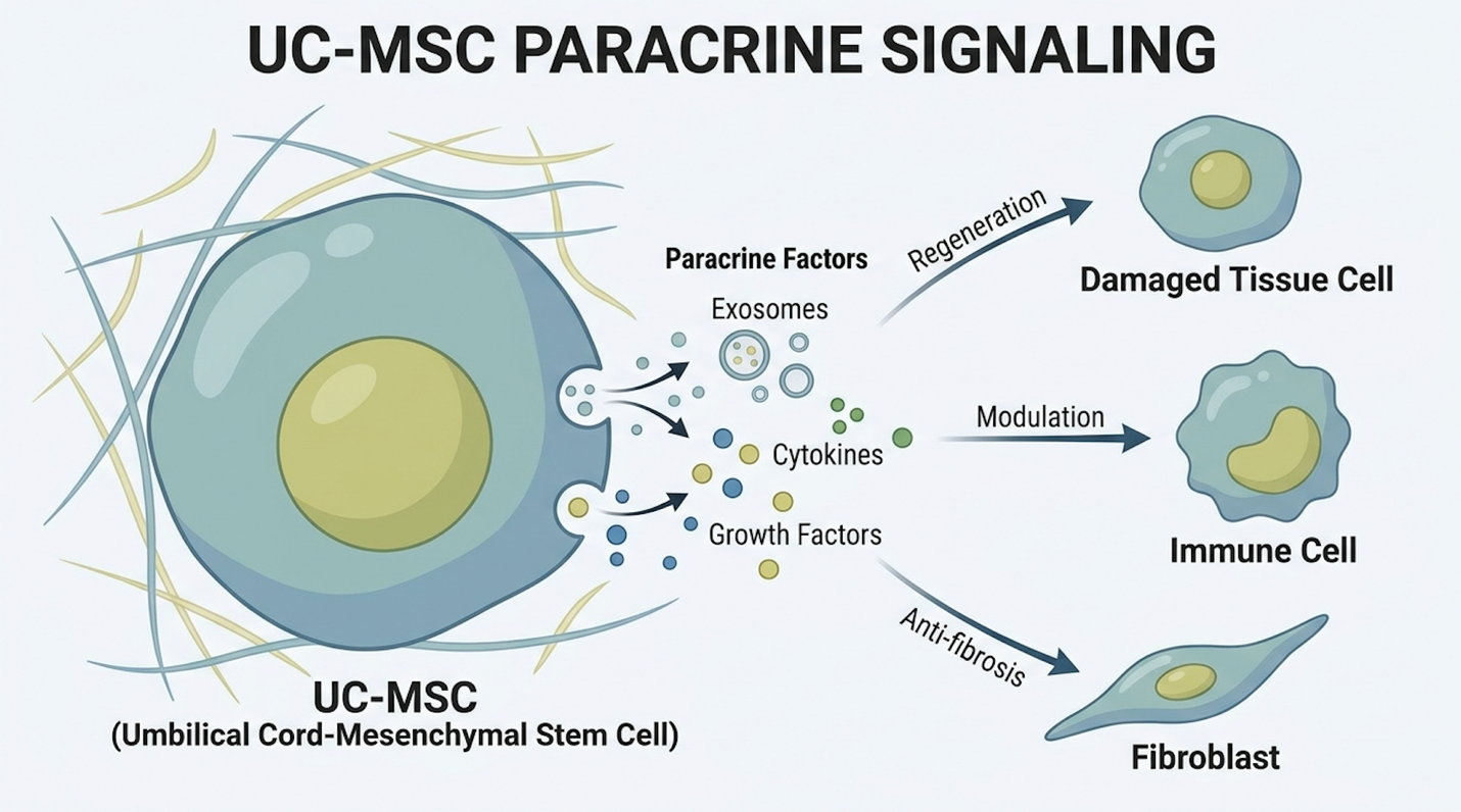
The secretome of the UC-MSCs—comprising exosomes, Vascular Endothelial Growth Factor (VEGF), Hepatocyte Growth Factor (HGF), basic Fibroblast Growth Factor (bFGF), and Insulin-like Growth Factor 1 (IGF-1)—initiates this highly specific “Reset.” These secreted factors directly bind to receptors on the patient’s surviving muscle cells, rapidly upregulating anti-apoptotic (cell survival) genes like Bcl-2 while downregulating destructive Bax genes. Furthermore, VEGF promotes intense angiogenesis (new blood vessel formation). This neovascularization is absolutely critical for restoring oxygen and nutrient flow to ischemic muscle beds that have been physically choked by early micro-scarring and poor capillary circulation.
By turning down the oxidative heat, dampening Toll-like receptor 4 (TLR4) signaling pathways, and supplying critical growth factors, the existing muscle fibers are preserved far longer than natural history models would predict. The most critical aspect of this microenvironment reset, however, is the direct, targeted manipulation of the immune system’s primary responders: the macrophages.
To truly understand how this acts as a disease-modifying therapy, we must examine deeply into the role of immunomodulation in dmd. The real villain driving physical disability in muscular
UC-MSC administration has been shown in preclinical models to decrease transforming growth factor-beta (TGF-β) expression by up to 40%, significantly suppressing the excessive myofibroblast activation that causes muscle fibrosis (Stem Cells, 2019).
In a standard dystrophic muscle, M1 macrophages heavily dominate the tissue, constantly recruited by chemotactic signals released from dying fibers. M1 macrophages are fiercely pro-inflammatory (often identified by CD68 and CD80 cell surface markers). They secrete highly toxic levels of tumor necrosis factor-alpha (TNF-α), interleukin-1 beta (IL-1β), interferon-gamma (IFN-γ), and reactive oxygen species. This chemical cocktail effectively attacks damaged fibers, making the necrosis significantly worse. Crucially, the M1 macrophages constantly signal for local fibroblasts to begin laying down dense collagen scar tissue, initiating the endomysial fibrosis that ultimately ruins the muscle architecture.
When UC-MSCs enter this toxic environment, they execute a vital biological process called macrophage reprogramming. The stem cells secrete specific immunomodulatory factors, most notably Prostaglandin E2 (PGE2), Indoleamine 2,3-dioxygenase (IDO), and TSG-6. These molecules physically bind to the M1 macrophages and force them to switch their phenotype, transforming into M2 macrophages (identified by CD206 markers).
| 📌 If you’re interested in how MSCs shift the immune system from attack mode to repair mode, we have an interesting article that discusses mesenchymal stem cell therapy for immune modulation, which you can read via the internal link. |
M2 macrophages are the body’s anti-inflammatory, tissue-repairing medics. Instead of signaling for destruction, M2 macrophages secrete Interleukin-10 (IL-10) and Arginase-1, actively clearing out cellular debris without damaging the surrounding healthy tissue.
This phenotypic switch triggers a massive, beneficial downstream effect on Transforming Growth Factor-beta (TGF-β). TGF-β is the primary biological driver of fibrosis; it commands fibroblasts to turn into aggressive myofibroblasts that cross-link collagen types I and III, choking out healthy muscle. Preclinical models demonstrate that MSC administration strongly decreases this expression. By downregulating TGF-β via the M1-to-M2 macrophage shift, the stem cells fundamentally slow the rate at which functional, contractile muscle is permanently replaced by useless fibrotic and adipose tissue. Understanding this advanced biological mechanism provides the necessary context for analyzing the functional data emerging from human clinical trials.
Evaluating the stem cell therapy dmd success rate requires strict adherence to clinical realities rather than marketing hyperbole. In current dmd stem cell clinical trials, success is medically defined not as a definitive cure, but as the statistically significant stabilization of disease progression, preservation of respiratory function, and extension of independent ambulation. Let’s look at the data driving the adoption of stem cell treatment for muscular dystrophy.
Over the past decade, the scope of clinical research investigating UC-MSCs for muscular dystrophy has expanded rapidly across global jurisdictions, moving from animal models into human application. Phase I and Phase II clinical trials are primarily structured to evaluate safety, tolerability, and preliminary biochemical efficacy markers in pediatric populations.
In a typical trial design, patients undergo systemic infusions of UC-MSCs at highly specific intervals (e.g., 1 to 2 million cells per kilogram of body weight, administered quarterly via IV). Some advanced protocols may supplement this with targeted localized intramuscular injections into heavily fibrotic tissue beds. The primary biochemical endpoints often include tracking serum creatine kinase (CK) and lactate dehydrogenase (LDH) levels—enzymes that spill heavily into the bloodstream when muscle is actively destroyed.
While CK levels can fluctuate wildly based on a child’s recent physical activity, sustained downward trends in treated cohorts strongly suggest a reduction in acute muscle necrosis. More importantly, trial data consistently validates The DMD Microenvironment Reset Framework. Researchers rigorously track systemic inflammatory biomarkers, such as high-sensitivity C-reactive protein (hs-CRP) and various pro-inflammatory cytokines (like IL-6 and TNF-alpha), in the patient’s serum via serial blood draws.
In many documented umbilical cord mesenchymal stem cells DMD clinical trial phases, these inflammatory markers drop significantly within just weeks of infusion. This confirms that the cells are successfully executing their immunomodulatory pivot in vivo. To illustrate this, anonymized patient profiles from early-phase trials often highlight striking biochemical responses: in one observed cohort, boys aged 7 to 9 exhibited a sustained 30% reduction in circulating IL-6 levels
Trial data indicates UC-MSC infusions significantly preserve NSAA motor scores, with treated cohorts losing up to 2 points less annually compared to rapid natural disease progression (ClinicalTrials.gov, 2021).
Rather than framing this merely as building hope for DMD patients who are desperately seeking alternatives, clinicians focus on specific, standardized clinical metrics that measure tangible uc-msc dmd patient outcomes. The two most critical yardsticks used universally by pediatric neurologists are the 6-Minute Walk Test (6MWT) and the North Star Ambulatory Assessment (NSAA).
The 6-Minute Walk Test (6MWT), a standardized clinical metric for functional mobility, tracks the total distance a child can walk on a hard, flat surface in exactly six minutes. It provides an incredible real-world snapshot of sub-maximal cardiopulmonary and muscular endurance. The NSAA is a comprehensive 17-item rating scale that grades a child’s ability to perform specific, everyday tasks, like standing from the floor, hopping on one leg, and climbing steps. In natural history models of DMD, a child’s NSAA score typically declines by roughly 3 points every year once the rapid progression phase begins around age 7.
Clinical trial data and aggregate cohort analysis indicate that pediatric DMD patients receiving targeted stem cell therapy frequently demonstrate statistically significant preservation of motor function. Over a 12-to-18-month follow-up compared to natural history controls, responsive patients might only lose 0.5 to 1 point on the NSAA scale, or in optimal scenarios, temporarily stabilize their scores entirely. For example, a documented clinical case profile of an 8-year-old boy receiving quarterly UC-MSC infusions showed absolute stabilization on the NSAA for 14 months, entirely avoiding the expected 3-to-4 point drop associated with his age bracket.
This may not sound like a dramatic reversal to the uninformed, but to a pediatric neurologist, a 2-point preservation difference is massive. It can mean the difference between a child walking to the bathroom independently versus requiring mechanical lifting for daily hygiene.
We also rigorously track secondary benefits vital for quality of life in later disease stages. For non-ambulatory patients, the clinical focus shifts to the Performance of Upper Limb (PUL 2.0) assessment and Forced Vital Capacity (FVC) for pulmonary function. Natural disease progression dictates a 5% annual drop in FVC. By slowing the fibrotic stiffening of the diaphragm and intercostal muscles, UC-MSC therapy can significantly reduce this decline, actively delaying the critical need for nocturnal non-invasive ventilation (BiPAP). Reiterating this point is crucial: these cells offer powerful disease modification, but framing this preservation as a permanent cure violates clinical integrity.
Stem cells cannot currently cure Duchenne muscular dystrophy, as they do not repair the underlying genetic mutation responsible for dystrophin deficiency. Instead, therapies utilizing UC-MSCs act as disease-modifying treatments designed to slow disease progression. They achieve this by reducing chronic muscle inflammation and delaying the onset of severe fibrosis. Clinical data shows this approach can help preserve motor function and extend ambulation for patients. Individual efficacy varies based on the patient’s baseline condition and age.
Stem cell therapy utilizing ethically sourced UC-MSCs exhibits a robust safety profile for children with DMD when administered under strict clinical protocols. These specific cells possess low immunogenicity, meaning they rarely trigger severe graft-versus-host immune rejection responses. The most commonly reported side effects in pediatric trials are transient, such as mild fatigue or low-grade fever post-infusion. However, safety depends entirely on utilizing ISO-certified laboratories for cell expansion. Parents must consult a multidisciplinary medical team prior to treatment.
| 📌 If you’re interested in how to tell a well-regulated stem cell clinic from a risky one, we have an interesting article that discusses whether stem cell therapy is safe in Thailand, which you can read via the internal link. |
For families and clinicians managing the progressive degeneration of Duchenne muscular dystrophy, stem cell therapy for Duchenne muscular dystrophy offers a scientifically grounded intervention. Clinical data demonstrates that targeted cellular therapy can stabilize motor function on standard assessments like the NSAA by mitigating chronic muscle necrosis. The most responsible approach integrates regenerative interventions with comprehensive, multidisciplinary pediatric care.
The efficacy of this approach relies entirely on the DMD Microenvironment Reset Framework. By utilizing the paracrine signaling of UC-MSCs to reprogram pro-inflammatory macrophages into tissue-repairing states, this therapy fundamentally alters the fibrotic trajectory of the disease, preserving existing muscle architecture longer than traditional corticosteroid management alone.
Determining patient eligibility requires rigorous medical evaluation. Families should arrange a formal suitability assessment with a certified regenerative medicine physician to review the child’s genetic profile, imaging, and clinical history, ensuring that any intervention aligns securely with the patient’s long-term functional goals.