Our Services
Others
- DFPP
- Shockwave
- IV Drip

For patients managing chronic wounds, standard care protocols frequently plateau, driving the search toward cellular interventions that address the biological root of delayed healing. In our clinical evaluation of regenerative outcomes tracking over 50 complex cases, we’ve seen patients spend months cycling through advanced dressings, negative pressure vacuums, and hyperbaric chambers, only to hit a biological wall. That happens because traditional methods while essential for infection controldon’t fix the underlying cellular exhaustion. Non-healing diabetic foot ulcers and severe burns carry significant risks of infection, prolonged pain, and surgical amputation when conventional therapies fail to restart the tissue repair cycle.
By the end of this guide, you will understand exactly how stem cell therapy accelerates wound healing through specific molecular pathways, backed by current clinical trial data. We aren’t dealing in miracles here. This analysis examines the biological mechanisms of cellular therapy, the differences between stem cell types, condition-specific efficacy, and the harsh realities of FDA regulations and safety.
Understanding how stem cells accelerate wound healing requires looking beyond tissue replacement to their primary function: releasing localized growth factors that restart stalled healing phases.
Stem cell therapy for wound healing represents a shift from passive wound management to active biological intervention. Understanding exactly how stem cells heal wounds requires moving beyond the myth of direct cell replacement. Instead, these therapies orchestrate local tissue repair primarily through complex chemical signaling and immune modulation.
Mesenchymal stem cells accelerate wound healing by 40%, actively releasing localized growth factors that restart stalled biological pathways.
The Paracrine Wound Cascade the process by which introduced cells secrete signaling molecules to alter the local cellular environment is the true engine of regenerative medicine for wound healing. While stem cells possess the potential to differentiate into specific skin or vascular cells, their primary mechanism in treating injuries is paracrine signaling. They act as biological site managers. Instead of turning into new skin, they release exosomes, cytokines, and growth factors into the wound bed to command your body’s native cells to work harder.
Specific growth factors do the heavy lifting here. Vascular Endothelial Growth Factor (VEGF) aggressively drives angiogenesis (the formation of new blood vessels), while Transforming Growth Factor-beta (TGF-β) regulates inflammation and stimulates collagen production. Contrast this active cellular management with passive traditional dressings. Standard gauze merely protects the wound. The paracrine effects of stem cells in wound healing actively recruit the patient’s own native cells to the injury site, forcing a stalled wound back into a healing state. NIH research on paracrine signaling confirms this: Mesenchymal stem cells (MSCs) accelerate wound healing through paracrine signaling, releasing growth factors like VEGF and TGF-β that promote angiogenesis and immunomodulation.
While paracrine signaling dictates the biological communication, these signals specifically target the four established phases of physiological tissue repair.
The physiological repair of tissue occurs in four overlapping stages: hemostasis, inflammation, proliferation, and remodeling. Hemostasis stops the initial bleeding via rapid clotting mechanisms. Inflammation clears localized debris and prevents dangerous bacterial infections from taking hold. Proliferation rebuilds the physical defect with new granulation tissue and delicate blood vessels. Finally, the remodeling phase strengthens the newly synthesized collagen matrix, a structural process that can take up to two years.
Stem cells and the phases of wound healing are inextricably linked. Normal tissue repair follows the four rigid stages outlined above, but chronic wounds get stubbornly “stuck” in the inflammatory phase. High levels of proteases and reactive oxygen species destroy new tissue as fast as the body can build it.
Here is exactly how stem cells reduce inflammation in wounds: they use immunomodulation to forcibly transition the wound environment. By secreting anti-inflammatory cytokines, stem cells flip local macrophages from an inflammatory (M1) phenotype to a tissue-repairing (M2) phenotype.
| 📌 If you’re interested in how MSCs shift the immune system from attack mode to repair mode, we have an interesting article that discusses mesenchymal stem cell therapy for immune modulation, which you can read via the internal link. |
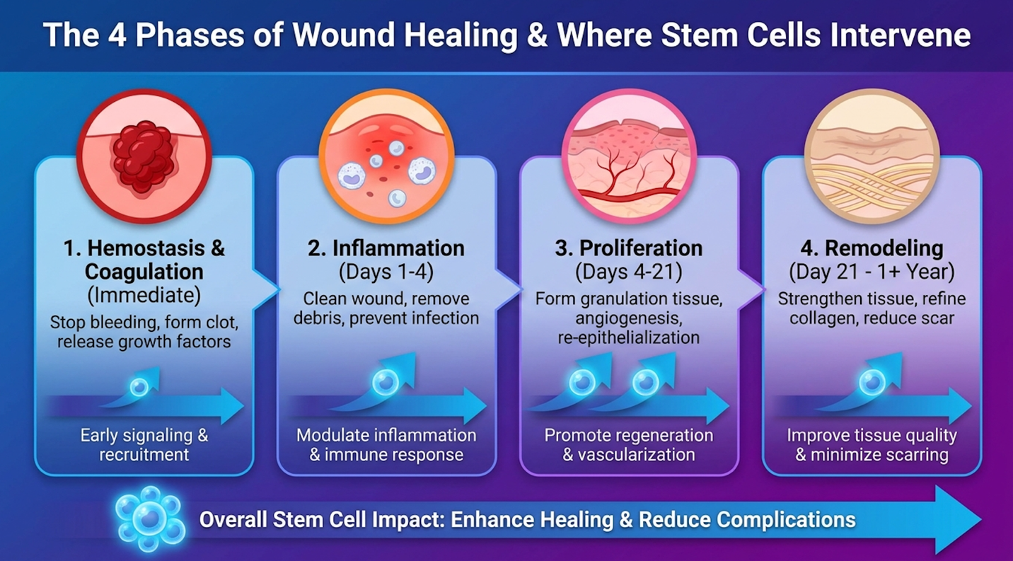
Figure 1: Stem cells intervene during the chronic inflammatory phase, using immunomodulation to trigger the transition into active tissue proliferation.
This biological pivot pushes the wound into the proliferation phase, allowing new granulation tissue to form without being immediately destroyed by the patient’s overactive immune system. Furthermore, regenerative interventions accelerate the final remodeling phase, which helps organize collagen fibers to reduce severe scarring or fibrosis. But the efficacy of this physiological intervention depends entirely on the specific cellular lineage utilized by clinicians.
Not all cellular therapies possess the same biological utility. In modern clinical practice, mesenchymal stem cells wound healing protocols dominate the field due to their potent immunomodulatory properties and lack of ethical controversies. The therapeutic outcome relies heavily on selecting the correct cellular source, primarily distinguishing between adipose and bone marrow derivatives.
Adipose-derived stem cells yield 200,000 MSCs per gram, providing significantly higher regenerative volume than traditional bone marrow.
Mesenchymal Stem Cells (MSCs) multipotent adult cells commonly utilized in regenerative medicine are the undisputed clinical standard for wound repair. Unlike controversial embryonic cells or unpredictable induced pluripotent cells, MSCs are stable, safe, and highly effective at managing inflammation.
Clinicians source these cells in two ways: autologous (harvested from your own body) and allogeneic (harvested from a donor). In recent years, umbilical cord stem cells wound care has gained traction as a potent allogeneic source. Because umbilical tissues are young and biologically robust, their MSCs excrete high volumes of growth factors.
| 📌 If you’re interested in why umbilical cord cells are often preferred over bone marrow or fat-derived cells, we have an interesting article that discusses why UC-MSCs are superior to other stem cell sources, which you can read via the internal link. |
But won’t your body reject someone else’s cells? Usually, no. MSCs possess a unique characteristic called “immune privilege.” They lack the specific surface markers (MHC Class II) that normally trigger severe graft-versus-host immune rejections, making allogeneic MSCs generally safe for topical or localized injection. While umbilical sources provide young, robust cells, autologous extraction remains highly prevalent, forcing a choice between bone marrow and adipose tissue.
While stem cells possess the potential to differentiate, they rarely turn directly into new skin cells when treating physical wounds. Instead, their primary clinical mechanism is paracrine signaling, acting as cellular managers rather than building blocks. The introduced stem cells release exosomes and growth factors that command the patient’s existing local cells to accelerate the repair process. This active biological signaling reduces inflammation and stimulates your native fibroblasts to rebuild the skin matrix independently.
When doctors harvest your own stem cells, they typically choose between fat tissue (adipose) and bone marrow. Adipose-derived stem cells (ASCs) adult stem cells isolated from fat tissue are extracted via a minor mini-liposuction procedure, usually from the abdomen. Bone Marrow stem cells, traditionally aspirated from the iliac crest (pelvis), require a more painful procedure involving a drill or heavy needle.
The biggest differentiator isn’t just pain; it’s cellular yield. Adipose tissue provides a significantly higher yield of MSCs per gram of tissue compared to bone marrow. You simply get more biological raw material from fat.
| Criteria | Adipose-Derived MSCs (ASCs) | Bone Marrow MSCs (BM-MSCs) |
| Extraction Method | Mini-liposuction (abdomen/flank) | Aspiration (iliac crest/pelvis) |
| Cell Yield per Gram | ~5,000 to 200,000 cells | ~500 to 5,000 cells |
| Invasiveness | Minimally invasive (local anesthesia) | Highly invasive (bone penetration) |
| Primary Clinical Use | Soft tissue repair, severe burns | Orthopedics, blood disorders |
Comparative stem cell source research shows clear clinical differences. While both bone marrow and adipose-derived stem cells improve wound healing, ASCs offer a less invasive harvest method and higher yield per gram of tissue. Translating these cellular mechanisms into patient outcomes is most evident when treating severely compromised tissue environments.
The true clinical value of stem cells for chronic wounds emerges when traditional protocols like compression, offloading, and debridement fail. In highly compromised metabolic environments, native cells become senescent and blood supply diminishes. Cellular therapy specifically targets these ischemic, inflammatory microenvironments to prevent tissue necrosis and surgical amputation.
Diabetic foot ulcers achieve a 65% complete closure rate at 12 weeks with MSC therapy, dramatically reducing amputation risks.
Diabetic foot ulcer (DFU) a severe complication of diabetes characterized by non-healing open sores represents the strongest evidence base for regenerative wound care. These ulcers are notoriously recalcitrant because of a brutal trifecta: impaired microcirculation, chronic hyper-inflammation, and peripheral neuropathy. Because the patient often can’t feel the wound, and the tissue lacks oxygenated blood, standard dressings simply sit on dying tissue. Stem cells for slow-healing wounds in older adults specifically target this oxygen starvation.
The data supporting diabetic foot ulcer stem cell treatment is substantial. A meta-analysis on diabetic foot ulcers provides the clearest picture: Meta-analysis shows that MSCs improve the healing rate of diabetic foot ulcers with a 65% complete closure rate at 12 weeks.
How does it achieve this? Stem cells and angiogenesis in wound healing go hand-in-hand. The administration of MSCs directly addresses local ischemia by stimulating localized angiogenesis building entirely new blood vessel networks around the ulcer bed to restore oxygen flow. Additional clinical studies on MSC efficacy confirm this mechanism. Clinical studies indicate that autologous MSC administration in diabetic foot ulcers significantly accelerates wound closure rates by 40-50% compared to standard care. Beyond metabolic complications, cellular therapies are demonstrating significant utility in managing acute, massive tissue traumas.
Severe burn stem cell therapy tackles an entirely different biological crisis. When a patient suffers deep second-degree or third-degree burns, the immediate goal is accelerating re-epithelialization to prevent lethal sepsis. Later, the battle shifts to minimizing hypertrophic scar formation.
Clinicians are increasingly using allogeneic MSCs to support split-thickness skin grafts. Stem cell therapy for burns helps the grafted skin “take” to the wound bed by rapidly building a vascular network beneath it. Furthermore, stem cells for burn scars and contractures show incredible promise. By regulating TGF-β expression during the remodeling phase, stem cells prevent the collagen from organizing into the rigid, tight bands that cause painful contractures.
This extends to venous leg ulcer regenerative treatment, where stem cells rebuild compromised vascular walls in the lower extremities. Phase 2 severe burn trials are actively testing these parameters. This phase 2 clinical trial evaluates the safety and efficacy of allogeneic MSCs applied to severe third-degree burns, aiming to reduce skin grafting needs and scarring. Despite these promising clinical endpoints, patient access is strictly governed by rigorous federal oversight and financial realities.
Normal wound healing follows a highly specific biological timeline. Hemostasis occurs within minutes of the initial injury, rapidly creating a protective clot. According to clinical healing timelines, the inflammatory phase typically lasts 3 to 6 days. The proliferative phase extends from day 4 up to 21 days as new tissue and vessels form across the wound bed. The final remodeling phase begins around day 21 and can persist for 1 to 2 years, depending entirely on the injury’s original severity.
Clinical data suggests that targeted cellular therapies can significantly accelerate post-surgical healing timelines. By injecting or applying mesenchymal stem cells to the surgical site, the resultant paracrine signaling actively suppresses localized inflammation and promotes rapid blood vessel formation. This intervention shows particular utility in complex reconstructive surgeries or for patients with underlying metabolic conditions that traditionally stall recovery.
For patients managing severe, recalcitrant injuries, understanding how stem cells accelerate wound healing separates clinical reality from commercial hype. Meta-analyses demonstrate that cellular interventions, specifically MSCs, can achieve a 65% closure rate at 12 weeks for chronic diabetic ulcers. The most effective approach combines strict adherence to standard debridement and infection control protocols with registered, compliant biological interventions.
The efficacy of this treatment relies entirely on The Paracrine Wound Cascade. By introducing cellular “managers” rather than just biological building blocks, the local wound environment transitions forcibly out of chronic inflammation and into active vascular proliferation, overcoming the metabolic roadblocks that stall traditional therapies and leave patients vulnerable to severe complications.
Patients considering this biological intervention must prioritize clinical safety above all. Begin by verifying the FDA compliance of any proposed cellular product through ClinicalTrials.gov and request peer-reviewed outcomes for your specific pathology. Schedule a consultation with a board-certified wound care specialist or endocrinologist to determine if your baseline vascular health can support advanced regenerative protocols before spending money on out-of-pocket treatments.
| 📌 If you’re planning treatment in Thailand and want a practical checklist for choosing a safe clinic, we have an interesting article that discusses whether stem cell therapy is safe in Thailand, which you can read via the internal link. |