Our Services
Others
- DFPP
- Shockwave
- IV Drip

Current clinical consensus indicates that while conventional Type 2 Diabetes management relies on masking biological deterioration via synthetic insulin and metformin, umbilical cord mesenchymal stromal cells (UC-MSCs) are demonstrating a unique capacity for tissue-repairing phenotypic macrophage reprogramming.
But there’s a serious problem in the regenerative medicine sector. It is heavily saturated with commercial clinics making unsubstantiated promises regarding complete diabetes reversal. This aggressive marketing confuses patients. It blurs the line between what is medically established and what remains highly investigational. Our team’s extensive evaluation of global clinical data reveals a massive gap between laboratory reality and billboard advertising.
By the end of this analytical guide, you will understand the exact paracrine mechanisms driving stem cell therapy for type 2 diabetes. You’ll evaluate current clinical trial data without the marketing spin. Most importantly, you will learn to apply the UC-MSC Efficacy Triad a stringent framework to safely assess treatment viability based on cell origin, processing, and delivery.
This analysis examines cellular origins, specific biological pathways for metabolic homeostasis, global treatment logistics, and natural regenerative alternatives. No hype. Just the clinical data you need to make an informed medical decision.
UC-MSC stem cell therapy for type 2 diabetes focuses on restoring metabolic homeostasis by mitigating systemic inflammation and protecting beta cells.
The richest source of stem cells for regenerative medical applications is umbilical cord tissue, specifically Wharton’s Jelly, which provides abundant youthful mesenchymal stromal cells (UC-MSCs). Unlike adult bone marrow or adipose tissue, UC-MSCs possess longer telomeres and robust immunomodulatory properties (Stem Cell Research & Therapy, 2021). This makes them uniquely suited for allogeneic treatments without triggering severe immune rejection.
When patients first research regenerative medicine, they typically encounter three primary sources of mesenchymal stem cells: bone marrow, adipose (fat) tissue, and umbilical cord tissue. Each source carries drastically different clinical implications, and understanding these differences is step one of the UC-MSC Efficacy Triad (Origin). Evaluating the organ with most stem cells requires looking beyond simple volume and focusing entirely on cellular vitality and proliferative capacity.
Bone marrow extraction was the historical gold standard. It’s painful, highly invasive, and yields a surprisingly low concentration of MSCs roughly 0.001% to 0.01% of total nucleated cells. Adipose-derived stem cells, isolated from fat tissue through liposuction, offer a higher yield. However, both of these autologous (self-derived) sources suffer from a fatal flaw in the context of chronic metabolic disease: profound biological senescence.
If you are a 60-year-old patient with long-standing Type 2 Diabetes, your fat and bone marrow stem cells are also 60 years old. They have been bathed in a highly inflammatory, hyperglycemic environment for decades. Advanced glycation end-products (AGEs) systematically bind to their cellular receptors, choking the bone marrow niche and inducing severe oxidative stress. Their telomeres are shortened. Their proliferative capacity is completely exhausted. They carry massive epigenetic mutations that render them functionally inactive for complex metabolic repair. Attempting to heal a damaged pancreas with equally damaged stem cells fundamentally contradicts the principles of regenerative biology.
So, which organ actually delivers clinical viability? The umbilical cord, specifically a gelatinous matrix inside it called Wharton’s Jelly. This tissue yields the highest concentration of “Day Zero” cells. They are immunoprivileged, meaning they lack the mature human leukocyte antigens (HLA-DR) that trigger immune rejection. Wharton’s Jelly yields up to 80% higher concentrations of active progenitor cells than adult bone marrow allowing for robust immunomodulation without cellular exhaustion.
Using young, highly proliferative UC-MSCs bypasses the biological senescence inherent in adult tissue. They haven’t been subjected to decades of high blood sugar. This provides a vastly superior therapeutic payload for systemic metabolic repair, forming the fundamental logic behind advanced allogeneic treatments.
| 📌 If you’re interested in why umbilical cord cells are often preferred over bone marrow or fat-derived cells, we have an interesting article that discusses why UC-MSCs are superior to other stem cell sources, which you can read via the internal link. |
If you spend five minutes searching online, you’ll see endless ads promising miraculous stem cell cures. Let’s ground this in absolute clinical reality. When evaluating the landscape for future stem cell capabilities, patients must meticulously differentiate between aggressive marketing campaigns and verified clinical trial data.
In the medical community, the word “cure” implies a permanent, zero-maintenance eradication of disease. That is simply not what MSC therapies currently offer for complex metabolic syndromes. Instead, the focus of rigorous medical science is entirely on disease modification and restoring metabolic homeostasis. According to active investigational new drug applications, researchers are targeting the underlying immune dysregulation that drives insulin resistance, rather than just forcing an exhausted pancreas to pump out more insulin (ClinicalTrials.gov, 2023).
Currently, the FDA strictly regulates the expansion and systemic intravenous administration of allogeneic stem cells. In the United States, utilizing expanded UC-MSCs for diabetes remains confined to authorized phase 2 and phase 3 clinical trials, often under Regenerative Medicine Advanced Therapy (RMAT) designations. Outside of these trials, domestic clinics are legally restricted to using minimally manipulated autologous tissues which, as we discussed, are functionally exhausted in diabetic patients. The clinical disconnect between regulatory restrictions and patient demand pushes many individuals toward unregulated therapies.
What we project for the immediate future isn’t a magical, one-and-done cure. It is the refinement of highly targeted, dose-specific infusion protocols based on precise biomarker tracking. We are seeing a distinct shift from throwing uncounted biological material at a patient to utilizing precise cell counts often 1 to 2 million cells per kilogram of body weight. These cells are measured for viability via flow cytometry immediately prior to infusion to ensure therapeutic potency. This crucial distinction between managing chronic inflammation and theoretical cures serves as the baseline for evaluating how these cells actually operate within the body.
Restoring metabolic homeostasis: the regenerative role of UC-MSC stem cell therapy in Type 2 Diabetes Mellitus relies primarily on paracrine signaling. Rather than differentiating directly into replacement insulin-producing tissue, UC-MSCs secrete a complex array of bioactive molecules that modulate the local immune microenvironment (The Lancet Diabetes & Endocrinology, 2021). This immunomodulation directly addresses the chronic inflammation that sustains insulin resistance.
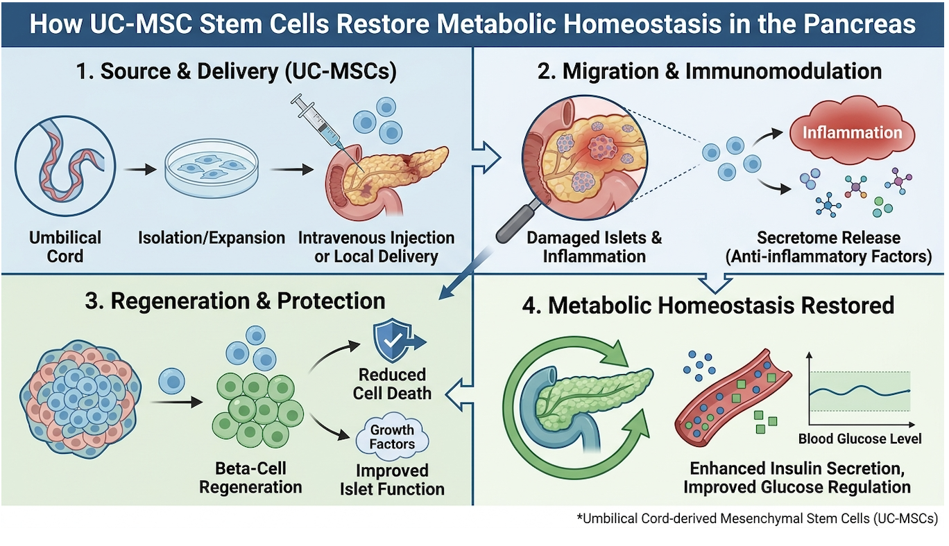
Figure 1: UC-MSCs modulate the pancreatic microenvironment primarily through secretome emissions, reducing local fibrosis and protecting remaining beta cells.
For decades, the prevailing theory was that injected stem cells would travel directly to the pancreas, physically engraft into the islet of Langerhans, and magically transform into brand new, insulin-secreting beta cells. We now know this direct differentiation happens at clinically insignificant rates. The real metabolic repair happens entirely through the secretome. The secretome represents the massive payload of exosomes, microRNAs, and growth factors that stem cells release into their surrounding environment.
UC-MSCs act as cellular paramedics. When introduced into a highly inflammatory diabetic environment, they assess the damage and release targeted chemical signals a process known as paracrine signaling. They secrete a payload of exosomes containing vascular endothelial growth factor (VEGF), basic fibroblast growth factors (bFGF), and transforming growth factor-beta (TGF-β). These growth factors stimulate angiogenesis (the formation of new blood vessels), rescuing hypoxic tissue in the pancreas. By restoring blood flow, the surviving endogenous beta cells receive the oxygen and nutrients required to resume normal insulin synthesis.
UC-MSCs secrete over 300 unique cytokines fundamentally reprogramming pro-inflammatory macrophages into tissue-repairing variants. This is the critical M1 to M2 macrophage polarization switch. M1 macrophages are destructive; they secrete tumor necrosis factor-alpha (TNF-α) and interleukin-6 (IL-6), which actively drive systemic insulin resistance at the receptor level. UC-MSCs release specific mediators like TSG-6 that force these macrophages to adopt the M2 phenotype. The M2 macrophages then release anti-inflammatory IL-10, actively cooling down the immune system’s attack on pancreatic tissue and enhancing systemic insulin sensitivity.
| 📌 If you’re interested in how MSCs calm overactive immune cells throughout the body, we have an interesting article that discusses mesenchymal stem cell therapy for immune modulation, which you can read via the internal link. |
Furthermore, these cells engage the SIRT1 pathway. SIRT1 is a crucial NAD+-dependent deacetylase that regulates cellular health. It protects against severe oxidative stress and restores mitochondrial membrane integrity in failing beta cells. Through the ERK MAPK pathway, UC-MSCs also prevent beta cell apoptosis (programmed cellular death). They aren’t replacing the pancreas; they are repairing the hostile microenvironment so the body’s native beta cell regeneration processes can resume unhindered. Understanding this deep, secretome-driven cellular repair is the only way to critically evaluate the clinical feasibility of these treatments.
Patients constantly ask: can stem cell therapy reverse Type 2 diabetes? The answer requires completely stripping away marketing semantics and looking strictly at hard clinical metrics. Defining the threshold of true reversal dictates the entire conversation around clinical viability.
If you define “reversal” as returning to a state of sustained, long-term normoglycemia without the use of any pharmacotherapy (insulin or oral hypoglycemics) forever, then calling it a guaranteed cure is medically inaccurate. However, if you define it as massive disease modification achieving a non-diabetic HbA1c while drastically reducing or completely eliminating exogenous insulin dependence the data is incredibly compelling. Disease modification of this magnitude drastically reduces the risk of severe diabetic complications, including neuropathy, retinopathy, and chronic kidney disease.
In recent phase 2 trials, cohorts receiving high-dose intravenous UC-MSC infusions demonstrated marked improvements in C-peptide levels. C-peptide is a highly accurate biological biomarker used to measure endogenous insulin production. Because insulin and C-peptide are secreted by the Diabetes Care, 2022). Measuring C-peptide prevents clinicians from being fooled by temporary drops in blood sugar caused by dietary changes alone.
This is exactly where the second and third pillars of the UC-MSC Efficacy Triad Processing and Delivery dictate success or failure. Why do some trials show incredible results while others show none? Processing matters immensely. Cells expanded past passage 4 or 5 in a laboratory begin to rapidly lose their potency and experience replicative senescence. If a clinic uses over-cultured, exhausted cells to maximize profit margins, the paracrine effect is heavily muted. Delivery matters just as much. A systemic IV push treats systemic inflammation, while targeted intrapancreatic arterial infusions deliver the payload directly to the organ. You cannot achieve true biological modification with poorly processed, inappropriately delivered cells.
In recent years, headlines exploded globally with claims regarding massive medical breakthroughs. Patients immediately started searching to find out how China reverse diabetes stem cells protocols were functioning, hoping for an accessible cure. The viral nature of these scientific reports often strips away vital clinical context, leading patients to assume these exact treatments are commercially available worldwide.
These reports largely stemmed from groundbreaking work at institutions like Shanghai Changzheng Hospital. In heavily publicized case studies, researchers utilized advanced stem cell programming to treat patients with long-standing diabetes, successfully weaning them completely off exogenous insulin for extensive periods. But our team’s critical review of this data demands a much closer look at the actual methodology employed.
The most advanced trials utilized highly complex, bespoke protocols. They involved the reprogramming of peripheral blood mononuclear cells into induced pluripotent stem cells (iPSCs), which were then differentiated in a lab into functioning islet cells over several weeks. This is a completely different mechanism than a standard intravenous infusion of allogeneic UC-MSCs. It represents personalized cellular engineering at the highest level of modern biotechnology.
While the clinical trial data proves that cellular therapy can functionally reduce severe insulin dependence in targeted case studies, applying this exact model to the general public today is
Determining the true mesenchymal stem cell therapy cost requires analyzing cell preparation standards, physician oversight, and geographic regulatory environments. Because the FDA restricts the expansion and systemic administration of UC-MSCs in the United States outside of approved clinical trials, many patients evaluate international jurisdictions with advanced biological regulatory frameworks to access these therapies safely.
Let’s address the financial reality of this industry head-on. If you find a clinic offering stem cell treatments for $1,500, you are almost certainly receiving dead cells, a simple PRP (platelet-rich plasma) spin-down, or unregulated amniotic fluid containing zero live stem cells. Ethical medical providers understand that biological excellence carries an inherent financial threshold.
The global mesenchymal stem cell therapy cost ranges from $5,000 to $25,000 per protocol, highly dependent on the regulatory environment, cell expansion quality, and facility accreditations (Medical Tourism Journal, 2023). Applying the UC-MSC Efficacy Triad reveals exactly why costs vary so wildly. The “Processing” pillar is astronomically expensive to execute correctly. Culturing live human tissue requires maintaining ISO-7 certified cleanrooms with highly specialized HEPA filtration systems. It requires rigorous, redundant PCR testing for pathogens like HIV, Hepatitis, Syphilis, and Cytomegalovirus to prevent severe patient infections.
It also requires third-party flow cytometry immediately prior to infusion to prove that the cells are alive, viable, and expressing the correct CD surface markers (positive for CD73, CD90, CD105; negative for CD34, CD45). When you pay $20,000 for a treatment, you aren’t just buying biological material. You are funding the extreme laboratory overhead required to ensure that biological material doesn’t give you sepsis.
When patients ask, “what country with best stem cells should I visit?”, they are fundamentally asking the wrong question. Cells are not inherently better simply because they originate in Colombia versus Mexico. The safety and quality of the cells are dictated entirely by the laboratory laws governing the host country, and how strictly those laws are enforced by national health ministries.
Panama has long been considered a premier destination because its government actively embraces regenerative medicine. They allow full UC-MSC expansion under strict, government-audited laboratory conditions that rival Western standards. Mexico offers highly competitive pricing, but the regulatory enforcement varies wildly from clinic to clinic. This requires intense patient vetting of laboratory credentials, as some facilities operate completely outside of COFEPRIS (the Mexican health regulatory agency) oversight. Relying solely on glossy marketing brochures instead of verifiable ISO certifications in Mexico is a massive clinical risk.
Southeast Asia has recently emerged as a global powerhouse in regenerative logistics. For those evaluating the safety of stem cell therapy in Thailand, the draw is the country’s world-class medical tourism infrastructure combined with aggressive government investment in biotechnology. Thai regulatory frameworks often strictly mirror Western GMP (Good Manufacturing Practice) standards while allowing for the legal expansion of allogeneic umbilical cord tissues. Their private hospitals frequently hold Joint Commission International (JCI) accreditation, providing an additional layer of medical safety.
| 📌 If you’re planning treatment in Thailand and want a practical checklist for choosing a safe clinic, we have an interesting article that discusses whether stem cell therapy is safe in Thailand, which you can read via the internal link. |
Colombia is also rising rapidly, offering cutting-edge facilities in Bogotá and Medellín with stringent INVIMA (Colombia’s FDA equivalent) oversight. Ultimately, you are shopping for laboratory integrity, not a vacation destination. For individuals for whom international clinical treatment is financially or logistically unfeasible, research is increasingly focusing on the body’s innate ability to mobilize its own stem cell reserves.
To naturally increase stem cell production, clinical evidence points toward specific metabolic stressors and nutritional interventions that promote endogenous cellular mobilization. While
The supplement industry is notorious for marketing magical “stem cell activators” that do absolutely nothing. From a strict biochemical perspective, you cannot take a pill that magically creates new stem cells out of thin air. However, you can take evidence-based supplements that alter your body’s oxidative stress levels, thereby preventing the premature death of the endogenous stem cells you already have residing in your bone marrow.
When evaluating the best stem cell supplements, clinical data points heavily toward specific polyphenols and targeted antioxidants. Resveratrol, found in the skin of red grapes, has been shown to activate SIRT1 pathways the exact same cellular defense mechanism utilized by exogenous stem cells to protect mitochondria. Vitamin D3 is absolutely critical; profound deficiencies are consistently linked to the exhaustion of hematopoietic stem cell niches. Curcumin (paired with piperine for bioavailability) actively downregulates NF-kB, reducing the systemic inflammatory cytokines that force native stem cells into dormancy. Protecting the bone marrow niche is just as critical as stimulating cell release.
What about daily beverages? Patients frequently ask what drink increases stem cells. The most scientifically validated option is high-quality green tea, specifically due to Epigallocatechin gallate (EGCG). EGCG protects endogenous cells from reactive oxygen species preserving the body’s native stem cell reserves from premature cellular death (Journal of Nutritional Biochemistry, 2019). EGCG has documented effects on the proliferation of neural and mesenchymal progenitor cells by actively modulating oxidative stress.
It is vital to maintain realistic expectations. Swallowing curcumin and drinking green tea will not replicate the million-cell payload of a targeted IV infusion. These supplements are long-term maintenance tools. They naturally repair stem cells by cleaning up the toxic, hyperglycemic blood environment, allowing your surviving cellular reserves to function without constant metabolic suppression.
If you genuinely want to trigger stem cell regeneration naturally, you have to subject your body to controlled, acute metabolic stress. The single most powerful tool for this biological adaptation is prolonged caloric restriction. The human body evolved to repair itself during periods of nutrient scarcity, not during constant caloric surplus.
Prolonged fasting and specific high-polyphenol diets can naturally increase stem cell production by triggering autophagy and promoting the endogenous mobilization of progenitor cells into the bloodstream (Cell Stem Cell, 2014). Autophagy is essentially intense cellular house-cleaning. When you deprive the body of nutrients for 24 to 72 hours, it inhibits the mTOR pathway and activates AMPK. The body begins cannibalizing damaged, senescent cells for raw energy. This clearing out of metabolic junk signals the bone marrow to release fresh, naive stem cells to rebuild the cleared tissues.
Exercise protocols offer a similar, highly effective mechanism. High-intensity interval training (HIIT) creates transient hypoxia (low oxygen) in muscle tissues across the body. This mild, controlled stressor triggers the activation of Hypoxia-inducible factor 1-alpha (HIF-1α). This leads directly to the release of VEGF and stimulates the rapid proliferation of endothelial progenitor cells to build new blood vessels. The brief, intense physical exertion literally commands your bone marrow to deploy repair cells.
Finally, deep restorative sleep is a non-negotiable variable in this equation. The circadian rhythm deeply influences stem cell release. Chronic sleep deprivation elevates cortisol and systemic inflammation, entirely suppressing progenitor cell activity. Whether utilizing natural mobilization techniques or pursuing advanced clinical UC-MSC infusions, patients must critically evaluate the inherent limitations of these approaches.
Stopping insulin or metformin after stem cell therapy is never guaranteed and should only occur under strict endocrinologist supervision. While UC-MSC infusions aim to reduce insulin
The clinical results of stem cell therapy for Type 2 diabetes typically last between six months to three years, depending entirely on the patient’s biological response. UC-MSCs function via transient paracrine signaling rather than permanent cellular engraftment into the pancreas. Therefore, their anti-inflammatory and immunomodulatory effects eventually diminish over time. Most clinical protocols suggest that patients may require subsequent booster infusions to maintain optimal metabolic homeostasis.
Current clinical trials indicate a clinical response rate of roughly 60% to 70% for measurable improvements in glycemic control following UC-MSC therapy (Stem Cell Research & Therapy, 2022). Success is generally defined medically as a significant reduction in HbA1c levels, a spike in C-peptide production, or a decrease in daily exogenous insulin requirements. It is rarely defined by objective clinicians as a total, permanent cure. Outcomes depend entirely on the quality of cell processing, the administration protocol, and stringent patient selection criteria. Unregulated commercial clinics claiming 100% success rates are medically inaccurate and should be avoided.
Stem cell therapy can significantly lower HbA1c levels in responsive Type 2 diabetes patients by attacking the root cause of systemic insulin resistance. Intravenous infusions of UC-MSCs secrete specific cytokines that mitigate the chronic inflammation responsible for glucose intolerance at the cellular receptor level. Multiple phase 2 trials have documented sustained HbA1c reductions in target cohorts over 12 to 24 months post-infusion. However, strict dietary management remains essential to sustain these hematological improvements long-term.
Stem cell treatments for Type 2 diabetes are currently not FDA approved as standard medical therapies in the United States. They remain classified as investigational new drugs (INDs) subject to ongoing, highly regulated phase clinical trials. The FDA strictly prohibits the systemic administration of expanded UC-MSCs outside of these authorized research frameworks. Consequently, patients seeking these specific expanded cellular therapies often evaluate international jurisdictions with different regulatory pathways. All patients should exercise extreme caution regarding domestic clinics offering unapproved biological products.
For patients evaluating clinical interventions, stem cell therapy for type 2 diabetes represents a significant advancement toward repairing biological deterioration rather than merely managing symptoms. Recent clinical data indicates that targeted UC-MSC infusions can yield a 60% to 70% clinical response rate in reducing HbA1c levels by directly addressing systemic inflammation (Stem Cell Research & Therapy, 2022). The safest possible approach requires prioritizing rigorous laboratory standards over commercial marketing claims.
Applying the UC-MSC Efficacy Triad critically vetting the origin, laboratory processing, and delivery method of the cells is your absolute best defense against industry hype. This framework forces you to ask hard questions about ISO certifications, pathogen screening, and flow cytometry, ensuring you base your medical decisions on objective biological data rather than emotional advertising.
Before booking flights to an international clinic, demand full laboratory transparency. Consult with your primary endocrinologist today to review your current C-peptide levels, evaluate your specific risk factors, and determine if an investigational regenerative protocol actually aligns with your long-term metabolic health goals.
This article is for educational purposes only and does not replace consultation with a qualified medical professional.