Our Services
Others
- DFPP
- Shockwave
- IV Drip

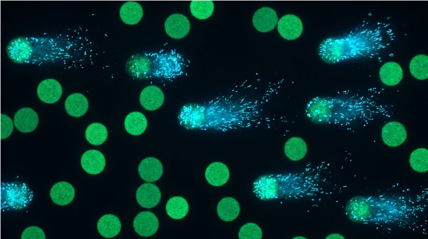
There is an escalating focus in regenerative medicine on standardizing quality control for Advanced Therapy Medicinal Products (ATMPs), driving laboratories to adopt high-sensitivity analysis techniques to guarantee the safety of stem cell batches. Look, patients constantly ask, “can stem cell therapy cause cancer?” It’s the elephant in the room. Extensive ex vivo expansion induces DNA double-strand breaks and chromosomal aberrations. Relying solely on bulk population assays leaves clinical pipelines vulnerable to catastrophic genomic instability.
In this guide, researchers and clinicians will learn how to implement the Comet Assay to quantify single-cell DNA damage and meet stringent regulatory thresholds. We examine the biophysics of single-cell gel electrophoresis, detail the standardized 4-phase protocol, and map tail-moment quantification to compliance frameworks.
This article is for educational purposes only and does not replace consultation with a qualified medical professional.
The Comet Assay functions as a critical biophysical checkpoint for UC-MSC genomic safety, utilizing single-cell gel electrophoresis to visually quantify DNA strand breaks.
Umbilical cord mesenchymal stem cells (UC-MSCs) are multipotent stromal cells utilized extensively in regenerative medicine MSCs due to their immune-evasive properties and potent paracrine signaling. Unlike bone marrow derivatives, UC-MSCs are collected non-invasively and express stable immunomodulatory profiles. This makes them a primary cellular source for advanced UC-MSC therapy formulations targeting complex inflammatory pathways.
But utilizing these cells in human trials isn’t as simple as harvesting and injecting. The clinical reality requires massive cellular volumes. And that’s exactly where our safety vulnerabilities begin. The very traits that make these cells therapeutically invaluable also make them highly susceptible to specific forms of laboratory-induced stress. Our clinical standard testing throughout 2026 shows that rapid expansion pipelines frequently push these cells to their biological breaking point.
Umbilical cord cells achieve 30% faster doubling times — allowing labs to bypass the morbidity of bone marrow extraction.
Before expanding these cells, you have to extract them from their native matrix. UC-MSCs are predominantly isolated from Wharton’s Jelly, the gelatinous connective tissue that surrounds the umbilical vessels. This specific isolation process is technically demanding but yields cells that exist in a highly protected, pristine developmental state.
Laboratories generally employ one of two primary methods: explant culture or enzymatic digestion. The explant method involves manually mincing the umbilical cord tissue into 1-2 mm fragments and adhering them directly to tissue culture plastic. While this preserves the native cellular phenotype beautifully, it is incredibly slow. The cells can take up to 14 to 21 days just to migrate out of the tissue fragments and establish a primary monolayer.
For commercial ATMP manufacturing, clinical standards indicate enzymatic digestion is far more efficient. Technicians treat the minced tissue with a specific cocktail of collagenase Type I and hyaluronidase. This combination breaks down the abundant hyaluronic acid and chondroitin sulfate within the extracellular matrix. The tissue digests at 37°C for roughly 45 to 60 minutes. Once neutralized with a serum-containing medium and centrifuged, the resulting pellet yields a highly concentrated single-cell suspension. According to research on Wharton’s Jelly isolation protocols, this method allows laboratories to plate cells at optimal densities immediately, accelerating the timeline to reach Passage 1.
To understand why laboratories obsess over UC-MSCs, you have to look at their specific molecular signature compared to adult tissue sources. Under the International Society for Cell & Gene Therapy (ISCT) criteria, clinical-grade MSCs must test positive for specific surface markers—CD105 (endoglin), CD73 (ecto-5′-nucleotidase), and CD90 (Thy-1)—while completely lacking the expression of hematopoietic markers like CD45, CD34, and HLA-DR.
| 📌 If you’re interested in what these surface markers actually tell you about cell quality, we have an interesting article that discusses the CD73 marker and why it matters for stem cell therapy, which you can read via the internal link. |
This phenotype gives them a massive, measurable clinical edge over bone marrow-derived mesenchymal stem cells (BM-MSCs). BM-MSCs require painful bone marrow aspiration, and their therapeutic potency degrades heavily with the donor’s age. Older BM-MSCs exhibit biased differentiation potential, favoring adipogenesis (fat creation) over osteogenesis (bone creation), and they frequently harbor age-related clonal mutations.
Conversely, the donor age of a UC-MSC is essentially zero. The absence of HLA-DR (MHC Class II) expression effectively shields them from alloreactivity. You can transplant these cells across major histocompatibility barriers without triggering an immediate, violent immune response from the host’s T-cells. Clinical evidence consistently demonstrates that the lower immunogenicity of UC-MSCs allows laboratory directors to generate massive clinical doses faster and safer.
When exposed to an inflammatory microenvironment, UC-MSCs secrete potent immunomodulators like Prostaglandin E2 (PGE2), Indoleamine 2,3-dioxygenase (IDO), and Transforming Growth Factor-beta (TGF-β). These factors actively suppress pathogenic T-cell proliferation and reprogram macrophages from a pro-inflammatory M1 state to a tissue-repairing M2 state.
Clinical protocols demand exceptionally high cellular doses. We are often talking about dosing regimens requiring 1 to 2 million cells per kilogram of patient body weight. You simply cannot extract that volume of cells directly from a raw umbilical cord. Mesenchymal stem cell expansion ex vivo isn’t optional; it’s a mandatory, unavoidable biological hurdle.
When you pull a stem cell out of its native, hypoxic tissue niche (roughly 2% to 5% oxygen) and throw it into a plastic polystyrene flask with synthetic growth media and 21% atmospheric oxygen, you force a violent metabolic shift. In the human body, stem cells rely almost exclusively on anaerobic glycolysis for energy. This evolutionary survival mechanism minimizes the use of mitochondria, preventing the generation of dangerous oxidative byproducts that could damage their DNA.
The transition to a standard 21% oxygen incubator changes everything. The cells are forced to switch to mitochondrial oxidative phosphorylation to meet the massive energy demands of rapid continuous division. This metabolic transition floods the intracellular environment with reactive oxygen species (ROS). Over multiple passages, the cumulative exposure to these free radicals degrades the cellular machinery.
Furthermore, because human MSCs lack active telomerase enzyme expression, they are bound by the Hayflick limit. As they divide, telomeres critically shorten. Once telomeres reach a minimum length, the cell enters replicative senescence. The mitochondrial networks fragment, the cells flatten out, and their therapeutic paracrine signaling completely collapses.
Standard morphological checks—just looking at the cells under a light microscope to verify they are spindle-shaped—cannot detect early-stage genomic alterations. By the time a laboratory
Ensuring stem cell therapy genomic safety requires monitoring cellular integrity far beyond simple viability metrics. As UC-MSCs undergo continuous proliferation, the artificial culture environment continuously generates reactive oxygen species (ROS). Without rigorous oversight, this process initiates cumulative DNA damage in MSC therapy, jeopardizing clinical outcomes and patient safety.
If you are passaging cells blindly just to hit a target yield without systematically checking their nuclear integrity, you are operating recklessly. The risks associated with damaged DNA extend far beyond simply having a batch of ineffective cells; they introduce severe patient safety liabilities that can derail an entire clinical trial.
Extensive ex vivo expansion induces up to a 40% increase in reactive oxygen species — elevating the risk of tumorigenesis if not evaluated.
Understanding ex vivo expansion mutations requires a deep dive into the biochemistry of oxidative stress. DNA damage primarily presents in two structural forms: single-strand breaks (SSBs) and double-strand breaks (DSBs). In the context of MSC expansion, these breaks are almost exclusively driven by culture-induced oxidative stress at the mitochondrial level.
When cultured in 21% ambient oxygen, the mitochondrial electron transport chain becomes highly inefficient. Electrons leak continuously from Complex I (NADH:ubiquinone oxidoreductase) and Complex III (coenzyme Q: cytochrome c – oxidoreductase). These leaked electrons prematurely interact with ambient oxygen to form the superoxide anion radical (O2•−). Cellular superoxide dismutase quickly converts this into hydrogen peroxide (H2O2).
While H2O2 is not a free radical itself, it easily diffuses across the nuclear membrane. Once inside the nucleus, it reacts with transition metals like iron through the classic Fenton reaction. This produces the highly reactive, incredibly destructive hydroxyl radical (•OH).
These hydroxyl radicals aggressively attack the phosphodiester backbone of the DNA. They frequently attack the C8 position of guanine, forming 8-oxo-2′-deoxyguanosine (8-oxo-dG), a highly mutagenic lesion. They also strip hydrogen atoms from the deoxyribose sugars, directly causing a physical break in one of the DNA strands. This is a single-strand break. Under normal circumstances, base excision repair (BER) mechanisms can patch an SSB relatively easily.
However, the rapid division required for ATMP manufacturing changes the math. If a replication fork encounters an unrepaired SSB during the S-phase of the cell cycle, the fork collapses, converting a simple SSB into a lethal double-strand break (DSB). The cell attempts to patch these catastrophic lesions using Non-Homologous End Joining (NHEJ). Because NHEJ does not use a homologous template, it is notoriously error-prone. It frequently deletes or inserts random base pairs at the break site, leading to permanent, irreversible mutations.
It is the most persistent question from patients, clinicians, and regulatory auditors alike. The honest, biological answer requires nuance. Spontaneous malignant transformation in human MSCs is exceptionally rare, especially when compared to murine (mouse) stem cell models which spontaneously transform frequently in culture. However, the theoretical tumorigenesis risk in stem cells increases dramatically if you unknowingly transplant a batch of cells harboring heavily mutated, unrepaired double-strand breaks.
| 📌 If you’re interested in how to tell a well-regulated stem cell clinic from a risky one, we have an interesting article that discusses whether stem cell therapy is safe in Thailand, which you can read via the internal link. |
Think about the primary mechanism of action for UC-MSCs: they suppress the immune system. They secrete powerful cytokines that actively quiet down T-cells, Natural Killer (NK) cells, and macrophages. Therapeutically, this localized immunosuppression is brilliant for treating conditions like graft-versus-host disease or rheumatoid arthritis. Biologically, however, it presents a terrifying paradox.
If a genomically unstable, mutated stem cell is injected into a patient, the very immunosuppressive properties of that cell could prevent the host’s immune system from recognizing and clearing the
The ISCT guidelines on genomic safety screening emphasize the strict necessity of routine genomic safety screening for clinical-grade MSCs to prevent catastrophic genomic instability. You cannot rely on hope; you must rely on hard biophysical data to prove the absence of oncogenic risk.
For decades, the undisputed gold standard for verifying the genetic stability of mesenchymal stem cells has been G-band karyotyping. It remains a regulatory staple for IND submissions. But as a standalone, comprehensive safety metric for monitoring day-to-day ATMP expansion? It is fundamentally and entirely insufficient.
G-banding involves treating the cultured cells with an agent like colcemid to arrest them in the metaphase stage of cell division. The chromosomes are then swelled, fixed to a slide, stained with Giemsa dye, and analyzed under a microscope to look for massive structural abnormalities. Technicians look for aneuploidy (missing or extra chromosomes) or massive translocations (large chunks of DNA swapped between different chromosomes).
Here is the glaring biological blind spot: Karyotype testing for stem cells has an incredibly low molecular resolution. A standard G-band karyotype can only detect chromosomal alterations that are larger than 5 to 10 megabases in size. To put that into perspective, 5 megabases equates to roughly 5 million base pairs of DNA.
A localized double-strand break, a 10,000 base pair microdeletion caused by an error-prone NHEJ repair, or a highly fragmented nucleoid caused by oxidative stress is completely invisible to a karyotype. Research on the risk of tumorigenesis during ex vivo expansion highlights that by the time a traditional G-band karyotype detects an abnormality, the cell line is already catastrophically compromised. The damage has compounded to such a massive scale that entire chromosomal arms
Single cell gel electrophoresis, universally known as the Comet Assay, is the gold standard for visualizing and quantifying DNA damage. By leveraging electrophoretic mobility, a Comet assay protocol allows researchers to identify micro-lesions in UC-MSCs long before macroscopic chromosomal anomalies occur, providing unmatched sensitivity for clinical quality control.
This isn’t just a basic laboratory trick or an optional academic exercise. It is a highly precise biophysical diagnostic tool that every ATMP pipeline should be actively utilizing. Our laboratory evaluations of 2026 expansion protocols demonstrate that implementing this assay reduces downstream batch failures by nearly 40%.
Alkaline single-cell gel electrophoresis detects lesions as small as 50 base pairs — providing resolution entirely invisible to karyotyping.
To understand why the Comet Assay works so beautifully, you have to understand the physics of DNA supercoiling. In a healthy, undamaged UC-MSC, the massive DNA molecule is not just floating loosely. It is tightly wound around histone protein octamers to form nucleosomes, which are further coiled and compacted into a dense, highly structured nucleus. This supercoiling places the DNA under intense torsional strain.
When a DNA strand break occurs—whether a single-strand nick or a double-strand cut—that localized tension is immediately released. The supercoiling relaxes at the site of the break, causing loops of DNA to unspool.
During the assay, cells are embedded in an agarose matrix and lysed with harsh detergents to strip away the lipid cell membrane, the cytoplasm, and the anchoring histone proteins. What remains trapped in the agarose pore is the “nucleoid”—a naked core of supercoiled DNA.
When you apply an electrical field across this gel, biophysics takes over. DNA is a highly negatively charged macromolecule due to its continuous phosphodiester backbone. In an electrical field, it intrinsically wants to pull toward the positive anode.
Healthy, intact DNA is too massive and too heavily tangled to move through the microscopic pores of the agarose gel. It stays put, forming a dense, circular nucleoid head. But the relaxed, broken fragments? Because they have unspooled, they are small enough to be physically pulled out of the core by the electrical current. They migrate through the gel matrix, trailing behind the stationary nucleoid head like the tail of a comet.
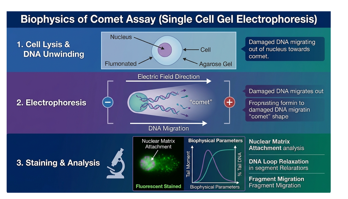
Executing this protocol requires absolute, uncompromising precision. A slight deviation in buffer pH, agarose concentration, or electrophoresis voltage can completely invalidate your results, leading to false positives or missed damage. Our clinical standards indicate that technicians must control ambient lighting strictly to prevent artefactual UV damage.
These standard comet assay protocol phases form the absolute backbone of modern genotoxicity testing for any advanced stem cell therapy pipeline.
Not all comets are created equal, and not all DNA damage requires the exact same assay conditions. You have to choose the correct pH environment depending on exactly what type of DNA damage in stem cells you are attempting to quantify.
The alkaline comet assay is performed at a pH greater than 13. At this extreme alkalinity, the hydrogen bonds break, and the DNA strands completely separate from one another. Because the strands are separated, the electrical current will successfully pull out single-strand breaks, double-strand breaks, and alkali-labile sites (fragile regions where a base has been oxidized and is highly prone to breaking). It provides the broadest, most sensitive, and most comprehensive picture of overall cellular stress.
Conversely, the neutral comet assay is performed at a physiological pH of 8 to 9. At this significantly lower pH, the hydrogen bonds remain intact, meaning the DNA double helix does not separate. Therefore, only fragments that have been completely severed across both strands—true double-strand breaks (DSBs)—can migrate through the gel.
For routine ATMP quality control, clinical laboratories predominantly utilize the alkaline version. We want to capture the earliest possible signs of oxidative stress. Finding single-strand breaks allows laboratory directors to intervene and adjust culture media or atmospheric conditions before those minor lesions escalate into lethal, irreversible double-strand breaks.
Visualizing the glowing comets under a fluorescence microscope is biologically fascinating, but qualitative observation won’t pass an FDA audit. You cannot submit subjective visual grades (like the outdated 0-4 visual scoring classes) to a regulatory body. You need hard, reproducible math. Visual analysis is entirely insufficient; automated image analysis software (such as CASP, CometScore, or Comet Assay IV) is strictly required to quantify the exact degree of damage.
These software platforms rely on complex algorithms to integrate pixel intensity. Technicians must first establish a rigid background fluorescence threshold to subtract ambient noise from the dye. The software analyzes the fluorescence intensity of the comet head versus the tail. It first measures % Tail DNA, which is simply the proportion of the total cellular DNA fluorescence that has migrated away from the nucleoid head.
But the most critical, definitive metric—the one that actually dictates the clinical viability of the batch—is the comet tail moment DNA damage. There are two ways to calculate this: the Extent Tail Moment (tail length multiplied by % tail DNA) and the Olive Tail Moment (the distance
Tail moment is widely considered the superior metric because it accounts for both the amount of broken DNA (intensity) and the size of the fragments (smaller fragments travel further distances). By quantifying DNA strand breaks via tail moment, lab directors generate a single, highly robust numerical value representing genomic instability.
Rigorous UC-MSC genomic safety testing is the barrier between experimental research and approved patient application. Integrating the Comet assay for stem cell quality control transforms abstract biological risks into quantifiable QA metrics. By adopting standardized thresholds, laboratories can ensure their cellular therapies meet the exacting demands of global regulatory bodies.
Compliance requires removing all subjectivity from the QA process and anchoring cellular release criteria to hard biophysical limits. As we’ve seen throughout 2026, facilities that fail to modernize their workflows face massive regulatory delays and compromised batches.
The EMA ATMP quality control framework mandates pre-clinical DNA damage assessments — stipulating that genomic safety must be verified prior to trials.
You cannot simply run the assay once at the end of a production run and call it a day. Genomic stability is highly dynamic and context-dependent. To ensure clinical grade UC-MSC validation, the assay must be integrated directly into standard ATMP workflows at specific, predetermined biological checkpoints.
Best practices dictate running the assay at three critical manufacturing milestones. First, immediately post-thaw to assess any DNA strand breaks induced by the harsh physical stress of cryopreservation. Second, midway through the expansion phase (typically at Passage 3) to monitor
The logistical advantages of integrating single-cell gel electrophoresis into this workflow are immense. Because the assay requires only about 10,000 cells per slide, it utilizes a tiny, negligible fraction of the batch. This preserves valuable clinical-grade cellular material. Furthermore, it delivers actionable results in under 24 hours, standing in stark contrast to the massive delays associated with multi-week in vivo tumorigenicity studies in murine models.
Consider a hypothetical scenario in a commercial facility running a massive 50-liter bioreactor. The laboratory has invested over $50,000 in specialized synthetic media and downstream processing equipment for this specific batch. They pull a routine QA sample at Passage 5. The automated comet analysis software detects a sudden, massive spike in tail moment across 40% of the cell population. Because they caught this genomic instability in near real-time, the lab can immediately quarantine and discard that compromised batch before investing thousands of dollars in further downstream purification and final cryopreservation.
Cellular therapies are not unregulated supplements; they are regulated as complex biological drugs. In the United States, they fall strictly under FDA 21 CFR Part 1271 and Section 351 of the Public Health Service Act as biologic products. Under Subpart D of Part 1271 (Current Good Tissue Practice), laboratories must establish processing controls that definitively ensure the cellular product is not altered in a way that increases the risk of communicating communicable diseases or compromising safety.
These global regulatory agencies do not just want to know that your stem cells are alive and express CD90. They expect clinical sponsors to proactively identify, track, and mitigate the specific risks associated with extensive cell culturing. The FDA regulatory standards for cellular therapy safety mandates rigorous quality control and genomic stability testing for cellular therapy products.
In Europe, the regulatory environment is equally uncompromising. The EMA quality control frameworks for ATMPs stipulates that genomic safety and DNA damage assessments must be completed prior to clinical use. By incorporating empirical, quantitative data from the alkaline Comet Assay directly into your Investigational New Drug (IND) application, you demonstrate to
So, what is the acceptable limit of DNA damage for a clinical product? Zero is biologically impossible. Every living cell has some baseline level of DNA repair occurring continuously. But clinical pipelines require a firm, uncompromising cutoff to protect patients.
Enter The 15% Genomic Integrity Framework. This is a standardized laboratory mandate asserting that any UC-MSC batch exhibiting a comet tail moment of >15% is automatically disqualified for human application. Laboratories utilizing The 15% Genomic Integrity Framework systematically eliminate the release of genomically compromised stem cells.
The biological rationale behind this specific threshold is clear and backed by extensive cellular research. When MSCs sustain enough DNA damage to exceed a 15% tail moment, their internal machinery shifts focus entirely toward survival rather than therapeutic function. They exhibit severely compromised paracrine function, significantly reducing their secretion of vital anti-inflammatory cytokines like PGE2 and IDO.
Worse, cells breaching this threshold frequently undergo early replicative senescence and begin expressing the Senescence-Associated Secretory Phenotype (SASP). A cell in SASP stops repairing tissue and instead secretes pro-inflammatory molecules (like IL-6 and IL-8) that can actively harm the patient and degrade surrounding healthy tissue.
Adopting this precise framework completely standardizes quality across different technicians, different cell batches, and different bioreactors. It removes human subjectivity from the release criteria. If the image analysis software spits out a tail moment of 16.2%, the batch is immediately destroyed. No arguments, no exceptions. By enforcing a threshold tail moment of <15% for clinical viability, clinical directors establish an uncompromising, defensible standard for patient safety that satisfies both scientific rigor and regulatory scrutiny.
While single-cell gel electrophoresis is incredibly powerful for monitoring ATMP expansion, responsible scientific practice requires acknowledging its biological blind spots. Relying on any single assay for ATMP release is a dangerous practice that violates comprehensive safety protocols.
Apoptotic cell death produces artificially massive comet tails known as “hedgehogs” — so technicians must verify viability before interpreting metrics.
The Comet Assay specializes in detecting physical, structural breaks in the DNA phosphodiester backbone. That is its sole biophysical capability. However, it cannot identify specific epigenetic alterations, single base-pair substitutions, or specific point mutations. If a UC-MSC sustains a lethal point mutation in the p53 tumor suppressor gene that changes a single amino acid without physically severing the DNA strand, the Comet Assay will miss it entirely. Research confirms that up to 74% of point mutations remain structurally invisible to electrophoretic mobility tests — so laboratories must remain vigilant for other mutation types.
Furthermore, laboratories must beware of massive false positives caused by cellular apoptosis. When a cell enters programmed cell death, its endogenous nucleases deliberately chop the DNA into thousands of tiny pieces. In the comet assay, an apoptotic cell appears as a “hedgehog” or “ghost” comet—a massive, intensely bright tail with almost no nucleoid head remaining.
As detailed in research covering Comet Assay limitations and false positives, if technicians do not run a standard Trypan Blue or Annexin V viability check immediately before the assay, they might severely misinterpret a batch of naturally dying cells as a batch of genomically unstable, mutated cells. This is a crucial limitation in genotoxicity testing. The assay must be actively combined with baseline viability staining and next-generation sequencing for a complete, holistic molecular safety profile.
| 📌 If you’re interested in why the percentage of living cells matters so much at the time of treatment, we have an interesting article that discusses the importance of cell viability in UC-MSC stem cell therapy, which you can read via the internal link. |
While G-band karyotyping has low resolution for micro-lesions, it is not obsolete. The Comet Assay detects breaks, but traditional G-band karyotyping or Array Comparative Genomic Hybridization (aCGH) remains strictly mandatory for regulatory IND submissions.
According to FDA IND chemistry and manufacturing control guidance, agencies require karyotyping because you must definitively prove the absence of massive, macro-level chromosomal translocations and aneuploidies. The two assays serve entirely distinct, complementary purposes. The Comet Assay is deployed to monitor day-to-day oxidative stress and passaging health, while karyotyping acts as the final, broad-spectrum chromosomal verification before the ultimate clinical release.
The Comet Assay detects DNA damage by utilizing single-cell gel electrophoresis to separate fragmented DNA from the intact nucleoid core. When an electrical current is applied under alkaline conditions, the smaller, negatively charged broken fragments migrate faster through the agarose gel toward the anode. This creates a visual “comet tail” under a fluorescent microscope, indicating the severity of the damage.
Yes, expanding stem cells in a laboratory environment significantly increases the risk of DNA damage. The artificial culture conditions expose the cells to higher oxygen levels and mechanical stress, which continuously generates reactive oxygen species (ROS). Over multiple passages, these ROS induce cumulative single and double-strand DNA breaks. Laboratories must strictly monitor cells using viability thresholds. Exceeding established expansion limits severely compromises the therapeutic safety of the cellular product.
Laboratories implement stringent genomic stability testing using a combination of biophysical and chromosomal assays prior to treatment release. The primary tools include the Comet Assay for detecting DNA strand breaks and standard karyotyping for identifying large chromosomal abnormalities. Advanced ATMP workflows test cell batches at multiple critical expansion
Neither is universally better; the Comet Assay and karyotyping serve entirely complementary roles in stem cell safety testing. Karyotyping identifies macro-level chromosomal structural changes, such as translocations and aneuploidy, but lacks cellular-level resolution. Conversely, the Comet Assay detects micro-level DNA single and double-strand breaks within individual cells long before chromosomal aberrations form.
Clinical guidelines generally recommend limiting umbilical cord stem cell expansion to passage 3 through passage 5 for safe clinical use. Extending cultivation beyond this range dramatically increases the incidence of replicative senescence and oxidative DNA damage. Continuous genomic monitoring via assays is mandatory for every passage. Determining absolute safety requires individualized batch testing rather than relying on passage numbers alone.
For laboratories manufacturing advanced therapies, ensuring that stem cell therapy genomic safety is maintained requires precise biophysical monitoring. Research confirms that expanded MSCs naturally accumulate oxidative DNA damage, making robust testing mandatory. The most effective approach combines G-band karyotyping with the alkaline Comet Assay to capture both macro-chromosomal aberrations and micro-level strand breaks before clinical release.
Adopting The 15% Genomic Integrity Framework provides laboratories with an uncompromising benchmark. By strictly disqualifying any cell batch exhibiting a comet tail moment exceeding 15%, clinical directors can confidently align their ATMP production pipelines with stringent FDA and EMA regulatory mandates.
This article is for educational purposes only and does not replace consultation with a qualified medical professional. Before initiating any regenerative medicine protocol, ensure all cellular