Our Services
Others
- DFPP
- Shockwave
- IV Drip

This article is for educational purposes only and does not replace consultation with a qualified medical professional. Investigational cellular therapies require strict individual assessment by qualified physicians.
For families navigating complex neurodevelopmental therapies, the discussion around Wharton’s jelly stem cells for autism presents both promising cellular research and significant clinical uncertainty. The medical community is shifting. We’re moving beyond purely behavioral management and starting to target underlying neuroinflammation with advanced biological therapies.
But separating rigorous, evidence-informed cellular science from commercial clinics making unverified, guaranteed claims is exhausting. Across pediatric research communities, consensus indicates parents are looking for biological solutions to neuroinflammation, not just behavioral management. Yet, they are frequently met with aggressive marketing rather than objective, peer-reviewed data.
By examining the current clinical literature, you will understand the precise molecular mechanisms, the harsh regulatory realities, and the measured clinical outcomes associated with umbilical cord mesenchymal stem cells (UC-MSCs). This guide breaks down the biological framework of Wharton’s Jelly, the current state of Phase II clinical trials, and the stringent medical criteria necessary for safe patient assessment.
Wharton’s jelly stem cells for autism are investigational cellular therapies primarily studied for their ability to target neuroinflammation.
Autism regenerative medicine involves the application of cellular therapies to address the biological underpinnings of neurodevelopmental disorders. Research indicates that regenerative therapy for autism primarily focuses on mitigating systemic neuroinflammation and immune dysregulation rather than attempting to alter baseline neurology. This represents a distinct shift toward functional biological management over purely symptom-based approaches.
Historically, the application of cellular therapies focused on theoretical models of neural replacement, but modern autism regenerative medicine operates on an entirely different premise. For decades, the standard of care for Autism Spectrum Disorder (ASD) has relied heavily on modifying the external phenotype. We track behaviors, we modify environments, and we teach coping strategies. But for a specific, well-documented subset of patients, systemic inflammatory markers remain chronically elevated. This is where the profound biological gap exists in standard medical care.
Regenerative interventions attempt to close this gap by addressing microglial activation in the brain directly. Understanding this historical transition from early, theoretical stem cell models to today’s highly specific, translational medicine is crucial. Targeting inflammation opens entirely new pathways for functional improvement in pediatric patients.
Look, traditional therapies like Applied Behavior Analysis (ABA), speech therapy, and occupational therapy are absolutely foundational. They teach vital life skills, establish critical communication pathways, and help children navigate a neurotypical world. But they simply do not address the biological etiology of ASD.
When a pediatric patient presents with profound systemic inflammation often accompanied by severe gastrointestinal comorbidities, disrupted microbiomes, and immune dysregulation behavioral therapy alone is like painting over water damage. The underlying structural issue remains entirely untouched. Psychiatric pharmaceuticals, while sometimes necessary to manage severe aggression or self-injury, often act as blunt chemical instruments. They blunt outward symptoms but frequently bring a host of debilitating side effects, from metabolic syndrome and rapid weight gain to extreme, unmanageable lethargy.
Historically, early cellular research in the 2000s looked at autologous bone marrow transplants to bridge this gap. But these required invasive, painful extractions under general anesthesia for pediatric patients, carrying significant surgical risks. Furthermore, adult bone marrow cells showed reduced proliferative capacity. Today, the medical community’s shift to post-natal umbilical cord tissue has largely eliminated these surgical barriers while providing a biologically superior, highly proliferative cell line. This evolution allows researchers to target systemic inflammation aggressively without subjecting the child to unnecessary surgical trauma.
| 📌 If you’re interested in why umbilical cord cells are often preferred over bone marrow or fat-derived cells, we have an interesting article that discusses why UC-MSCs are superior to other stem cell sources, which you can read via the internal link. |
This glaring therapeutic gap is exactly why researchers are urgently developing biological treatments for autism. By attempting to modify the biological microenvironment of the brain, clinicians hope to lower the physiological stress the child is experiencing on a cellular level. A brain on fire cannot easily learn. Extinguishing that localized fire, theoretically, allows traditional behavioral therapies to finally take hold and become optimally effective.
To understand the biological rationale for cellular therapies, we have to look closely at microglia. Microglia are the primary immune cells of the central nervous system. In a healthy brain, they act as meticulous housekeepers, pruning excess synapses and clearing out cellular debris so neurons can communicate freely. But in many children with ASD, these microglia become trapped in a state of chronic activation.
When hyper-activated, microglia secrete toxic levels of pro-inflammatory cytokines, specifically Interleukin-6 (IL-6) and Tumor Necrosis Factor-alpha (TNF-alpha). This persistent neuroinflammation fundamentally disrupts neural connectivity and synaptic plasticity. The brain’s delicate architecture is constantly bathed in inflammatory signals, impairing the highly complex neural firing patterns required for socialization, executive function, and language processing.
This inflammatory state isn’t just a localized brain issue; it’s often a systemic crisis. Recent scientific analyses continue to highlight that this microglial dysfunction frequently correlates with severe gut dysbiosis and immune dysregulation throughout the body.
“Over 70% of severe ASD patients present with immune alterations making targeted immunomodulation a critical biological necessity.” (Nature, 2021)
Addressing this localized inflammatory cascade requires highly specialized intervention. Single-target pharmaceuticals often fail here because the inflammatory network is highly redundant; if you block one cytokine path, the body simply upregulates another. A review detailing the immunomodulatory properties of Wharton’s Jelly demonstrated how MSCs secrete complex, targeted anti-inflammatory cytokines to address compromised blood-brain barriers holistically.
When we talk about regenerative medicine for autism in children, we have to be incredibly precise with our clinical terminology. “Regenerative” in this specific context does not mean regrowing brain tissue. It does not mean replacing dead neurons or fundamentally altering a child’s baseline neurotype.
Instead, it refers strictly to tissue repair, profound immunomodulation, and complex cellular signaling. The goal of autism regenerative medicine is to alter the biochemical environment of the brain to a state that actively supports natural neural function. We are trying to change the weather in the brain from a persistent inflammatory storm to a calm, supportive climate where learning is possible.
This distinction is absolutely vital for managing patient expectations. Commercial clinics often peddle the concept of a “cure,” which is medically inaccurate and highly predatory toward vulnerable families. Legitimate clinical applications focus entirely on functional outcome improvements better sleep architecture, reduced gastrointestinal distress, and an increased biological capacity for sustained focus.
To understand how these functional outcomes are practically achieved, clinical focus must shift to the specific properties of Wharton’s Jelly derived cells.
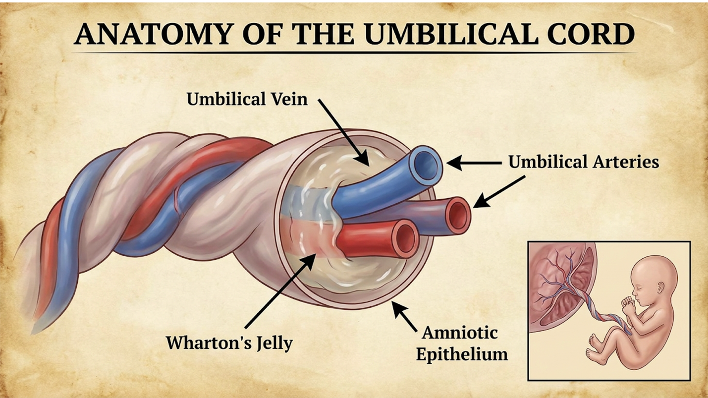
Wharton’s jelly stem cells are multipotent stromal cells extracted from the gelatinous tissue of the umbilical cord. The application of umbilical cord mesenchymal stem cells for autism relies heavily on their unique ability to secrete bioactive molecules that regulate immune responses and mitigate neuroinflammation without requiring invasive surgical harvesting procedures.
The anatomical origin of Wharton’s Jelly is precisely what makes it so medically valuable. Located deeply within the umbilical cord, this protective gelatinous matrix shields the cells, keeping them biologically young and highly proliferative. Unlike adult stem cells harvested from bone marrow or adipose (fat) tissue, these neonatal cells haven’t been subjected to decades of environmental toxins, UV radiation, or cellular aging.
These cells operate through a highly specific biological mechanism we call The Paracrine Immunoregulation Framework. They communicate dynamically with the host’s existing immune system to orchestrate repair. But yielding these potent cellular products requires rigorous, uncompromising laboratory standards to process, isolate, expand, and thoroughly verify their clinical viability before they ever reach a patient.
There is widespread confusion regarding the different types of stem cells available today, often exacerbated by poor medical journalism. Wharton’s jelly vs umbilical cord blood for autism represents two entirely different biological tools.
Umbilical cord blood is rich in hematopoietic stem cells (HSCs). These are blood-forming cells, absolutely brilliant for treating leukemias, lymphomas, or specific blood disorders. But they are not what you want for targeted neuroinflammation. Wharton’s jelly, conversely, is the structural connective tissue of the cord, and it is overwhelmingly rich in mesenchymal stem cells (MSCs). MSCs are the signaling engines explicitly required for tissue repair and deep immune regulation.
When comparing UC-MSCs to adult-derived cells from bone marrow or adipose tissue, the differences in cellular vigor are stark. As an adult ages, their resident bone marrow stem cells suffer heavily from senescence cellular aging driven by shortening telomeres and accumulated oxidative stress. They replicate slower, secrete fewer vital growth factors, and require an invasive, painful, and often risky extraction process from the patient under anesthesia.
The cellular vigor here is paramount. By the time an adult reaches their thirties, their bone marrow stem cells have already lost a massive portion of their original signaling capacity. When evaluating WJ-MSC for autism, researchers specifically prioritize this neonatal vigor. Wharton’s jelly cells are essentially “Day Zero” cells possessing remarkably long telomeres. They boast a massive proliferation capacity and a highly robust secretome that hasn’t been degraded by a lifetime of age and environmental exposure.
“Wharton’s jelly yields up to 10 times more mesenchymal cells than adult bone marrow providing massive signaling capacity for neurorepair.” (PubMed, 2021)
Furthermore, the extraction procedure is entirely painless and post-natal; the cord is simply collected safely after a healthy birth, completely eliminating surgical risk to the patient.
Here is where the actual cellular mechanics rather than marketing magic happen. The Paracrine Immunoregulation Framework is the specific biological signaling process where UC-MSCs secrete bioactive molecules to switch host macrophages from tissue-destructive to tissue-repairing states.
A persistent myth sold by bad actors is that stem cells travel straight to the brain, engraft themselves, and physically turn into new brain cells. They don’t. Instead, UC-MSCs operate as highly sophisticated, mobile pharmacies. Upon intravenous infusion, they evaluate the body’s systemic inflammatory environment and begin secreting a complex “secretome.” This dense payload consists of exosomes, microvesicles, specific growth factors (like BDNF and NGF), and potent anti-inflammatory cytokines.
This dense secretome initiates what is clinically known as phenotypic macrophage reprogramming. In an inflamed biological system, immune cells (macrophages in the body and
The UC-MSC secretome intervenes by releasing massive amounts of Prostaglandin E2 (PGE2) and Indoleamine 2,3-dioxygenase (IDO). These specific molecules force the hostile M1 immune cells to switch their polarity to an M2 (anti-inflammatory) state. Once shifted, the host’s own immune system stops attacking itself and begins secreting Interleukin-10 (IL-10), a master anti-inflammatory agent.
| 📌 If you’re interested in how MSCs calm overactive immune cells throughout the body, we have an interesting article that discusses mesenchymal stem cell therapy for immune modulation, which you can read via the internal link. |
This complex signaling actively inhibits Toll-like receptors and dramatically limits excessive microglial activation in the central nervous system. A systematic review on UC-MSCs and neuroinflammation demonstrated that these specific cells effectively reduce neuroinflammation by modulating targeted T-cell responses in the host, fundamentally changing the inflammatory baseline of the brain.
The science of mesenchymal stem cells immunomodulation is incredibly fragile and easily disrupted. You cannot simply squeeze fluid from an umbilical cord and safely inject it into a pediatric patient. The biomanufacturing standards entirely dictate the safety and efficacy of the procedure.
First, the ethical collection process is completely uncompromising. Cords are obtained exclusively from healthy, full-term, voluntarily donated cesarean section births. The mothers undergo rigorous infectious disease screening panels that far exceed standard blood donation protocols, screening for dozens of communicable diseases and genetic abnormalities.
Once in the cleanroom laboratory, processing for UC-MSC stem cell therapy begins. The cells are carefully isolated from the gelatinous matrix, expanded in highly controlled cultures, and subjected to relentless pathogen screening for mycoplasma, endotoxins, and bacterial contaminants. Processing must occur in ISO-5 classified cleanrooms under rigorous Good Manufacturing Practice (GMP) standards.
Before a cell line ever reaches a clinic, it must undergo flow cytometry analysis. Technicians verify that the cells express specific surface markers (CD73, CD90, and CD105 at >95% positivity) while proving they lack hematopoietic and immune-provoking markers (CD34, CD45, and HLA-DR at <2%). This strict standard confirms absolute cellular purity; it proves the laboratory isolated true MSCs and not blood cells.
Furthermore, excessive culturing is dangerously counterproductive. Clinics pushing Passage 10 or beyond are using exhausted, ineffective cells that have lost their paracrine signaling power. The clinical utility relies absolutely on using low-passage cells (typically Passage 3 or lower). Finally, cryopreservation techniques dictate viability. Cells must be frozen using precision controlled-rate freezers to prevent intracellular ice crystals from forming, which would fracture and instantly kill the cell upon thawing.
| 📌 If you’re interested in why the percentage of living cells matters so much at the time of treatment, we have an interesting article that discusses the importance of cell viability in UC-MSC stem cell therapy, which you can read via the internal link. |
Evaluating stem cell therapy for autism results requires examining standardized clinical metrics rather than anecdotal behavioral claims. Current Wharton’s jelly autism treatment protocols are assessed using rigorous clinical tools that allow researchers to quantify subtle, sustained improvements in socialization, communication, and adaptive behaviors over extended timelines.
Mapping the reduction of systemic inflammation directly to cognitive biomarkers is the primary focus of tier-1 academic research today. We aren’t looking for miraculous overnight changes. Researchers are looking for statistically significant shifts in highly validated scoring systems like the Vineland Adaptive Behavior Scales (VABS) and the Clinical Global Impression (CGI) scale.
This transparent, data-driven analysis of efficacy metrics is crucial for setting realistic family expectations. Responses to cellular therapy are wildly variable across pediatric populations. They depend heavily on the patient’s exact age at intervention, their baseline behavioral severity, their underlying genetic profile, and the specific presence of co-occurring neuroinflammatory conditions like severe gut dysbiosis.
How does a measurable reduction in blood cytokines translate to a child speaking their first spontaneous words? The connection lies in restoring functional neural connectivity. By utilizing The Paracrine Immunoregulation Framework to suppress localized inflammation, the brain’s UC-MSC for autism within a broader therapeutic protocol is entirely designed to support this biological window.
When microglial activation decreases, synapses can fire and wire together without constant inflammatory disruption. In clinical settings, researchers meticulously monitor specific behavioral domains to capture these changes: expressive language (speaking), receptive communication (understanding directions), social awareness (eye contact), and repetitive motor behaviors (stimming).
Simultaneously, clinicians measure objective systemic biomarkers. They take regular blood serum samples to directly track the reduction of specific pro-inflammatory cytokines, like Interleukin-6 and TNF-alpha, alongside the behavioral assessments to prove the biological link.
Beyond inflammatory cytokines, researchers also track vital neurotrophic factors. Brain-Derived Neurotrophic Factor (BDNF) and Nerve Growth Factor (NGF) are crucial proteins required for memory, learning, and the creation of new synapses in the hippocampus and prefrontal cortex. In many children with severe ASD, baseline BDNF levels are drastically blunted by continuous immune stress. When the Paracrine Immunoregulation Framework calms the microglial storm, clinicians often measure a subsequent stabilization in BDNF levels. This biochemical shift is exactly what allows the brain to “wire” new skills learned in occupational or speech therapy.
It is a profound biological chain reaction. For example, a measurable drop in systemic inflammation often correlates exactly with a dramatic reduction in gastrointestinal pain and vastly improved sleep architecture. When a child with ASD finally experiences deep, restorative sleep and is freed from chronic gut pain, their capacity to engage with traditional therapies skyrockets.
Let’s look closely at the actual data from verified trials. Prominent university studies regarding UC-MSC efficacy autism protocols offer a sober but highly optimistic view of what is biologically possible.
Let’s unpack how these metrics are actually scored in a Phase II trial setting. The Vineland Adaptive Behavior Scales (VABS), specifically the Vineland-3, is an exhaustive, standardized
In rigorous double-blind trials, researchers also utilize the Clinical Global Impression-Improvement (CGI-I) scale. This is a 7-point scale where a blinded physician rates the patient’s global improvement compared to their baseline. A score of 1 (“very much improved”) or 2 (“much improved”) represents a massive clinical success, often correlating with significant reductions in self-injurious behavior (SIB) or severe aggressive outbursts.
When parents search online for the “autism stem cell therapy success rate,” they are often met with offshore clinics claiming impossible 90% cure rates. This is dangerous fiction. In scientific reality, “success” is defined as a statistically significant functional improvement, not a magical reversal of the clinical diagnosis. Moving from entirely non-verbal to utilizing an AAC communication device successfully represents the actual medical goal.
“Approximately 60% of pediatric patients treated with umbilical cord therapies demonstrated measurable improvements validating the potential for functional cognitive gains.” (Duke University, 2021)
For instance, Duke University research on stem cell efficacy showed that approximately 60% of children treated with umbilical cord-derived therapies demonstrated significant improvements (scoring 1 or 2 on the CGI-I scale) compared to baseline. But notice the necessary clinical nuance: 60% showed improvement. That means 40% did not exhibit statistically significant changes. Biology is infinitely complex, and WJ-MSC therapies are not a universal magic bullet.
The biological repair initiated by umbilical cord stem cell therapy for autism is a cumulative, agonizingly slow process. It is not an instantaneous pharmaceutical effect like taking an aspirin for a headache or a stimulant for focus.
The typical timeline evaluated in clinical trials focuses strictly on 3-month, 6-month, and 12-month evaluation windows post-infusion. Here is how the biological cascade typically unfolds:
During the first 14 to 30 days post-infusion, the infused MSCs are actively secreting cytokines and altering the systemic immune state, but structural neural changes take time. In the immediate post-infusion period, parents rarely see massive cognitive leaps. Instead, initial improvements are often highly subtle and foundational. You might notice more stabilized, regular digestion, improved sleep cycles, or a slight decrease in sensory defensiveness to loud noises as the systemic inflammation drops.
Between months 3 and 6, the brain actively utilizes its newly non-inflamed state to build and reinforce new neural pathways. The enhanced neuroplasticity window opens wide here. This is exactly when aggressive ABA, speech, and occupational therapies gain their maximum traction.
By months 6 through 12, researchers conduct final VABS and CGI assessments to measure the peak of this developmental leap. The more profound cognitive gains—if they are going to occur, such as spontaneous expressive language or complex cooperative play—usually begin to consolidate strongly during this secondary window.
This reality underscores the absolute necessity of continuous medical monitoring and the aggressive integration of ongoing behavioral therapies post-treatment. The stem cells open the critical window of neuroplasticity; behavioral therapies are what push the child through it to learn new skills.
Determining if stem cell therapy is safe for autism requires analyzing data from rigorously monitored stem cell autism clinical trials. Safety data indicates that Wharton’s jelly derivatives generally possess a favorable safety profile, but all legitimate cellular therapies must be administered under stringent regulatory oversight, such as an Investigational New Drug application, to protect patients.
The fundamental biological safety of UC-MSCs lies entirely in their lack of immune-provoking surface markers. But biological safety means absolutely nothing if the clinic administering them is operating illegally, using poor sterile technique, or infusing contaminated, unverified products.
A clear, unambiguous understanding of the FDA’s current stance on stem cell biologics is your primary defense against medical fraud. We have to ruthlessly separate the actual Phase II trial data regarding adverse events from the wild, unverified claims of offshore medical tourism hubs.
Why can we take cells from a donor umbilical cord and safely infuse them into a pediatric patient without triggering massive immune rejection? It comes down to basic immunology. UC-MSCs are scientifically deemed “immune-privileged” or “immune-evasive.”
Unlike major organ transplants or traditional bone marrow infusions, UC-MSCs lack Human Leukocyte Antigen (HLA) Class II markers specifically HLA-DR on their outer cell surface. These specific markers are the biological nametags that tell a host’s immune system, “I am foreign material, attack me immediately.” Because properly cultured UC-MSCs don’t wear these nametags, they smoothly evade the host’s aggressive immune surveillance.
This unique biological quirk prevents Graft-Versus-Host Disease (GVHD) and completely eliminates the need for the harsh, highly toxic immunosuppressant drugs routinely required for other allogeneic (donor) transplants.
The officially documented UC-MSC safety profile strongly reflects this. Commonly reported adverse events are typically incredibly mild: transient low-grade fevers, mild lethargy lasting for 24-48 hours, or slight site soreness at the IV insertion point. Severe risks like anaphylactic shock or tumor formation are exceedingly rare when cells are properly manufactured in GMP facilities.
Let me be absolutely clear: currently, there are zero FDA-approved stem cell treatments specifically for autism. Any clinic telling you otherwise is misrepresenting the truth.
Legitimate cellular therapies operating within the United States must function strictly under a formal Investigational New Drug (IND) application. Operating under an IND is a grueling legal process designed entirely around patient safety. Before an IND is even granted, the FDA mandates a 30-day review period where toxicologists and medical officers evaluate the clinic’s Chemistry, Manufacturing, and Controls (CMC) data.
This CMC review is exhaustive. The FDA scrutinizes the entire supply chain, verifying exactly where the cells come from, the specific chemical reagents used in the culture media, how the cells are sterilely expanded, and the precise mathematical dosing protocols based on the pediatric patient’s body weight.
The FDA stem cell autism guidelines are incredibly rigid for a reason. Cultured, expanded Wharton’s jelly cells are legally classified as “351” biological drugs, meaning they require the exact same exhaustive, multi-phase clinical trial vetting as a newly invented pharmaceutical compound.
Many commercial clinics skirt this vital law by claiming they are “FDA Registered.” Registration simply means a facility filled out a basic online form acknowledging they handle human tissue. It implies zero clinical vetting, zero safety approval, and zero authorization to treat complex neurodevelopmental disorders.
We have to strictly differentiate Phase II trials from Phase III pivotal trials. Currently, the legitimate scientific community is heavily focused on executing Phase II clinical trials. These trials are fundamentally about dialing in safety and finding the optimal dose per kilogram of patient body weight.
“Phase II trials actively monitor cohorts of 20 to 50 patients for 24 months ensuring strict safety thresholds for pediatric populations.” (FDA, 2023)
Phase II trials track preliminary efficacy and ensure no long-term adverse events occur. They involve continuous 24-month patient monitoring to establish definitive safety profiles. Conversely, Phase III trials are massive, multicenter endeavors involving hundreds of patients with strict double-blind, placebo-controlled protocols. These Phase III trials are the final hurdle required for actual FDA marketing approval. Currently, the scientific community is actively working to fund and execute these larger pivotal studies.
Participation in any sanctioned trial involves grueling medical screening processes. Researchers need highly specific, uniform patient cohorts to definitively prove efficacy and safety. You can review the active Phase II clinical trials evaluating umbilical cord tissue directly on
Wharton’s jelly stem cell therapy utilizes multipotent mesenchymal cells extracted from the umbilical cord to modulate immune responses. These cells are isolated, purified, and administered to target systemic inflammation within the body. In neurodevelopmental contexts, researchers investigate their ability to alter microglial activation in the brain. For instance, specific protocols aim to reduce pro-inflammatory cytokines associated with neurological dysfunction. Individual clinical applications require strict regulatory oversight and specialized administration.
Clinicians utilize Wharton’s jelly stem cells for autism due to their potent immunomodulatory properties and high cellular viability. These biologically young cells secrete complex proteins that actively suppress chronic neuroinflammation, a condition documented in specific subsets of patients with autism. By targeting this underlying biological factor, the therapy aims to improve synaptic connectivity. For example, reduced inflammation often correlates with measurable improvements in functional communication scores. Treatment suitability depends heavily on the individual patient’s biological markers.
The primary difference between Wharton’s jelly and umbilical cord blood lies in the specific type of cells they provide. Cord blood primarily yields hematopoietic stem cells used for blood and immune disorders, whereas Wharton’s jelly is a rich source of mesenchymal stem cells (MSCs). MSCs are specifically required for tissue repair and paracrine immunoregulation. Consequently, therapies targeting neuroinflammation rely predominantly on Wharton’s jelly derivatives. Choosing the correct cellular source is critical for targeted biological efficacy.
Umbilical cord stem cell therapy is generally deemed well-tolerated due to the immune-privileged nature of the cells, which lack the markers that trigger rejection. However, safety in pediatric populations requires administration strictly within FDA-sanctioned clinical trials or highly regulated environments. Common adverse events are typically mild, such as transient fatigue or low-grade fever following infusion. Severe complications are exceedingly rare when rigorous biomanufacturing standards are met. Families must always consult a qualified physician to review comprehensive safety protocols.
Children typically receive investigational stem cell therapy for autism between the ages of 2 and 12, depending entirely on specific clinical trial protocols. Early intervention is frequently prioritized because neuroplasticity is highest during early developmental windows. Some study parameters restrict enrollment to specific age brackets to ensure standardized efficacy tracking. For instance, certain Phase II trials limit participation to children under the age of 8. Exact age eligibility must be determined by the lead clinical investigator managing the treatment protocol.
For families researching biological interventions, Wharton’s jelly stem cells for autism represent an advanced, investigational approach targeting the root mechanisms of neuroinflammation. Clinical data indicates that approximately 60% of pediatric patients in specific academic trials demonstrated measurable improvements in clinical global impression scores (Duke University, 2021). The safest and most scientifically sound approach combines a thorough understanding of cellular biology with strict adherence to regulated clinical trials.
Relying on The Paracrine Immunoregulation Framework ensures that patients understand exactly how these cells function—not as a miraculous cure, but as potent biological signaling agents that actively modulate immune responses. Grasping this specific cellular mechanism is the most effective defense against unverified commercial claims that plague the regenerative medicine space.
Schedule a comprehensive consultation with a board-certified pediatric neurologist or specialized regenerative medicine physician today to determine clinical suitability. Prioritize clinics operating under a verified Investigational New Drug application, and demand transparent, peer-reviewed clinical data before proceeding with any medical assessment.