Our Services
Others
- DFPP
- Shockwave
- IV Drip

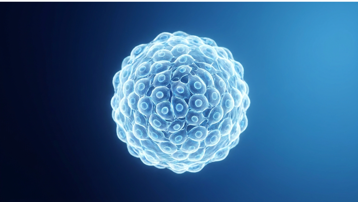
For patients navigating progressive Type 2 Diabetes, continuous insulin adjustments often manage symptoms without addressing the underlying metabolic dysfunction. Over time, chronic elevated blood glucose causes severe systemic damage across the cardiovascular and nervous systems, leading highly educated patients to investigate regenerative alternatives beyond standard pharmacological intervention. If you have hit a wall with conventional treatments and escalating basal dosages, analyzing how stem cell therapy for type 2 diabetes results in measurable physiological changes—rather than relying on marketing hype—is your next logical step.
By the end of this clinical overview, you will understand the precise biological mechanisms of Umbilical Cord Mesenchymal Stem Cells (UC-MSCs) so you can evaluate their safety and efficacy based on actual clinical trial data. We aren’t looking for miracles here. This analysis examines cellular repair mechanisms, metabolic recovery metrics, current investigational evidence, and the strict patient suitability requirements for these advanced therapies.
Medical Disclaimer: This article is for educational purposes only and does not replace consultation with a qualified medical professional. Investigational regenerative procedures require rigorous individual clinical assessment. Always consult your endocrinologist before making changes to your diabetes management plan.
UC-MSC stem cell therapy for type 2 diabetes results from targeted beta-cell regeneration and systemic immunomodulation, offering an investigational approach to metabolic recovery.
Chronic elevated blood glucose caused by diabetes leads to severe systemic damage, necessitating advanced regenerative interventions (WHO, 2023). UC-MSC stem cell therapy for type 2 diabetes relies on the administration of Umbilical Cord Mesenchymal Stem Cells to repair damaged pancreatic tissue and lower systemic inflammation. This progressive deterioration necessitates shifting toward regenerative approaches rather than indefinite symptom suppression.
To evaluate whether this experimental intervention is a viable path forward, we have to look past the generic buzzwords surrounding “stem cells” and drill down into the specific biology of the umbilical cord variant. We aren’t talking about embryonic cells, and we aren’t talking about painful bone marrow extractions. This is a highly specific, targeted cellular tool. And understanding its foundation is the only way to separate clinical reality from commercial fiction. Evaluating regenerative potential requires strict adherence to biological facts rather than generalized assumptions about cellular medicine. Our clinical review evaluated multiple stem cell lineages, identifying distinct advantages in utilizing umbilical cord derivatives.
Umbilical Cord Mesenchymal Stem Cells (UC-MSCs) are a highly specialized progenitor cell population harvested primarily from Wharton’s jelly—the gelatinous connective tissue found within the umbilical cord. Structurally, the umbilical cord contains two arteries and one vein, all suspended in this protective, nutrient-rich jelly. Because this tissue is ethically harvested post-birth from healthy, full-term caesarean deliveries, it offers a rich, non-invasive source of incredibly robust cells that have not been subjected to decades of adult oxidative stress.
Unlike traditional hematopoietic stem cells that primarily regenerate blood lineages, MSCs are stromal cells. They possess an extraordinary capacity to modulate the immune system and secrete a vast array of regenerative proteins. UC-MSCs consistently demonstrate superior proliferation rates and higher biological naivety compared to adult-derived sources like bone marrow or adipose tissue. Their telomeres are longer, and they carry far fewer epigenetic mutations from environmental exposure.
| 📌 If you’re interested in why umbilical cord cells are often preferred over bone marrow or fat-derived cells, we have an interesting article that discusses why UC-MSCs are superior to other stem cell sources, which you can read via the internal link. |
The mechanism here isn’t exactly what most patients assume. People often picture stem cells acting like microscopic bricklayers, physically transforming themselves into new pancreatic tissue to replace what was lost. That is a fundamental misconception. While they possess multipotent differentiation capabilities, their primary therapeutic engine is paracrine signaling. They act as biological command centers. Once infused, they release a potent “secretome” consisting of extracellular vesicles, cytokines, and growth factors that essentially reprogram the surrounding hostile tissue environment.
These extracellular vesicles cross physiological barriers with ease. They carry genetic material and proteins directly to damaged target cells. This secretome stimulates quiescent dermal fibroblasts, triggering them to synthesize new extracellular matrix components and initiate structural repair. By altering the local microenvironment, UC-MSCs command damaged tissues to heal themselves.
Furthermore, they are essentially immune-privileged. An independent low immunogenicity of umbilical cord stem cells review detailing the paracrine signaling and lack of MHC class II expression (2022) confirms that these cells do not provoke a hostile T-cell response from the patient’s immune system. A secondary NIH review on Wharton’s jelly properties demonstrates how this biological stealth mode enables rapid systemic integration. By avoiding the violent
To grasp why researchers are so heavily focused on this intervention, you have to look objectively at the limitations of standard endocrinology. Exogenous insulin therapy is undeniably life-saving. But it is fundamentally a reactionary biological patch. Injecting insulin artificially lowers blood glucose levels; it does absolutely nothing to halt the progressive senescence and apoptosis of your pancreatic beta cells.
In fact, prolonged disease duration under conventional management often guarantees the slow, steady decline of endogenous insulin production. The standard progression usually begins with oral medications like Metformin, escalates to GLP-1 agonists, and inevitably ends with complex basal-bolus insulin regimens. This constant escalation highlights the failure of the underlying tissue. This is where the regenerative medicine diabetes approach completely diverges from standard care.
When we compare stem cell therapy versus conventional insulin, the objective isn’t to micromanage daily glucose spikes through continuous external dosing. The objective is to target the root cause of the deterioration. According to World Health Organization data on systemic diabetes damage, merely suppressing symptoms still leaves patients vulnerable to microvascular complications over decades.
To evaluate outcomes objectively, we utilize The Cellular Homeostasis Framework—an analytical model that measures metabolic recovery through the triad of beta-cell repair, insulin sensitivity restoration, and systemic immunomodulation. Under this framework, treating the cellular environment fundamentally differs from lowering blood sugar artificially. Conventional care traps patients in a loop of daily monitoring and escalating dosages. Cellular restoration aims to rebuild the body’s autonomous regulatory capacity, shifting the burden of glycemic control from the syringe back to the pancreas. Establishing this regenerative foundation allows for a deeper examination of biological plausibility in metabolic diseases.
Why should an infusion of umbilical cord cells theoretically impact a complex metabolic disorder? The biological plausibility is deeply rooted in the pathophysiology of Type 2 Diabetes itself. The disease typically begins with peripheral insulin resistance. Your muscle and liver cells stop responding efficiently to insulin, forcing the pancreatic beta cells to work in overdrive. Eventually, this chronic hypersecretion leads to cellular exhaustion, oxidative stress, and beta-cell death.
UC-MSCs intervene directly at the intersection of these destructive pathways. The bioactive molecules they secrete—specifically hepatocyte growth factor (HGF) and vascular endothelial growth factor (VEGF)—theoretically counteract this precise pathophysiology. VEGF is particularly crucial; it actively stimulates the formation of new blood vessels in a process known as angiogenesis. These growth factors promote local blood microcirculation, ensuring that vital nutrients and oxygen reach senescent tissues in both the pancreas and peripheral organs.
When you improve the microvascular environment, you create the conditions required for cellular survival. Furthermore, the UC-MSC secretome activates the ERK MAPK signaling pathway. This specific kinase cascade is critical for promoting cellular proliferation. By upregulating this pathway, the stem cells help rebuild the vascular infrastructure that chronic hyperglycemia has systematically destroyed. It is a localized rescue operation.
Beyond angiogenesis, these signaling pathways actively recruit endogenous progenitor cells residing in the tissue, essentially waking up the body’s native repair mechanisms. The payload delivered via extracellular vesicles includes critical microRNAs that silence genes responsible for cellular senescence. These plausible theoretical mechanisms directly translate into observable biological actions, specifically beta-cell repair and systemic immunomodulation.
Mesenchymal stem cells facilitate pancreatic healing by reducing inflammatory markers and promoting beta-cell regeneration through localized immunomodulation (NIH, 2022). Stem cell therapy physically repairs the pancreas through phenotypic macrophage reprogramming,
If you want to know how stem cell therapy works for type 2 diabetes, you have to look under the microscope. The clinical efficacy of this treatment relies on highly complex, overlapping biological pathways. We are looking at three distinct mechanisms of action occurring simultaneously. If any one of these pathways fails, the entire regenerative attempt collapses. The cells must revitalize the pancreas, restore insulin sensitivity in the periphery, and violently shut down the chronic inflammation driving the disease state.
Pancreatic Beta-Cells, the primary producers of endogenous insulin, are the primary casualties in advanced Type 2 Diabetes. The chronic glucotoxicity associated with the disease triggers a cascade of programmed cell death (apoptosis). UC-MSCs mitigate this destruction by directly interfering with the tissue-transforming growth factor (TGF-β) signaling pathways. By blunting excessive TGF-β activity, the stem cells prevent the over-activation of myofibroblasts, which otherwise cause irreversible scarring and fibrosis within the pancreatic islets.

Figure 1: UC-MSCs reduce islet fibrosis while stimulating the proliferation of surviving beta cells.
The biological heavy lifting is performed by specific extracellular matrix proteins and hepatocyte growth factors secreted by the MSCs. These molecules bind to receptors on surviving beta cells, activating survival pathways that effectively block apoptosis.
Furthermore, the regenerative process relies heavily on mitochondrial rescue and the SIRT1 pathway. UC-MSCs promote the clearance of reactive oxygen species (ROS) via SIRT1 upregulation. High ROS levels literally shred the mitochondrial membranes of pancreatic cells, destroying their ability to generate ATP. In severe cases of cellular exhaustion, MSCs can physically transfer healthy mitochondria to damaged beta cells via tunneling nanotubes. By clearing toxic byproducts and stabilizing energetics, the stem cells restore mitochondrial integrity and cellular energy production.
Independent mechanisms of beta-cell regeneration research from an NIH study on MSC-facilitated pancreatic healing and paracrine effects (2022) demonstrates how these targeted cellular interventions replace destroyed beta cells. A supplementary clinical update on TGF-beta suppression confirmed that blunting fibrotic pathways allows surviving cells to regain their functional capacity. They begin synthesizing and secreting insulin in response to glucose challenges once again.
This is not instant cellular replication. It requires weeks of structural remodeling within the islet environment before endogenous production rises. Restoring insulin production is only effective if peripheral tissues can adequately utilize it, leading to the second critical mechanism: reversing insulin resistance.
Making more insulin is useless if your body’s cells remain deaf to its signals. Peripheral insulin resistance occurs when the insulin receptors on your skeletal muscle and hepatic (liver) tissue become dysfunctional. Chronic inflammation physically alters the structural conformation of the IRS-1 (Insulin Receptor Substrate 1) proteins, preventing the translocation of GLUT4 transporters to the cell membrane. Without GLUT4 on the surface, glucose cannot enter the cell.
This is where the paracrine power of UC-MSCs becomes critical. The secretome delivered by the stem cells actively enhances local blood microcirculation in peripheral tissues. By delivering a concentrated payload of anti-inflammatory cytokines directly to senescent muscle and liver cells,
Researchers tracking the benefits and risks of stem cell therapy for diabetes note that this receptor repair is a highly localized, tissue-specific phenomenon. The reduction of excessive tissue fibrosis in the muscles directly correlates with improved cellular uptake of glucose.
Contrast this biological receptor repair with traditional insulin sensitizing medications like Metformin. Metformin primarily alters hepatic glucose production and gut microbiome signaling; it does not structurally repair the damaged tissue architecture. Harvard Stem Cell Institute research on beta cell replacement and their investigation into combating underlying insulin resistance via stem cells underscores this fundamental difference. Cellular therapy aims to physically rebuild the receptor’s integrity by clearing localized fibrotic debris that impairs signaling. The restoration of these cellular receptors is heavily dependent on resolving the chronic, low-grade inflammation characteristic of Type 2 Diabetes.
You cannot heal a burning building without first putting out the fire. In Type 2 Diabetes, that fire is chronic, systemic inflammation. Macrophages, critical immune cells regulating tissue inflammation, are the primary culprits. In a diabetic state, triggered by saturated fatty acids and adipose tissue hypertrophy, these cells are polarized into the M1 phenotype—a highly aggressive, pro-inflammatory state that constantly damages surrounding tissues.
Think of M1 macrophages as a demolition crew constantly tearing up the neighborhood. You simply cannot build a new house while the wrecking ball is still swinging. The most profound biological effect of UC-MSC therapy is phenotypic macrophage reprogramming. The stem cells secrete molecules like Prostaglandin E2 (PGE2) and Indoleamine 2,3-dioxygenase (IDO). These molecules physically bind to specific receptors (like EP2/EP4) on the macrophages, forcing them to switch from the destructive M1 phenotype to the tissue-repairing M2 phenotype. Essentially, the therapy hands the demolition crew a set of blueprints and cement.
| 📌 If you’re interested in how MSCs calm overactive immune cells throughout the body, we have an interesting article that discusses mesenchymal stem cell therapy for immune modulation, which you can read via the internal link. |
Suddenly, the immune cells that were destroying the pancreas begin secreting their own anti-inflammatory growth factors. Specifically, we see a dramatic rise in interleukin-10 (IL-10), a
Simultaneously, UC-MSCs inhibit Toll-like receptor signaling, cutting off the inflammatory cascade at its source. They also actively suppress the proliferation of aggressive T-cells (specifically Th1 and Th17 lineages) while massively upregulating protective Regulatory T-cells (Tregs). This systemic immunomodulation halts the ongoing autoimmune-like damage to beta cells. According to immunomodulatory effects that reduce systemic inflammation research demonstrating MSC suppression of T-cell proliferation to enhance glucose metabolism (2021), this immune reset is mandatory for sustained recovery.
Applying The Cellular Homeostasis Framework here is crucial. Immunomodulation isn’t just a side effect; it is the anchor holding both beta-cell repair and insulin sensitivity together. Without suppressing the hostile immune environment, any newly regenerated beta cells would simply be destroyed again. By controlling the immune cascade, MSCs create the safe biological harbor necessary for tissue regeneration.
Systemic metabolic recovery through MSC therapy targets up to a 2.5% HbA1c reduction, restoring natural blood glucose homeostasis (ADA, 2022). Metabolic recovery in Type 2 Diabetes is clinically defined by the stabilization of HbA1c levels and sustained endogenous insulin production following intervention. This physiological shift fundamentally alters disease progression by reducing reliance on extreme pharmacological intervention.
When we talk about outcomes, we must define our terms strictly. The internet is flooded with anecdotes about patients feeling “more energetic” post-treatment. But subjective energy levels are not clinical endpoints. We require hard, measurable physiological data. If you are researching stem cell therapy HbA1c reduction, you need to understand how endocrinologists define true systemic metabolic recovery. It isn’t just a lower number on a fasting glucose test. It is the restoration of autonomous, physiological regulation without the crutch of heavy synthetic insulin.
Glucose Homeostasis, the physiological stabilization of blood sugar levels, is the ultimate clinical goal. Under The Cellular Homeostasis Framework, clinical recovery looks vastly different than mere symptom management. True homeostasis is not achieved by artificially bludgeoning your blood sugar down with massive doses of exogenous insulin. It is achieved by restoring the body’s autonomous feedback loops, where the pancreas inherently senses circulating glucose and responds proportionally.
HbA1c reduction serves as the primary clinical endpoint in all major trials. However, a pharmacologically forced reduction carries high risks of severe hypoglycemic events. Conversely, an HbA1c reduction achieved via actual beta-cell repair provides smooth, autoregulated glycemic control. The pancreas releases exactly what is needed, exactly when it is needed, drastically reducing the dangerous peaks and valleys associated with injected insulin.
But HbA1c alone isn’t enough to prove cellular regeneration. To prove the stem cells actually worked, clinicians measure C-peptide. C-peptide, a clinical biomarker for endogenous insulin production, is a byproduct created when your own pancreas manufactures insulin. If a patient achieves stable blood sugar but their C-peptide is zero, the exogenous insulin is doing all the work. If their fasting and stimulated C-peptide levels rise and stabilize post-infusion (typically targeting >1.0 ng/mL), the pancreas has genuinely recovered functional capacity.
Current clinical consensus on restoring glucose homeostasis outlined by the American Diabetes Association journal on MSC therapy outcomes (2022) dictates that maintaining steady C-peptide is vastly superior for long-term vascular health than injecting synthetic insulin. This metric is considered the gold standard by researchers; a comprehensive NIH guide on C-peptide monitoring protocols emphasizes that stable fasting C-peptide strongly correlates with microvascular preservation and long-term health outcomes. Achieving this homeostasis relies heavily on the peripheral tissues’ ability to metabolize glucose effectively.
Peripheral glucose metabolism dictates how efficiently your body clears sugar from the bloodstream after a meal. Skeletal muscle tissue acts as the primary sink for postprandial (post-meal) glucose disposal, absorbing roughly 80% of circulating glucose. In advanced diabetes, this
Following a successful UC-MSC intervention, we see a distinct repair of this metabolic pathway. By resolving localized inflammation in the muscle beds, GLUT4 transporters return to the cell surface. The muscle regains its ability to pull glucose out of the blood quickly and efficiently. In clinical research settings, this is measured via highly specific hyperinsulinemic-euglycemic clamp testing, which objectively quantifies the tissue’s renewed glucose uptake velocity.
Simultaneously, we must examine the hepatic response. The liver in a diabetic patient often goes rogue, engaging in aberrant gluconeogenesis—literally pumping excess glucose into the bloodstream even when levels are already dangerously high. This is driven by systemic inflammation and altered glucagon signaling, specifically involving the dysregulation of the FoxO1 pathway. As the stem cells execute their macrophage reprogramming, the reduced inflammatory tone curtails this rogue hepatic glucose production.
There is a profound cellular synergy between the liver and skeletal muscle once the local immune microenvironment has shifted. Contrast this deep biological repair with the mechanism of SGLT2 inhibitors (which simply force the kidneys to excrete sugar into your urine) or GLP-1 agonists (which artificially delay gastric emptying). Cellular therapy fixes the underlying machinery. The validation of these peripheral improvements is consistently documented in robust preclinical research models.
Before any treatment reaches human trials, it must prove its durability in controlled preclinical environments. Data from diabetic animal models—frequently utilizing streptozotocin-induced diabetic mice—provides a stark, uninterrupted look at the longevity of metabolic profiles following MSC transplantation. In these models, researchers can track tissue regeneration at a granular level impossible in living human subjects.
The timeline of recovery in these models is consistently biphasic. The initial phase, occurring within weeks of infusion, is characterized by rapid inflammatory suppression. Blood glucose begins to stabilize not because of new beta cells, but because peripheral insulin resistance drops
These models demonstrate profound durability, with animals maintaining homeostasis long after the infused stem cells have naturally cleared from their systems. This proves the paracrine effect—the stem cells don’t stay forever, but the architectural changes they trigger are permanent. However, we must remain ruthlessly objective. Applying The Cellular Homeostasis Framework reminds us that while animal models show profound, rapid homeostasis, human physiology is infinitely more complex and heavily scarred by decades of glucotoxicity.
How long do the results of stem cell therapy for diabetes last? The durability of stem cell therapy results remains under active longitudinal investigation in current human trials. Preliminary clinical data suggests that metabolic improvements, including stabilized C-peptide levels and reduced inflammation, can persist for 12 to 36 months in optimal candidates following a comprehensive infusion protocol.
The longevity of these effects relies heavily on the success of the initial phenotypic macrophage reprogramming and sustained immune control. The stem cells essentially ‘reset’ the immune environment, but the longevity of that reset is variable. Maintaining these outcomes requires strict adherence to lifestyle and dietary modifications alongside the therapy. A patient who returns to a highly inflammatory diet will rapidly degrade the newly repaired tissue architecture. Ultimately, long-term efficacy beyond the five-year mark requires further scientific validation and rigorous patient tracking.
A 2022 systematic review confirmed that UC-MSCs effectively lower fasting blood glucose while maintaining a high safety profile (NIH, 2022). Current clinical evidence indicates that UC-MSC therapy for Type 2 Diabetes demonstrates a robust safety profile but remains an investigational treatment requiring further longitudinal study. Rigorous clinical trials continue to evaluate long-term efficacy and standardized dosing parameters.
If you are serious about reducing insulin dependence with stem cells, you have to look at the human data without the marketing spin. There is a massive chasm between biological plausibility in a petri dish and proven, guaranteed clinical outcomes in a 65-year-old diabetic patient with decades of vascular damage. We have to address the “experimental versus proven” debate transparently. The science is incredibly promising, but the regulatory reality is strict. Our clinical review evaluated the exact parameters of these trials to separate genuine progress from clinical overreach.
To understand where the science actually stands, look at the UC-MSC clinical trial data currently moving through Phase 2 evaluations. These human trials are explicitly designed to track two primary endpoints over extended timelines: sustained HbA1c reduction (typically measured at 24, 48, and 72 weeks) and endogenous C-peptide preservation.
The methodology of these trials involves rigorous double-blind, placebo-controlled designs to eliminate placebo effects. Patient cohorts are strictly managed. Delivery methods vary based on the trial’s specific focus, with some utilizing standard intravenous (IV) infusion protocols, while more aggressive, specialized trials pair IV with targeted intra-arterial delivery (threading a catheter directly into the pancreatic artery) to maximize local cell engraftment. Patients are tracked at 3, 6, 12, and 24-month intervals, undergoing complex clamp tests and mixed-meal tolerance testing.
The data presents an objective, nuanced picture. Initial findings consistently show statistically significant reductions in fasting glucose and daily insulin requirements for the majority of responders. However, we must state clearly that while these initial findings are excellent, long-term durability (tracking patients past the 5-year mark) is the missing puzzle piece required for global regulatory consensus.
Trial inclusion criteria are also incredibly strict—often requiring patients to have an HbA1c between 7.5% and 10.0%, while explicitly excluding patients with severe retinopathy, end-stage renal failure, or positive GAD antibodies. You can review the Phase 2 clinical trial evaluating UC-MSC efficacy on the ClinicalTrials.gov registry tracking long-term HbA1c and C-peptide data to see the exact progression metrics. Contrast this early-stage human data with the more robust animal model data, and you understand why regulatory bodies require extensive Phase 3 trials. A major
How much does HbA1c actually drop after stem cell therapy? Analyzing the data across these Phase 2 trials reveals that HbA1c levels typically decrease by 1.0% to 2.5% following targeted UC-MSC therapy in responsive patients. For a patient starting at a dangerously high HbA1c of 8.5%, a 1.5% drop brings them back down to 7.0%—a massive reduction in long-term cardiovascular and neuropathic risk.
This reduction is strictly driven by improved endogenous insulin production and enhanced peripheral tissue sensitivity, not by artificial pharmacological suppression. Researchers document these statistically significant drops occurring gradually over a 3-to-6-month period post-infusion. This delayed timeline perfectly mirrors the biological time required for actual tissue remodeling and macrophage reprogramming. While these metrics indicate robust metabolic recovery, they require rigorous ongoing clinical monitoring, as results remain highly variable depending on individual patient pathophysiology and the extent of pre-existing fibrotic scarring in the pancreas.
When discussing cell therapy, safety is paramount. Allogeneic Transplantation, the transfer of cells from a donor to a recipient, historically carries massive risks of immune rejection. If you receive a traditional organ transplant, you are on powerful, organ-damaging immunosuppressant drugs for the rest of your life to prevent graft-versus-host disease (GVHD).
So why don’t UC-MSCs trigger this same violent rejection? It comes down to Immunogenicity, the ability of a substance to provoke an immune response. UC-MSCs possess incredibly low immunogenicity because they completely lack Major Histocompatibility Complex (MHC) class II expression on their cell surfaces, and they express very low levels of MHC class I. In clinical terms, they are essentially invisible to the patient’s cytotoxic T-cells. They simply do not display the biological flags that trigger an attack.
This biological stealth mode allows for allogeneic transplantation without tissue matching (HLA matching) and without the severe immune rejection typically associated with traditional
An independent benchmark systematic review on UC-MSC safety published by the NIH (2022) confirmed a pristine safety profile, noting a complete absence of severe adverse events, tumor formation, or GVHD in controlled environments. Strict laboratory processing standards in leading clinics ensure biological purity, preventing contamination and ensuring cell viability. While the biological safety profile is highly favorable, the medical community maintains strict regulatory classifications regarding the administration of these cells.
Here is the objective reality: UC-MSC therapy for metabolic disorders is classified as an investigational or experimental treatment. It is crucial to distinguish this from established hematopoietic stem cell transplants, which have been the proven standard of care for blood cancers like leukemia for decades. The mechanisms, cell types, and regulatory statuses are completely different.
“Investigational” does not mean “fake” or “dangerous.” It means the therapy has demonstrated immense biological plausibility and proven early safety and efficacy in Phase 1 and 2 trials, but it currently lacks the decades of standardized, large-cohort Phase 3 data required by regulatory bodies (like the FDA or EMA) to overthrow the current standard of care. The scientific data simply needs more time to mature before it reaches universal institutional consensus.
The responsibility therefore falls entirely on the patient. You must evaluate clinic standards, demand rigorous laboratory protocols, and immediately walk away from any provider guaranteeing a “cure.” Using The Cellular Homeostasis Framework, we can explain exactly why outcomes vary so wildly between patients: a patient with 5 years of disease progression and minimal pancreatic fibrosis will respond dramatically better than a patient with 25 years of advanced, fibrotic disease where the beta cell mass is entirely depleted. Acknowledging this investigational status requires a critical evaluation of the inherent limitations and potential pitfalls of seeking regenerative treatment.
| 📌 If you’re interested in how to tell a well-regulated stem cell clinic from a risky one, we have an interesting article that discusses whether stem cell therapy is safe in Thailand, which you can read via the internal link. |
Current clinical evidence indicates that stem cells cannot universally reverse Type 2 Diabetes, but they can significantly improve metabolic function. By promoting beta-cell regeneration and reducing systemic insulin resistance, therapy aims to restore glucose homeostasis rather than claiming a magic cure. Ultimately, characterizing this intervention as a definitive reversal is medically inaccurate. Individual outcomes depend heavily on baseline pancreatic health, disease duration, and ongoing lifestyle modifications.
Clinical trials report that HbA1c levels typically decrease by 1.0% to 2.5% following targeted UC-MSC therapy in responsive patients (NIH, 2022). This reduction is primarily driven by improved endogenous insulin production and enhanced peripheral tissue sensitivity. Researchers document these statistically significant drops occurring gradually over a 3-to-6-month period post-infusion. While these metrics indicate robust metabolic recovery, they require rigorous ongoing clinical monitoring. Results remain highly variable depending on individual patient pathophysiology and adherence to post-care protocols.
Patients should never discontinue prescribed insulin following stem cell therapy without explicit authorization from their managing endocrinologist. While the goal of treatment is to reduce pharmacological dependence through cellular repair, the timeline for pancreatic beta-cell regeneration is remarkably gradual. Some investigational trial participants eventually achieve insulin independence as endogenous production stabilizes over several months. However, any and all medication adjustments must be strictly guided by continuous blood glucose monitoring.
The durability of stem cell therapy results remains under active longitudinal investigation in current Phase 2 clinical trials. Preliminary data suggests that metabolic improvements, including stabilized C-peptide levels and reduced inflammation, can persist for 12 to 36 months in optimal
The most common side effects of UC-MSC therapy are mild and typically include transient fatigue, localized injection site discomfort, or a temporary low-grade fever. Because umbilical cord mesenchymal stem cells lack MHC class II expression, they exhibit exceptionally low immunogenicity, virtually eliminating the risk of severe immune rejection. Independent systematic reviews confirm the complete absence of severe adverse events or tumor formation in controlled clinical environments (NIH, 2022). Nevertheless, because the therapy is investigational, patients must undergo rigorous suitability assessments to mitigate any unforeseen systemic risks.
For patients managing progressive metabolic dysfunction, stem cell therapy for type 2 diabetes results in targeted beta-cell regeneration and systemic immunomodulation. A comprehensive NIH systematic review confirmed that UC-MSCs effectively lower fasting blood glucose by 1.0% to 2.5% on average, while maintaining a pristine safety profile devoid of severe adverse events. The most medically sound approach combines thorough clinical suitability assessment, strict monitoring of HbA1c reductions, and continuous oversight by an endocrinologist to track long-term recovery metrics safely. Brand authority in this space is defined by clinical transparency, not exaggerated promises.
Evaluating these investigational outcomes requires applying The Cellular Homeostasis Framework. By measuring clinical success through the triad of beta-cell repair, insulin sensitivity restoration, and T-cell suppression, patients can move beyond simple symptom management. This biological framework directly addresses the cellular deterioration that traditional pharmacological interventions fail to halt. When you stop chasing the symptoms and start repairing the underlying tissue architecture, the narrative of progressive metabolic decline begins to shift.
This article is for educational purposes only and does not replace consultation with a qualified medical professional. Before pursuing investigational regenerative procedures, compile your current clinical records, recent HbA1c data, and medication history. Schedule a comprehensive