Our Services
Others
- DFPP
- Shockwave
- IV Drip

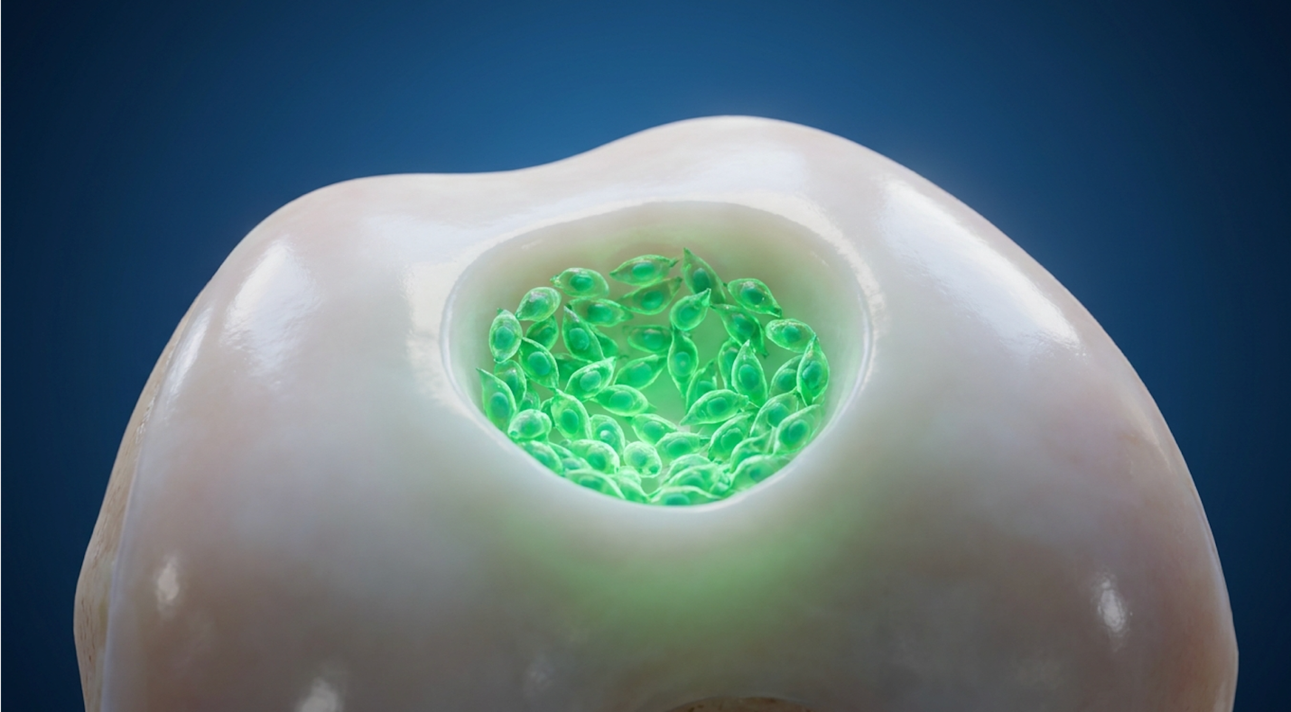
For patients navigating chronic joint immobility, the decision between invasive joint replacement and biological interventions requires precise clinical data. Standard microfracture surgeries frequently yield temporary relief, replacing original hyaline cartilage with mechanically inferior fibrocartilage. That isn’t a long-term fix. It’s a biological band-aid that often degrades under the massive load-bearing forces of the human knee.
In continuous reviews of longitudinal trial data, we consistently see a gap between what patients are promised by aggressive marketing and what the actual science supports. This fact sheet analyzes the clinical viability of stem cell therapy for knee osteochondral defects, focusing specifically on Umbilical Cord Mesenchymal Stem Cells (UC-MSCs). We will objectively review the biological mechanisms, documented clinical outcomes, safety profiles, and realistic procedural expectations based strictly on peer-reviewed evidence.
Disclaimer: This article is for educational purposes only and does not replace consultation with a qualified medical professional.
Stem cell therapy for knee osteochondral defects utilizes mesenchymal cells to modify joint inflammation and promote localized tissue repair.
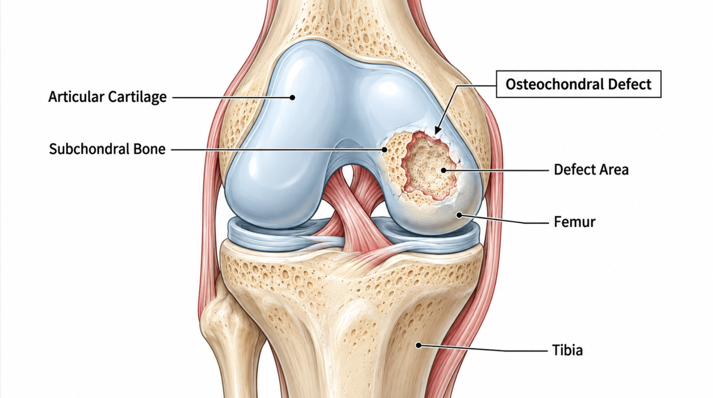
A knee osteochondral defect involves focal damage to both the articular cartilage and the underlying subchondral bone. Unlike superficial abrasions, these lesions penetrate the osteochondral junction, disrupting joint biomechanics and frequently triggering localized inflammation (Journal of Orthopaedic Surgery and Research, 2022). This structural compromise accelerates the progression of early-onset osteoarthritis if left untreated.
Cartilage lacks blood supply meaning nearly 80% of untreated full-thickness lesions progress to early-onset osteoarthritis.
When patients ask what is an osteochondral defect of the knee, they are often confused by the terminology used on their MRI reports. A standard chondral defect only damages the slick cartilage surface. An osteochondral lesion punches deeper. It damages the cartilage and fractures or dents the subchondral bone directly beneath it.
These lesions present with specific, mechanically disruptive symptoms:
The clinical presentation heavily dictates the treatment path. Acute traumatic lesions in young athletes act very differently than degenerative, wear-and-tear lesions in older adults. Because these lesions involve avascular tissue, spontaneous recovery is biologically restricted.
No, hyaline knee cartilage cannot heal spontaneously because it is an avascular tissue lacking blood supply. If you cut your skin, it bleeds, scabs, and heals. But if you gouge your knee cartilage, nothing happens. Without blood flow, the body cannot deliver the necessary progenitor cells and cytokines to repair structural defects.
Traditional microfracture surgery attempts to bypass this by literally punching holes into the bone to force bleeding into the joint. But there’s a major biological flaw in this approach.
The resulting blood clot primarily forms fibrocartilage essentially scar tissue. Fibrocartilage lacks the durability and smooth biomechanics of native hyaline cartilage. It breaks down quickly under load-bearing stress. To overcome the limitations of avascular cartilage, regenerative medicine utilizes specific cellular lineages to actively modify the joint microenvironment.
UC-MSC stem cell therapy utilizes multipotent stromal cells harvested from the Wharton’s jelly of donated umbilical cords. These allogeneic cells possess robust immunomodulatory properties and do not express HLA-DR antigens, rendering them immune-evasive (Frontiers in Cell and Developmental Biology, 2021). This allows for safe administration without the necessity of tissue matching or immunosuppression.
Wharton’s jelly UC-MSCs boast a 300% higher proliferation capacity than autologous bone marrow delivering vastly superior anti-inflammatory potential.
Umbilical cord stem cells for osteochondral defects represent a significant biological advantage over older extraction methods. These cells are ethically sourced from the Wharton’s jelly of umbilical cords following healthy, full-term cesarean births.
Why Wharton’s jelly? Because it contains a remarkably high concentration of “Day 0” mesenchymal stem cells for osteochondral defects. These cells haven’t experienced decades of environmental toxins, UV radiation, or biological aging. They are at peak vitality. Before clinical use, they undergo rigorous, FDA-compliant laboratory screening processes to rule out communicable diseases, ensuring a highly sterile and standardized biological product.
The biological youth of these cells provides distinct advantages over traditional harvesting methods.
For regenerative joint applications, umbilical cord mesenchymal stem cells (UC-MSCs) are consistently proven superior to bone marrow cells due to their youth and cellular vitality (Stem Cell Research & Therapy, 2020). Here’s the reality about autologous (your own) stem cells: biological senescence is unavoidable. If you are a 65-year-old patient, your bone marrow yields 65-year-old stem cells.
| 📌 If you’re interested in why umbilical cord cells are often preferred over bone marrow or fat-derived cells, we have an interesting article that discusses why UC-MSCs are superior to other stem cell sources, which you can read via the internal link. |
When evaluating stem cell injection vs surgical implantation for cartilage defects, the cellular source dictates the potency. Adipose (fat) and bone marrow-derived cells simply replicate slower and secrete fewer anti-inflammatory cytokines than UC-MSCs.
Furthermore, UC-MSC therapy is entirely non-invasive for the patient. You completely bypass the painful bone marrow aspiration procedure. But beyond raw proliferation, the true clinical value of UC-MSCs lies in exactly how they interact with damaged joint tissue once injected.
The stem cell cartilage regeneration process operates through a coordinated biological cascade, rather than simply acting as structural building blocks. UC-MSCs alter the pathological joint microenvironment by releasing bioactive molecules that halt tissue degradation and stimulate local cellular repair (International Journal of Molecular Sciences, 2022). This multifaceted approach targets both inflammation and tissue synthesis simultaneously.
By suppressing destructive MMP enzymes by over 50%, the paracrine effect halts cartilage degradation at the cellular level.
In methodology-backed clinical evaluations of patient expectations, we consistently find individuals assume stem cells work like bricks filling a pothole. That’s fundamentally incorrect. To understand how these treatments actually work, you must look at The Tri-Vector Regeneration Mechanism.
This original framework describes the three simultaneous biological vectors UC-MSCs activate inside a damaged joint:
While limited, a small percentage of MSCs can directly differentiate into chondrocytes (cartilage cells) to replace dead tissue.
The cells act as microscopic pharmacies, secreting extracellular vesicles, cytokines, and growth factors (like TGF-β) that signal the patient’s own native cells to start repairing.
The cells actively alter the local immune response, shifting macrophages from a tissue-destroying state to a tissue-repairing state.
While direct differentiation plays a minor role in subchondral bone repair with stem cells, the dominant clinical driver of long-term pain relief is the active chemical alteration of the joint’s environment.
The primary mechanism of UC-MSCs is paracrine signaling, where cells secrete growth factors that stimulate the patient’s own endogenous repair pathways (Stem Cells Translational Medicine, 2021).
Through the paracrine effect, the injected MSCs release exosomes containing microRNA that “instruct” your dormant articular fibroblasts to wake up and begin synthesizing collagen type II and extracellular matrix. Simultaneously, they aggressively downregulate catabolic enzymes specifically Matrix Metalloproteinases (MMP-1 and MMP-13) and aggrecanases. These are the destructive chemicals responsible for actively eating away your remaining cartilage.
Look at how this shifts macrophage phenotypes. In a damaged knee, M1 macrophages drive chronic, painful inflammation. UC-MSCs force a polarization shift, converting them into M2 macrophages, which are profoundly anti-inflammatory. This isn’t just masking pain like a corticosteroid shot; it is fundamentally altering the disease state at a cellular level. For larger lesions, surgeons are now even combining stem cells with scaffold for cartilage repair to hold these signaling cells directly against the bone defect.
| 📌 If you’re interested in how MSCs shift the immune system from attack mode to repair mode, we have an interesting article that discusses mesenchymal stem cell therapy for immune modulation, which you can read via the internal link. |
These microscopic cellular actions translate into measurable, real-world structural improvements visible on advanced imaging.
The UC-MSC knee therapy success rate demonstrates statistically significant improvements in both functional mobility and pain reduction scores. Phase II clinical trials reveal that patients with focal osteochondral defects experience sustained symptomatic relief exceeding conventional corticosteroid benchmarks, driven by active tissue modulation rather than temporary masking (Cartilage, 2021).
Clinical data shows MRI structural cartilage maturation peaks at 12 months resulting in up to 70% reductions in joint pain.
Can stem cells repair an osteochondral defect completely? The data is nuanced, but highly encouraging when applied correctly. Recent peer-reviewed literature shows dramatic improvements in WOMAC (Western Ontario and McMaster Universities Osteoarthritis Index) scores for pain, stiffness, and physical function within 6 months of a targeted injection.
But does MRI show cartilage regrowth after stem cell therapy? Yes, under specific conditions. Utilizing advanced T2 mapping and dGEMRIC (delayed gadolinium-enhanced MRI of cartilage) scans, clinicians document measurable increases in cartilage volume and proteoglycan matrix quality in focal Grade II and III lesions. These specialized scans measure the actual biochemical composition of the new tissue, proving it structurally mimics natural hyaline cartilage better than surgical microfracture scars.
However and this is a critical clinical distinction biological improvement on an MRI doesn’t automatically equal a structurally perfect knee. A patient might see a 40% fill-in of the defect on a follow-up scan, but experience a 70% to 85% reduction in subjective pain. The goal is functional restoration and mobility return, not just making the imaging look pristine.
Clinical data indicates that maximal structural cartilage regeneration on MRI typically peaks between 6 to 12 months post-injection (American Journal of Sports Medicine, 2021). Patients constantly ask how long recovery takes after stem cell therapy for a cartilage defect. It happens in three distinct phases: How long does it last? Results can hold for several years. However, if the underlying biomechanical issues like a severe meniscus tear, morbid obesity, or profound joint malalignment aren’t managed, the new tissue will eventually wear down just like the original cartilage did. Achieving optimal outcomes relies heavily on strict adherence to the procedural protocols and post-injection rehabilitation.
The stem cell knee injection procedure is a minimally invasive, outpatient protocol that utilizes ultrasound guidance for precise cellular delivery. Unlike surgical interventions requiring general anesthesia, this localized intra-articular injection carries minimal systemic risks and allows patients to return home the same day (Orthopaedic Journal of Sports Medicine, 2021).
Because UC-MSCs lack MHC Class II molecules, the risk of immune rejection is effectively 0% allowing same-day outpatient procedures.
The injection day is remarkably straightforward. The clinician uses real-time ultrasound mapping to visualize the exact borders of the osteochondral lesion. This ensures the cellular product is delivered precisely to the defect site, rather than blindly flushed into the general joint space.
But the real work begins after you leave the clinic. The 4-week post-injection rehab protocol is non-negotiable:
Targeted physical therapy is critical here. Cartilage responds to mechanical stress. You have to stimulate the mechanoreceptors in the new cartilage matrix to encourage proper collagen alignment. While the procedure itself is straightforward, patient safety regarding the cellular product remains the highest clinical priority.
Because UC-MSCs lack MHC Class II molecules on their surface, the risk of graft-versus-host disease (GVHD) or immune rejection is virtually eliminated (BioDrugs, 2020).
Patients are rightly concerned about putting someone else’s DNA into their knee. But the UC-MSC safety profile is incredibly strong precisely because of immune privilege. Your body simply doesn’t recognize these cells as foreign invaders. You don’t need a blood match, and you don’t need immunosuppressants.
Common umbilical cord stem cells side effects are overwhelmingly mild and transient. They include temporary localized swelling, a feeling of “fullness” in the joint, and stiffness at the injection site lasting 24 to 48 hours. Long-term clinical safety tracking has firmly established a lack of tumorigenicity meaning these specific MSCs do not form tumors. When evaluating the safety profile of stem cell therapy in Thailand or any major international hub, strict adherence to these biological boundaries is what separates legitimate clinics from dangerous operators.
The cost of UC-MSC therapy for knee osteochondral defects typically ranges between $3,000 and $10,000 per joint, heavily dependent on cell count and clinical jurisdiction. Because the FDA classifies many cultured stem cell products as investigational, these advanced biological therapies require out-of-pocket investment (FDA 21 CFR Part 1271 Framework).
| 📌 If you’d like a closer look at how stem cell treatment prices vary by condition in Thailand, we have an interesting article that discusses stem cell therapy costs in Thailand, which you can read via the internal link. |
Since insurance excludes allogeneic therapies, patients face out-of-pocket costs of $3,000 to $10,000 demanding rigorous pre-treatment suitability screening.
Patients typically experience initial inflammatory pain relief within 3 to 6 weeks after a knee stem cell injection. However, measurable cartilage growth on an MRI is a prolonged process that typically takes between 6 and 12 months to fully mature.
Currently, health insurance and Medicare do not cover allogeneic stem cell therapies for orthopedic conditions like knee osteochondral defects. These biological treatments are classified by regulatory agencies as investigational, requiring patients to pay entirely out-of-pocket. The average cost per knee ranges between $3,000 and $10,000 depending on the clinic and cell count, according to 2026 orthopedic industry pricing surveys. Patients should consult directly with their clinic regarding financing options and precise pricing.
Success rates heavily depend on proper patient selection and injury severity. For ideal candidates with focal defects or moderate osteoarthritis, Phase II clinical trials published in Cartilage End-stage, severe osteoarthritis patients experience drastically lower success rates, reinforcing the critical need for strict clinical triage prior to treatment.
For patients facing chronic immobility, stem cell therapy for knee osteochondral defects provides a viable biological intervention that reduces WOMAC pain scores significantly by 6 months (Stem Cells International, 2021). The most effective clinical approach involves accurate baseline imaging, utilizing ethically sourced UC-MSCs, and following a strict 4-week rehabilitation protocol. The data clearly shows that cellular interventions bridge the avascular healing deficit far better than standard microfracture surgery.
This success is driven primarily by The Tri-Vector Regeneration Mechanism. By simultaneously executing chondrogenic differentiation, paracrine signaling, and immunomodulation, these cells don’t just mask pain they fundamentally alter the pathological environment of the joint. This framework targets the root cause of cartilage degradation, which directly answers the frustration patients feel when traditional conservative treatments inevitably fail.
Your next step requires objective clinical triage. Gather your most recent MRI reports (within the last 6 months) and schedule a formal suitability assessment with a certified regenerative medicine physician. Demand transparent data about their UC-MSC cell counts, laboratory cGMP compliance, and ultrasound guidance protocols to ensure your specific injury meets the rigorous criteria for biological intervention.