Our Services
Others
- DFPP
- Shockwave
- IV Drip

Knee Osteoarthritis (OA) is a progressive, whole-joint pathological disorder characterized by the gradual degradation of articular hyaline cartilage, chronic synovial inflammation, abnormal subchondral bone remodeling, and osteophyte formation. Standard palliative care focuses primarily on temporary symptom relief through corticosteroid injections or systemic anti-inflammatory drugs, both of which lack disease-modifying potential and fail to slow extracellular matrix (ECM) breakdown.
Consequently, allogeneic Umbilical Cord-Derived Mesenchymal Stem Cells (UC-MSCs) from Wharton’s jelly have emerged as a high-potential regenerative strategy. The real-world clinical success of these cell-based interventions depends directly on the purity, identity, and metabolic activity of the cell line. These qualities are determined using Cluster of Differentiation (CD) surface antigen profiling.
This review provides a deep analysis of the specific molecular roles of positive surface markers (CD73, CD90, and CD105) and negative lineage markers (HLA-DR, CD34, and CD45) in managing knee joint tissue. We break down how these surface proteins regulate cell survival in low-oxygen joint environments, shift inflammatory joint macrophages from destructive to healing states, and activate direct cartilage repair pathways.
Finally, we detail the laboratory quality control benchmarks and advanced imaging techniques required to translate these cellular mechanisms into reliable clinical success within Thailand’s premier medical networks.
1. The Complex Pathobiology of Knee Osteoarthritis: The Catabolic Synovial Fluid Loop
To evaluate why specific surface markers matter, we must analyze the chronic, multi-system inflammation that destroys an osteoarthritic knee joint. Modern clinical science rejects the early theory that knee OA is simply a mechanical “wear-and-tear” condition of old age. Instead, it is recognized as a complex, active disruption of joint homeostasis driven by immune-mediated pathways.
[Mechanical Shear Stress & Unstable Load Distribution]
│
▼
[Production of Joint Tissue Cartilage Fragments]
│
▼
[Activation of Toll-Like Receptors on Synoviocytes]
│
▼
[Synovial Macrophages Locked in a Pro-Inflammatory M1 State]
│
┌───────────────────┴───────────────────┐
▼ ▼
[Catabolic Proteolysis Activation] [Chondrocyte Functional Arrest]
(MMP-13 & ADAMTS-4/5 Production) (Suppression of TGF-β & Sox9)
│ │
▼ ▼
[Cleavage of Collagen Type II] [Accelerated Cellular Apoptosis]
(Loss of Joint Elasticity) (Complete Structural Exposure)
The degenerative loop begins with abnormal mechanical shear stress or joint trauma, which sheds microscopic tissue fragments into the synovial space. These tissue fragments bind to Toll-like receptors on tissue-resident synoviocytes, prompting synovial macrophages to polarize into a pro-inflammatory M1 state.
These overactivated M1 macrophages release high baseline concentrations of destructive cytokines, primarily Interleukin-1 Beta () and Tumor Necrosis Factor-Alpha (). This constant cytokine exposure forces local articular chondrocytes into a state of structural arrest, where they stop producing cartilage and upregulate two catastrophic catabolic enzymes:
Matrix Metalloproteinase-13 (MMP-13): Specifically targets and cuts the tightly woven triple-helix framework of Type II collagen fibers.
ADAMTS-4 and ADAMTS-5: Aggressive proteases that chop up aggrecan protein networks, stripping the cartilage of its water-binding capacity and resistance to compression.
Concurrently, this inflammatory environment suppresses the canonical TGF-/Smad survival pathway, accelerating chondrocyte cell death and thinning the protective hyaline matrix until the underlying subchondral bone is completely exposed.
2. Epitope Profiling Matrix of Wharton’s Jelly UC-MSC Stem Cell Therapy: The ISCT Characterization Standard
To ensure clinical safety and predictable joint repair, the cellular therapies introduced into this catabolic environment must meet strict quality standards. This validation is achieved through Cluster of Differentiation (CD) surface epitope profiling, utilizing laser-based flow cytometry to map specific protein tags on the cell membrane.
The international guidelines established by the International Society for Cell & Gene Therapy (ISCT) define the baseline requirements for a cell population to be classified as functional, uncompromised Mesenchymal Stem Cells:
The Positive Selection Target ()
Flow cytometry data must confirm that at least 95% of the cell population strongly expresses three crucial positive surface proteins: CD73, CD90, and CD105. These surface markers are not generic labels; they are active functional engines that regulate cell survival, homing capacity, and cartilage repair pathways inside the damaged joint space.
The Negative Lineage Exclusion Filter ()
To guarantee absolute safety, the cell pool must exhibit a near-complete absence () of specific hematopoietic and immunological markers, including CD34, CD45, and HLA-DR.
Excluding these surface proteins filters out unwanted blood elements and ensures the cells maintain an immune-privileged status. This unique biological property allows allogeneic (donor-derived) UC-MSC stem cell therapy to navigate the host immune system safely without triggering joint rejection or requiring dangerous immunosuppressive medications.
3. Functional Mechanisms: How Positive Surface Markers Re-Engineer the Joint Environment
The positive surface markers identified during laboratory screening govern the key biological pathways that counter the progress of knee osteoarthritis.
CD73 (Ecto-5′-Nucleotidase): The Anti-Inflammatory Adenosine Engine
CD73 is a membrane-bound enzyme that plays a critical role in controlling joint inflammation. When cells are introduced into a damaged, low-oxygen joint environment, they encounter high levels of extracellular adenosine triphosphate (ATP), which acts as a powerful pro-inflammatory danger signal.
CD73 initiates a vital two-step enzymatic reaction, converting pro-inflammatory AMP directly into extracellular adenosine. This newly synthesized adenosine binds with high affinity to and receptors located on local host immune cells and synoviocytes.
This binding cascade suppresses the activation of the NF-B pathway, shifting synovial macrophages out of the destructive M1 state and into the anti-inflammatory, healing M2 state. These newly polarized M2 macrophages produce high levels of Interleukin-10 (), effectively dampening the chronic inflammation driving cartilage breakdown.
CD90 (Thy-1): Regulating Cell Homing and Adhesion Dynamics
CD90 is a highly glycosylated cell-surface protein that serves as the primary navigation system for the delivered cells. It regulates how the cells interact with surrounding tissues, controls cytoskeleton organization, and governs cell migration.
When a cell suspension is delivered into the knee joint, CD90 interacts directly with localized adhesion molecules and integrins expressed on damaged cartilage surfaces. This molecular tethering prevents the cells from being prematurely cleared by synovial fluid flow, tracking them directly to areas of mechanical stress and tissue injury.
Furthermore, CD90 signaling actively balances cell differentiation pathways, ensuring the cell line retains its strong capacity to stimulate tissue repair rather than forming unwanted fibrous scars or fatty deposits within the joint recess.
CD105 (Endoglin): Activating Chondrogenic Matrix Resynthesis
CD105, also known as endoglin, functions as an essential part of the receptor complex for the Transforming Growth Factor-Beta (TGF-) superfamily. It interacts directly with signaling receptors to regulate tissue repair.
When exposed to localized healing cues, CD105 directs the phosphorylation of intracellular effector proteins Smad2 and Smad3. The activated Smad complex translocates into the nucleus, binding to specific promoter regions to upregulate Sox9—the primary master transcription factor governing cartilage synthesis.
Sox9 directly activates the transcription of the COL2A1 and ACAN genes, triggering a steady production of fresh Type II collagen and hydrophilic aggrecan molecules. This targeted synthesis reinforces the damaged matrix, restores joint elasticity, and cushions the joint against vertical mechanical loads.
4. Analytical Precision: Flow Cytometry Characterization in Laboratory Practice
To guarantee the quality of every therapeutic batch, processing laboratories must verify cell marker distribution profiles using advanced, multi-color flow cytometry before clinical release.
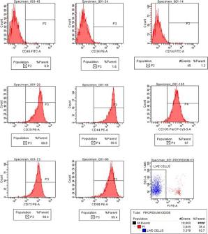
Quantitative Flow Cytometry Analysis and Surface Epitope Validation of Purified UC-MSC stem cell therapy. Source: ResearchGate
As shown in the laboratory assay panels above, the cells must display clear, uniform shifts in fluorescence intensity to confirm a highly purified population. The positive markers (CD105, CD73, and CD90) must show a clean, high-density distribution shifting well beyond the 95% threshold.
Concurrently, the negative lineage markers (CD45 and CD34) must remain securely locked within the baseline negative controls (). This strict separation proves the cell line is completely free from contaminating blood elements or endothelial fragments, confirming a uniform, highly therapeutic product ready for joint delivery.
5. Clinical Delivery Protocols, Targeting Logistics, and Post-Injection Progression
Translating this verified cell profile into reliable structural repair requires an optimized clinical sequence designed to preserve high cell viability and ensure precise anatomical placement within the joint cavity.
Controlled Thawing and Viability Assessment
Cryogenic Cold-Chain Transition
The cryopreserved Wharton’s jelly UC-MSC stem cell therapy suspension is removed from liquid nitrogen vapor storage () and transitioned rapidly using a specialized water bath. Automated cell counters must confirm a baseline living cell threshold exceeding prior to clinical compounding.
Arthrocentesis and Joint Space Decompression
Synovial Fluid Cleansing
Under strict sterile conditions, the target knee joint undergoes careful arthrocentesis to clear any excess inflammatory fluid. Decompressing the joint space reduces the concentration of host catabolic cytokines (, ) and prepares the microenvironment for optimal cell function.
Ultrasound-Guided Intra-Articular Injection
High-Resolution Visualization
Using live musculoskeletal ultrasound guidance, the clinician navigates a 21G/22G needle into the targeted intra-articular joint space or directly adjacent to the perimeniscal recess. The validated, marker-verified cell suspension is delivered slowly to maximize cell localization around areas of cartilage injury.
Kinetic Care and Joint Offloading
Early Tissue Stabilization
The patient undergoes a short period of supervised, non-weight-bearing passive joint mobilization to distribute the delivered secretome evenly across the cartilage surfaces. Specialized offloading braces are utilized to minimize mechanical shear stress during the early phases of cell-mediated repair.
6. Patient Stratification: Mapping Joint Degradation and Success Probabilities
Achieving reproducible functional recovery requires strict patient selection based on validated radiological and tissue degradation scales.
Joint Structural Suitability Matrix
| Diagnostic Tier | Kellgren-Lawrence (K-L) Grade | Cartilage Structural Integrity | Expected Clinical Outcome |
| Tier 1: Optimal Candidate | Grade I – II (Early osteophyte formation, minimal joint space narrowing). | Localized fissuring, superficial thinning of the hyaline layer, mild subchondral bone stress. | High probability of long-term pain resolution, functional recovery, and potentially measurable structural matrix stabilization. |
| Tier 2: Moderate Candidate | Grade III (Moderate joint space narrowing, multiple osteophytes). | Significant partial-thickness cartilage defects, localized focal erosions down to the subchondral layer. | Favorable pain reduction and functional recovery; typically used alongside offloading physical therapy to maximize outcomes. |
| Tier 3: Poor Candidate | Grade IV (Severe joint space narrowing, advanced bone-on-bone friction). | Complete loss of functional hyaline thickness, widespread subchondral bone friction and remodeling. | Palliative functional and anti-inflammatory support only. Structural tissue regeneration is highly limited; joint replacement surgery remains the primary indication. |
Absolute Contraindications and Safety Controls: Patients are excluded from intra-articular cell therapy if they present with active septic arthritis, intra-articular crystal arthropathies (unmanaged gout or pseudogout), severe structural axial malalignment (varus/valgus deformities requiring corrective osteotomy), active localized malignancies, or systemic autoimmune joint conditions (e.g., rheumatoid arthritis) that are not managed under rheumatological care.
7. Multi-Dimensional Clinical Endpoint Monitoring
To accurately separate true cell-mediated tissue repair from temporary symptomatic improvement, clinical protocols track patient outcomes across strict post-injection intervals (Months 3, 6, and 12):
Validated Functional Scaling (PROMs)
WOMAC (Western Ontario and McMaster Universities Osteoarthritis Index): Tracks changes across three distinct subscales: Pain intensity, joint stiffness, and physical functionality during daily tasks. Clinical protocols target a minimum 30–50% reduction in total WOMAC scores within 24 weeks.
KOOS (Knee Injury and Osteoarthritis Outcome Score): Evaluates comprehensive progress across five domains, including daily living requirements, high-impact sport/recreation mobility, and knee-related quality of life.
Advanced Radiographic Tracking via Quantitative MRI (qMRI)
T2 Relaxation Time Mapping: Quantitative MRI (qMRI) measures changes in water distribution and collagen fiber alignment within the healing hyaline layer, tracking structural organization and tissue quality over time.
dGEMRIC (Delayed Gadolinium-Enhanced MRI of Cartilage): Bypasses standard visual limitations by measuring absolute glycosaminoglycan (GAG) density within the extracellular matrix, providing clear proof of structural synthesis.
8. Biosafety Assurance and Regulatory Compliance Frameworks in Thailand
Because regenerative medicine is expanding rapidly throughout Thailand, processing laboratories and clinical networks must adhere to strict international safety and processing standards to ensure patient safety and maintain regulatory compliance.
Cell Viability and Sterility Assay Validation
Every therapeutic cell batch must be certified completely free from bacterial, fungal, or mycoplasma contamination using automated culture tracking and quantitative PCR assays. Endotoxin levels must be explicitly confirmed to be safely below via standard Limulus Amebocyte Lysate (LAL) testing to prevent localized joint inflammation or systemic reactions.
Cryopreservation and Cold-Chain Logistics
The long-term therapeutic viability of allogeneic UC-MSC stem cell therapy depends on strict temperature management. Cells are preserved using controlled-rate freezers in specialized cryoprotectants and stored in liquid nitrogen vapor at .
Any thermal fluctuation during storage or transport across Thailand will compromise cell membranes, causing immediate cellular death. Injecting damaged cells degrades a premium regenerative therapy into a non-viable introduction of cellular debris, causing localized immune stress rather than target-specific immunomodulation.
Regulatory Alignment within Thailand
Practitioners must operate in strict alignment with the regulatory frameworks established by the Thailand Food and Drug Administration (TFDA) and the Medical Council of Thailand. Administering fully validated cell products within certified clinical trial frameworks or verified Good Tissue Practices (GTP) ensures clear compliance.
Medical teams are ethically obligated to maintain complete scientific honesty with patients—framing UC-MSC stem cell therapy not as a standalone miracle cure that reverses aging overnight, but as an advanced biological booster designed to complement and maximize the outcomes of traditional physical therapy and structured orthopedic care.
Conclusion
Epitope profiling of Wharton’s jelly-derived UC-MSC stem cell therapy via specific surface antigen characterization is essential for ensuring safety and predictable outcomes in managing knee osteoarthritis. By confirming high expression of positive markers (CD73, CD90, and CD105) alongside the complete exclusion of negative lineage markers, clinical teams can ensure they are deploying a highly pure, immune-privileged cell line.
These certified cells actively suppress chronic synovial inflammation, halt enzymatic cartilage breakdown, and stimulate the synthesis of authentic hyaline matrix. When guided by precise patient stratification, ultrasound-guided delivery, and quantitative imaging endpoints, UC-MSC stem cell therapy serves as a premier, compliant treatment framework within Thailand’s modern orthopedic and regenerative medicine landscapes.