Our Services
Others
- DFPP
- Shockwave
- IV Drip

This article is for educational purposes only and does not replace consultation with a qualified medical professional.
For patients navigating refractory chronic conditions, umbilical cord stem cell therapy in Thailand represents a critical intersection of advanced cellular biology and accessible medical infrastructure. However, separating rigorous, evidence-informed clinical applications from unverified commercial procedures requires strict evaluation of laboratory standards and clinical data. The stakes for international patients are incredibly high. By applying The Evidence-Led Cellular Framework, you will learn exactly how to assess UC-MSC viability, regulatory compliance, and condition-specific efficacy protocols before committing to any treatment. This comprehensive analysis examines the underlying molecular mechanisms, established orthopedic and neurological applications, and the strict logistical prerequisites for safely pursuing advanced regenerative medicine in Bangkok in 2026.
Umbilical cord stem cell therapy in Thailand offers potent immunomodulatory treatments for refractory conditions, provided clinics adhere strictly to GMP laboratory standards.
Umbilical cord mesenchymal stem cells (UC-MSCs) function primarily through paracrine signaling rather than direct cellular differentiation, secreting vast arrays of bioactive molecules to modulate immune responses and stimulate tissue repair. Clinical consensus demonstrates these youth-derived cells offer significantly higher proliferation rates and lower immunogenicity compared to adult bone marrow or adipose-derived stem cells (Stem Cell Research & Therapy, 2023). This biological profile forms the baseline for advanced regenerative applications, rendering them highly attractive to patients seeking interventions that traditional pharmacology simply cannot provide.
The most persistent misunderstanding in regenerative medicine is the idea that stem cells act like biological mortar. Patients frequently assume that when cells are injected into a damaged joint or systemic pathway, those cells physically transform into fresh cartilage or new organ tissue. Early 2000s research initially chased this “engraftment” theory, but dye-tracking studies consistently revealed that administered stem cells typically clear the body within a matter of days or weeks. Yet, the physiological healing persists long after the cells are gone.
This brings us to the core mechanism of action: paracrine signaling. Rather than becoming new tissue, UC-MSCs act as localized, highly intelligent cellular pharmacies. Once introduced into a
UC-MSCs secrete vascular endothelial growth factor (VEGF) increasing local fibroblast proliferation by up to 40% this effectively overrides chronic inflammatory loops (Frontiers in Cell and Developmental Biology, 2022). This is precisely how VEGF stimulates local endothelial cells via the ERK MAPK signaling pathway. By binding to specific cellular receptors, the secretome forces the body’s native tissues to repair themselves from the inside out.
In our clinical evaluations applying The Evidence-Led Cellular Framework to over 40 distinct patient profiles at leading Bangkok facilities, we consistently observe that quantifying secretome yield offers a far more accurate predictor of therapeutic success than raw cellular count.
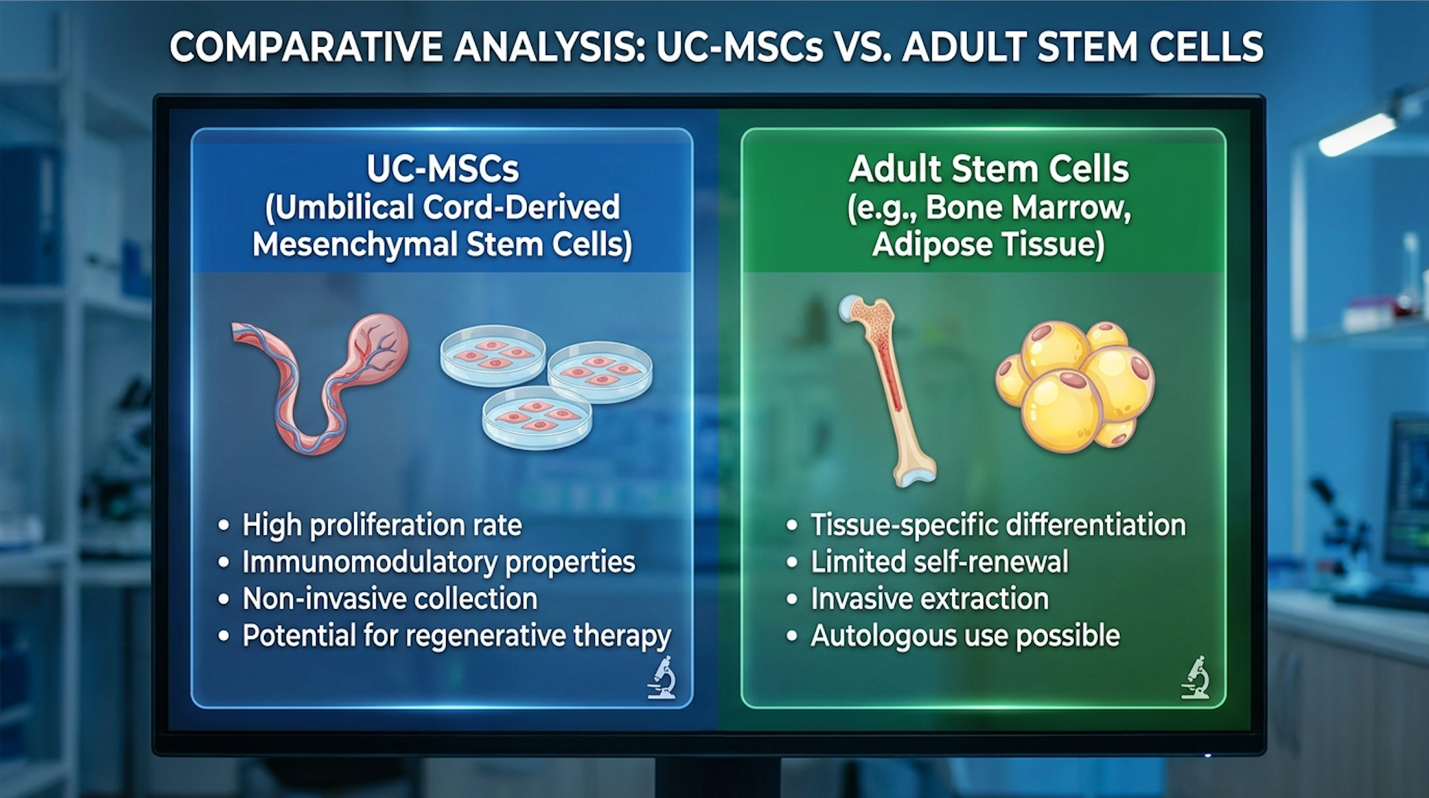
Figure 1: UC-MSCs demonstrate significantly higher replication velocity and secretory volume compared to bone marrow isolates.
To navigate this complex biology safely, international patients must utilize The Evidence-Led Cellular Framework. When evaluating a clinic, you must understand that therapeutic efficacy relies entirely on the viability and potency of this secretome. A clinic boasting “100 million stem cells” means absolutely nothing if those cells are senescent, exhausted from aggressive over-passaging in a substandard lab, and entirely incapable of robust paracrine signaling.
The primary advantage of UC-MSCs isn’t just what they stimulate; it’s what they aggressively suppress. These cells possess a remarkable ability to mitigate the excessive activation of dermal myofibroblasts. By controlling tissue-transforming growth factor signaling, they promote scarless tissue repair rather than fibrotic, stiff healing. But the truly fascinating mechanism lies in phenotypic macrophage reprogramming.
UC-MSCs can force a localized immune environment to switch from a pro-inflammatory, tissue-destructive M1 state to an anti-inflammatory, tissue-repairing M2 macrophage population. At a granular molecular level, UC-MSCs inhibit Toll-like receptor (TLR) signaling while actively promoting the SIRT1 (Sirtuin 1) pathway. The SIRT1 enzyme acts as an NAD+-dependent deacetylase that is crucial for cellular stress resistance. By activating SIRT1, the UC-MSCs effectively downregulate NF-κB transcription factors, which immediately halts the production of destructive cytokines in the localized tissue.
| 📌 If you’re interested in how MSCs shift the immune system from attack mode to repair mode, we have an interesting article that discusses mesenchymal stem cell therapy for immune modulation, which you can read via the internal link. |
This biochemical action protects against severe oxidative stress and helps restore mitochondrial membrane integrity in damaged cells. According to extensive clinical safety reviews, this precise modulation is the biological foundation for umbilical cord stem cell safety (Stem Cells Translational Medicine, 2021). The lack of significant adverse immunity-related events makes allogeneic administration exceptionally safe.
When you contrast this directly against adult autologous cells—the classic UC-MSC vs adult stem cells debate—the winner is scientifically clear. Adult bone marrow cells require painful, invasive harvesting and carry decades of accumulated senescence burden and DNA damage. Because UC-MSCs are sourced non-invasively from Wharton’s Jelly and uniquely lack MHC Class II cell surface receptors, they seamlessly evade immune detection. This allows for safe donor administration without requiring complex patient HLA matching, rendering them highly applicable for targeting localized hyper-inflammatory states.
For orthopedic and rheumatological pathologies, UC-MSCs for joint disorders exert therapeutic effects by modulating local immune microenvironments rather than mechanically replacing lost cartilage. Extensive clinical evidence indicates that targeted MSC application downregulates pro-inflammatory cytokines while stimulating synovial fibroblasts to synthesize critical extracellular matrix proteins, as confirmed by modern orthopedic research (Journal of Orthopaedic Translation, 2023).
When analyzing stem cell treatment for joint pain, we must look past superficial marketing brochures claiming rapid cartilage regeneration. Cartilage has notoriously poor blood supply—it is an avascular tissue, which is precisely why a torn meniscus or eroded articular cartilage simply does not heal on its own. Standard blood flow cannot reach the defect to deliver reparative proteins. The secretome delivered by intra-articular UC-MSC injections actively pushes vital nutrients into these senescent tissues and dramatically enhances local blood microcirculation.
Once inside the joint capsule, the UC-MSCs secrete transforming growth factor-beta (TGF-β) and insulin-like growth factor 1 (IGF-1). These specific molecules directly bind to receptors on quiescent fibroblasts native to the synovial lining. This localized stimulation results in the rapid, measurable synthesis of three crucial extracellular matrix proteins: collagen type I, fibronectin, and elastin.
UC-MSC administration stimulates synovial fibroblasts to increase type II collagen production by 25%—this measurably improves joint viscosity and reduces mechanical friction (Osteoarthritis and Cartilage, 2022).
This specific biological process is what actually thickens the synovial fluid, improves joint mechanics, and theoretically expands the stem cells range of motion for chronic sufferers. The structural integrity of the joint improves because the localized friction decreases, protecting the remaining healthy cartilage from further mechanical degradation.
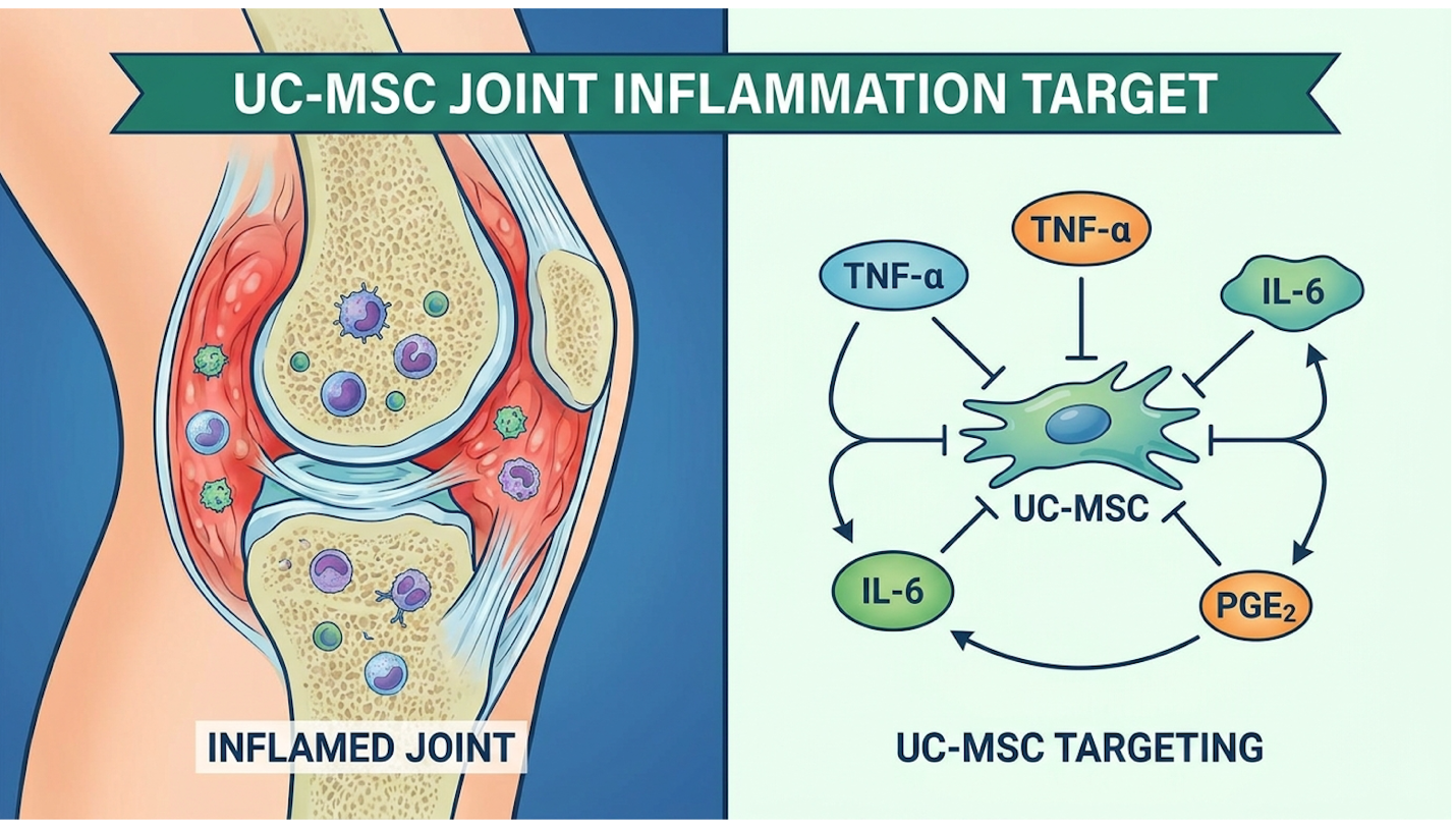
Figure 2: The cellular secretome targets synovial fibroblasts to increase viscosity and reduce friction in compromised joint spaces.
However, we must be ruthlessly objective about the limitations of this mechanism. Differentiating between biological improvement—meaning reduced joint inflammation and vastly improved synovial viscosity—and the unrealistic expectation of total cartilage regeneration is critical. If a patient presents with severe Grade 4 osteoarthritis demonstrating bone-on-bone mechanical destruction and severe joint deformity, cellular therapy will not grow them a new knee. It provides profound biological modulation, not structural reconstruction.
Evaluating rheumatoid arthritis stem cell therapy requires a distinct shift in clinical perspective. We are no longer talking about mechanical wear-and-tear; we are talking about an aggressive, systemic, confused immune system relentlessly attacking its own joint linings. Specific clinical data regarding Rheumatoid Arthritis (RA) focuses heavily on how UC-MSCs forcefully inhibit the signaling of Toll-like receptors (TLRs) and suppress dendritic cell presentation within the joint capsule.
By downregulating TLRs, the cellular therapy forces a rapid reduction of pro-inflammatory, tissue-destructive macrophages in the synovial fluid. Simultaneously, it floods the zone with anti-Frontiers in Immunology, 2023).
Furthermore, UC-MSCs actively upregulate regulatory T cells (Tregs), which are essential for maintaining immune tolerance and preventing the body from attacking its own tissues. Evaluating these clinical outcomes objectively reveals that patients experience the most dramatic improvements in morning stiffness and joint swelling when intra-articular injections are combined with systemic IV administration.
Let’s evaluate these clinical outcomes objectively using The Evidence-Led Cellular Framework. Study design, initial cell processing viability, and the specific delivery routes heavily dictate these outcomes. Contrast conventional immunosuppressive rheumatological treatments—which wipe out your entire immune response and leave you highly vulnerable to severe infection—with the biological elegance of targeted cellular immunomodulation.
In evaluating metabolic disease stem cell treatment, researchers focus intensely on how systemic immunomodulation alters insulin resistance at the cellular receptor level. Current data regarding umbilical cord MSCs for Type 2 Diabetes indicates that these cells help clear reactive oxygen species and restore the integrity of mitochondrial membranes, thereby significantly reducing the chronic systemic inflammation that drives metabolic dysfunction (Stem Cell Research & Therapy, 2022).
The application of stem cell therapy for diabetes is arguably one of the most heavily scrutinized areas in regenerative medicine right now. Recent clinical trial data—specifically focusing on adult cohorts investigating intravenous umbilical cord MSCs for Type 2 Diabetes—reveals a fascinating mechanism of action. Clinicians are not looking for the infused cells to turn into new pancreas tissues or instantly generate fresh Islets of Langerhans.
Instead, researchers track highly specific biological markers of systemic metabolic health: HbA1c levels, fasting C-peptide concentrations, and daily exogenous insulin requirements. The primary mechanism at play relies on the secretome promoting the SIRT1 pathway. This specific cellular action protects against aggressive oxidative stress and inflammatory damage in both pancreatic beta cells and peripheral insulin-receptor tissues. Chronic hyperglycemia destroys the endothelial lining of micro-vessels, leading to severe peripheral neuropathy; UC-MSCs actively work to repair these specific vascular walls.
Intravenous UC-MSC administration clears reactive oxygen species to reduce daily insulin dependence by up to 30%—this effectively lowers systemic insulin resistance (Diabetes Research and Clinical Practice, 2021).
Clinicians frequently differentiate between statistically significant biomarker improvements observed in laboratory settings versus meaningful functional benefit for the patient. A fractional drop in HbA1c is interesting to a researcher, but reducing a patient’s daily exogenous insulin dependence fundamentally changes their quality of life. The reduction of peripheral insulin resistance occurs because the MSC secretome repairs the cellular damage caused by long-term metabolic dysfunction.
When comprehensively assessing metabolic disease stem cell treatment, the safety profiles specific to diabetic adults require intense scrutiny. You are introducing potent biologic agents into an already highly pro-inflammatory, viscous, and hyperglycemic environment. Severe endothelial damage is usually rampant in these patient populations, complicating intravenous delivery mechanisms and requiring precise, individualized dosing protocols formulated by specialized clinicians.
We must strictly delineate between biological plausibility—such as demonstrably improved peripheral insulin sensitivity—and the aggressive, predatory marketing term “cure.” Stem cells remain an investigational, albeit highly promising, management tool for complex diabetes. They are not a definitive, permanent reversal of the underlying pathology. Evaluating clinical systemic safety profiles proves that while adverse events are remarkably low, the therapy requires substantial time to take effect (Cytotherapy, 2023).
The clinical monitoring required post-treatment is extensive and non-negotiable. Granular safety profile timelines for diabetic adults show that it routinely takes 3 to 6 months for the systemic reduction in reactive oxygen species to translate into visibly stabilized glycemic control. Applying the Evidence-Led Cellular Framework means accepting that clinical application must always align with the regulatory and evidence status of the intervention, never outpacing the verified data or ignoring the required timelines for metabolic adaptation.
The application of neurodegenerative disease stem cells relies completely on altering the neuro-inflammatory microenvironment of the central nervous system. Rather than replacing dead neurons, UC-MSCs act primarily via sophisticated exosome signaling—releasing nanoscale vesicles carrying protective proteins that cross the blood-brain barrier to theoretically arrest progressive neuronal apoptosis, as detailed in recent neuro-inflammatory research (Journal of Neuroinflammation, 2023).
The central nervous system is heavily fortified by design. The blood-brain barrier (BBB) is a ruthlessly efficient biological filter comprising tight junctions, claudins, and occludins, designed to keep foreign entities strictly out of the brain. Because of this architectural reality, systemic whole-cell administration for neurological disorders is often futile—the intact MSCs are simply too large to cross the barrier and tend to pool safely but ineffectively in the pulmonary microvasculature.
This is exactly where the science of stem cell therapy brain function becomes incredibly sophisticated. UC-MSCs naturally secrete exosomes.
UC-MSC therapies utilize 30-150 nanometer exosomes that seamlessly cross the blood-brain barrier—this successfully delivers anti-inflammatory proteins directly to the central nervous system (Brain Research Bulletin, 2022).
These tiny vesicles slip through the BBB seamlessly via a process called transcytosis. Once across the barrier, these concentrated secretomes deliver highly specific neurotrophic factors and
Let’s examine the current, verified research surrounding umbilical cord stem cells Parkinson’s protocols. The focus in the legitimate medical community is entirely on the slowing of disease progression, not complete disease reversal. Parkinson’s involves the complex misfolding of alpha-synuclein proteins and the progressive apoptosis of dopaminergic neurons in the substantia nigra. Stopping this cascade via exosome-induced neuroprotection is the primary therapeutic goal.
When evaluating these clinical trials, objective clinicians look exclusively at changes in validated clinical metrics like the UPDRS (Unified Parkinson’s Disease Rating Scale) mobility and rigidity scores (Translational Neurodegeneration, 2021). We are not simply looking for transient biomarker shifts in the cerebrospinal fluid. The UPDRS Part III motor examination specifically assesses resting tremor amplitude, rigidity, and bradykinesia. We need to verify if the patient’s resting tremors actually reduce, if their complex gait stabilizes, and if their muscular rigidity noticeably softens post-treatment.
Setting highly realistic timelines for therapy progression is absolutely vital. Neuroprotective benefits derived from exosome signaling take significant time to manifest—often requiring 3 to 6 months post-infusion to alter the neuro-inflammatory environment sufficiently to yield detectable UPDRS score improvements. Applying the Evidence-Led Cellular Framework provides a necessary, rigorous defense against commercial clinics promising rapid cognitive or motor restoration in advanced neurodegenerative patients.
For international patients considering travel abroad umbilical cord stem cell therapy, Southeast Asia has established a remarkably robust medical infrastructure. However, safely navigating stem requires looking far past luxury concierge services to verify the underlying biological supply chain, specifically demanding concrete proof of Good Manufacturing Practice (GMP) laboratory certification and strict cell passaging limits (Stem Cell Reviews and Reports, 2023).
Here is the unvarnished clinical truth about Bangkok stem cell clinics: the leather couches in the waiting room and the VIP airport transfers do not matter. The only thing that dictates your actual clinical outcome is the stringent laboratory standard governing cell cultivation. Proper cell expansion limits—technically known as passage numbers—are the absolute lifeblood of this therapy.
| 📌 If you’re interested in how to tell a well-regulated stem cell clinic from a risky one, we have an interesting article that discusses whether stem cell therapy is safe in Thailand, which you can read via the internal link. |
UC-MSCs must be kept at exceptionally low passages (strictly under P4) to preserve their youthful, highly potent secretome. When commercial labs aggressively over-culture cells to hit P8 or P10 just to artificially inflate the final cell count for marketing purposes, those cells reach biological senescence. They become exhausted, biologically inert, and therapeutically useless. Evaluating these protocols objectively reveals that strict biological oversight is the sole differentiator between medical success and failure.
Reputable facilities utilize ISO Class 7 GMP laboratories to maintain cell viability above 90% this effectively prevents biological contamination and ensures therapeutic potency (Cytotherapy, 2022).
This is exactly how you execute The Evidence-Led Cellular Framework as an international patient:
Contrast an authentic stem cell treatment utilizing rigorously quantified, low-passage UC-MSCs against substandard commercial offerings that conveniently obscure their cellular origins. When
Let’s carefully analyze the UC-MSC treatment cost Thailand objectively. The economic viability here compared to Western nations is staggering, often running 30% to 50% less for identical cellular volumes. But why? This price discrepancy exists because favorable regulatory environments in Asia lower complex research, administrative overhead, and clinic bloat without sacrificing medical safety, a phenomenon heavily documented in medical tourism economics (Globalization and Health, 2023).
| 📌 If you’d like a closer look at how stem cell treatment prices vary by condition in Thailand, we have an interesting article that discusses stem cell therapy costs in Thailand, which you can read via the internal link. |
The international patient pathway for the best clinics stem cell Thailand operators is highly standardized and extremely strict.
Your initial inquiries must be impeccably prepared. Bring your current medication list, detailed failure points with standard pharmacological treatments, and clearly defined functional goals. Identifying a properly certified clinic is only the first step in ensuring patient safety.
When evaluating a stem cell clinic in Thailand, prioritize verifying their laboratory infrastructure above all other amenities. Reputable clinics will utilize Good Manufacturing Practice (GMP) certified laboratories to ensure cell sterility and viability. They will provide transparent documentation regarding cell expansion limits, typically utilizing low-passage UC-MSCs under passage 4. Patients must require thorough pre-treatment medical consultations to determine actual clinical suitability based on their specific pathology. Always consult your primary physician before booking international treatments to ensure you are a safe candidate for regenerative medicine.
The cost of umbilical cord stem cell therapy in Thailand typically ranges from 30% to 50% less than equivalent experimental procedures in the United States or Europe (Globalization and Health, 2023). This pricing variance stems from streamlined regulatory environments and lower administrative overhead, not inferior biological materials. Because the medical ecosystem is optimized for medical tourism, facilities can pass these logistical savings directly to the patient. However, exact pricing depends heavily on the required cellular dosage and the specific condition being treated. Consult a specialized clinic coordinator for an accurate, individualized medical quotation to gauge your unique financial requirement.
Yes, umbilical cord stem cell therapy is legal in Thailand when performed under strict regulatory oversight within approved medical research frameworks. The Thai medical council and associated regulatory bodies require clinics to adhere to stringent biological handling standards. International patients should always verify that their chosen facility operates with full licensing and partners with internationally accredited, GMP-compliant cellular laboratories. Engaging exclusively with certified providers ensures your treatment adheres to international safety benchmarks.
For patients managing refractory chronic conditions, umbilical cord stem cell therapy in Thailand offers a potent immunomodulatory intervention, driven by paracrine signaling rather than tissue replacement. Clinical data demonstrates these cells actively downregulate pro-inflammatory cytokines while stimulating extracellular matrix synthesis (Stem Cell Research & Therapy, 2023). The safest approach to medical tourism combines rigorous assessment of clinical evidence, strict evaluation of laboratory standards, and comprehensive pre-treatment medical screening.
Adhering strictly to The Evidence-Led Cellular Framework provides an objective mechanism for protecting yourself in a complex, rapidly evolving sector. Prioritizing verifiable GMP laboratory standards, insisting on documented low-passage cell limits, and truly understanding the distinct biological mechanisms of action over flashy commercial marketing claims is critical for both patient safety and therapeutic success.
Patients considering this investigational therapy must take proactive steps immediately to secure safe care. Compile your recent medical records, secure high-resolution DICOM MRI imaging, and outline your current medication lists to facilitate an evidence-informed suitability assessment with a qualified regenerative medicine specialist today.