Our Services
Others
- DFPP
- Shockwave
- IV Drip

For patients and clinical researchers evaluating interventions for Spinocerebellar Ataxia (SCA), the distinction between established symptom management and investigational biological treatments requires rigorous objective analysis. We aren’t dealing with a simple condition here. While conventional physical therapies offer temporary functional support, the relentless progression of cerebellar neurodegeneration demands disease-modifying approaches. However, the commercial proliferation of unverified cellular therapies creates significant patient risk. Clinical specialists frequently observe families draining their savings on unproven international clinics without understanding the basic pharmacokinetics of what they are buying.
This clinical review evaluates the exact pathophysiology of SCA, the role of conventional management, and the objective clinical trial data surrounding Umbilical Cord Mesenchymal Stem Cells (UC-MSCs). We will systematically analyze disease progression, standard lifestyle interventions, and the evidence comparing intrathecal versus intravenous stem cell administration for spinocerebellar ataxia.
This article is for educational purposes only and does not replace consultation with a qualified medical professional. Investigational treatments carry inherent risks.
Investigational stem cell therapy for spinocerebellar ataxia using UC-MSCs targets neuro-inflammation, with clinical trials indicating potential to temporarily suppress disease progression.
Spinocerebellar ataxia progression involves the gradual neurodegeneration of the cerebellum, spinal cord, and brainstem. While exact SCA life expectancy depends heavily on the specific genetic subtype (ranging from SCA1 to SCA40+), disease duration typically spans one to three decades (ClinicalTrials.gov registry data, 2024). Understanding this trajectory is critical for timing clinical interventions.
To understand what triggers spinocerebellar ataxia, you have to look directly at the genetic architecture of the brain. Spinocerebellar ataxia is triggered by inherited genetic mutations, most commonly abnormal polyglutamine (polyQ) expansions in highly specific genes. In a healthy human genome, a specific CAG sequence repeats a normal number of times usually between 10 and 30. But in SCA patients, this sequence stutters and expands wildly, sometimes reaching 40, 60, or even over 100 repeats.
This genetic stutter translates into a mutant protein called ataxin. Normal ataxin plays a vital role in RNA regulation and intracellular transport. Mutant ataxin? It’s a structural disaster. Because of the abnormally long polyglutamine tail, the protein misfolds. The brain’s internal garbage disposal system the ubiquitin-proteasome pathway desperately attempts to clear these misfolded proteins but quickly becomes overwhelmed by the sheer volume of toxic cellular waste.
Genetic analyses of SCA (2024) demonstrate that these protein aggregates clump together to form toxic intranuclear inclusions. These inclusions cause severe mitochondrial dysfunction inside the Purkinje cells of the cerebellum. The mitochondria begin leaking reactive oxygen species, causing oxidative stress to spike dramatically. Eventually, the cellular machinery fails completely, triggering apoptosis programmed cell death.
This cellular toxicity doesn’t happen at the same speed for everyone. Contrast the rapid, aggressive cellular toxicity seen in SCA1 with slower-progressing non-polyQ SCAs, like SCA6. In SCA1, the mutant ataxin localizes directly to the nucleus, disrupting gene transcription almost immediately. The phenomenon of “anticipation” further complicates matters; in subsequent generations, the CAG repeat length often expands, leading to earlier symptom onset and faster decline.
Because of this profound variability, securing comprehensive genetic testing is an absolute necessity. You simply can’t predict the speed of neurodegeneration without knowing the exact genetic subtype you’re fighting. Spinocerebellar ataxia genetic panels reveal a $2,000 median testing cost presenting a significant financial barrier to early intervention for many families. However, targeted testing for a known familial mutation is significantly cheaper than broad next-generation sequencing panels. While the genetic triggers operate at the cellular level, their macroscopic impact follows a predictable clinical staging.
Clinicians don’t just guess at spinocerebellar ataxia progression. We measure it rigorously using validated diagnostic instruments like the Scale for the Assessment and Rating of Ataxia (SARA). The SARA scale is an 8-item clinical rating tool that runs from 0 (no ataxia) to 40 (severe, wheelchair-bound ataxia). It specifically evaluates gait (0-8), stance (0-6), sitting (0-4), speech disturbance (0-6), finger chase (0-4), nose-finger test (0-4), fast alternating hand movements (0-4), and the heel-shin slide (0-4).
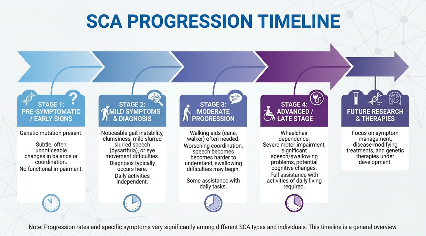
Progression moves through distinct, measurable clinical stages. The preclinical stage is silent. The patient is genetically positive but entirely asymptomatic, though advanced MRI volumetric analysis might show microscopic cerebellar volume loss. The early symptomatic stage kicks in when patients notice subtle gait instability bumping into doorframes, or struggling with tandem walking. SARA scores during this early phase typically hover between 3 and 8.
The middle stage is where functional independence starts slipping. Dysarthria (slurred speech) and dysphagia (difficulty swallowing) become clinically apparent. Patients usually require walking aids like canes or rolling walkers to prevent devastating falls. SARA scores climb between 10 and 20. Then we hit the end stages of cerebellar ataxia. Patients are generally wheelchair-bound, requiring complete assistance for basic activities of daily living, with severe bulbar dysfunction complicating both breathing and eating.
Longitudinal SARA progression data (2023) highlights a critical timeline for intervention. This brings us to a foundational concept in neuro-restoration: The Neuro-Inflammatory Suppression Window. Biological therapies including cell-based interventions hypothetically require administration during the early-to-middle stages. Why? Because you cannot rescue a dead neuron. The Neuro-Inflammatory Suppression Window is that precise period where Purkinje cells are deeply inflamed and metabolically distressed, but haven’t yet undergone irreversible apoptosis. If you wait until the end stages, the target tissue is simply gone. Leading clinical researchers emphasize that targeting this specific window is the only mathematically viable way to delay severe motor decline.
As the disease advances through these clinical stages, the focus inevitably shifts toward long-term prognosis, supportive care, and survival rates.
A frequent inquiry in clinical consultations is whether spinocerebellar ataxia is a terminal illness. The answer requires precise medical framing. SCA is incurable and undeniably life-limiting. But patients typically do not die from the ataxia itself. The ataxia doesn’t stop the heart. It doesn’t shut down the kidneys.
Instead, mortality is driven almost entirely by secondary complications resulting from the neurological decline. As bulbar dysfunction worsens, the muscles controlling the throat weaken and lose their intricate coordination. This leads to silent aspiration where food, liquid, or saliva slips past the vocal cords and down into the lungs. Aspiration pneumonia is the leading cause of mortality in end-stage SCA. Respiratory failure follows closely behind, driven by the inability to clear lung secretions due to a severely weakened cough reflex.
Median SCA survival spans 10 to 30 years post-onset, highlighting the urgent need for early intervention before bulbar dysfunction accelerates.
For example, SCA mortality studies evaluating large patient cohorts found that SCA1 tends to have a shorter survival median (around 10-15 years post-onset) due to earlier brainstem involvement. SCA6, on the other hand, is notoriously slow-moving, with many patients living a normal lifespan and eventually dying of unrelated age-associated causes. Machado-Joseph disease (SCA3), the most globally prevalent subtype, typically falls somewhere in the middle, heavily influenced by the specific CAG repeat length.
But here’s the thing that aggressive commercial clinics won’t tell you: meticulous conventional management dramatically alters this timeline. Rigorous pulmonary care, proactive swallowing assessments (like modified barium swallow studies), and early intervention with feeding tubes when dysphagia becomes severe can extend survival by years. The clinical priority must always remain focused on preventing respiratory infections while maintaining baseline nutrition. Given this relentless disease trajectory, patients must first optimize their baseline health through rigorous conventional management before pursuing experimental therapies.
Before exploring cellular therapies, establishing a baseline of cerebellar ataxia conventional management is medically mandatory. Clinical protocols prioritize neuro-rehabilitation to preserve motor function and strict dietary modifications, including specific foods to avoid with cerebellar ataxia, to minimize systemic neuro-inflammation and prevent excitotoxicity in already compromised neural pathways (National Institutes of Health guidelines, 2024).
Let’s talk about the neuro-inflammatory impact of diet. When dealing with a neurodegenerative disorder, your central nervous system is already on fire. Pouring metabolic gasoline on that fire accelerates Purkinje cell death.
First, consider gluten. Even in cases of genetically confirmed SCA (not just immune-mediated gluten ataxia), a strict gluten-free diet is often recommended to reduce baseline neurological inflammation. Circulating anti-gliadin antibodies can cross a compromised blood-brain barrier and exacerbate cerebellar astrogliosis. When the immune system is busy fighting dietary antigens, it amplifies the baseline inflammatory response within the central nervous system.
Then there’s ethanol. Alcohol is profoundly neurotoxic to the cerebellum even in entirely healthy brains. In an SCA brain, ethanol directly accelerates Purkinje cell apoptosis by disrupting calcium homeostasis. Alcohol is strictly, unequivocally contraindicated. Even small amounts can trigger severe, temporary exacerbations of ataxia symptoms that take days to resolve.
Neurological dietary impact studies (2024) also highlight the danger of high-glycemic foods and advanced glycation end products (AGEs). Chronic hyperglycemia spikes systemic oxidative stress, stripping the brain of its natural antioxidant reserves just when the cerebellum needs them most.
Here is the baseline clinical protocol for dietary management:
While dietary restrictions mitigate internal physiological stress, physical therapies address the external mechanical manifestations of the disease.
Clinical consensus is absolute: physical therapy is not optional. It is the single most effective intervention we currently have for maintaining independence. But general physical therapy isn’t enough. You need targeted neuro-rehabilitation designed specifically for ataxia.
The physiological goals are distinct. We aren’t building massive muscle strength. We are improving core stability, forcing the brain to recalibrate proprioception (your awareness of where your body is in space), and preventing muscular contractures. And this must be done without inducing severe central fatigue, which actually temporarily worsens ataxia symptoms.
What do safe exercises for people with cerebellar ataxia look like?
Continuous targeted physical therapy improves early-stage SARA scores by 1.5 points over 6 months proving movement functions as medicine.
Physical therapy programs focusing on balance disorders also heavily integrate Vestibular Rehabilitation Therapy (VRT). Because SCA often degrades the ocular-motor reflexes making it hard to track moving objects or stabilize vision while walking VRT helps retrain the brain to rely on alternative sensory inputs to prevent devastating fall-related trauma. In tandem with exercise, clinicians must rigorously monitor for secondary metabolic deficits that could masquerade as or accelerate disease progression.
Sometimes, neurodegeneration isn’t purely genetic. It’s metabolic. When diagnosing a progressive ataxia disorder, a good neurologist will immediately order a comprehensive metabolic panel to rule out acquired causes. If you have an underlying deficiency, it will dramatically accelerate the mechanical decline of your ataxia.
How exactly does a vitamin deficiency cause ataxia? The central nervous system is highly sensitive to oxidative stress. Vitamin E is a potent lipid-soluble antioxidant that protects neuronal cell membranes. In a specific genetic disorder called Ataxia with Vitamin E Deficiency (AVED) caused by mutations in the TTPA (alpha-tocopherol transfer protein) gene the body cannot retain Vitamin E. Without this protection, the spinocerebellar tracts degenerate rapidly, mimicking Friedreich’s ataxia perfectly.
Similarly, severe Vitamin B12 deficiency attacks the myelin sheath the protective insulation around nerves. B12 is essential for the function of methylmalonyl-CoA mutase. When this fails, toxic metabolic byproducts build up. This leads to subacute combined degeneration of the spinal cord. The dorsal columns fail, wiping out proprioception, which manifests clinically as a profound sensory ataxia rather than a true cerebellar ataxia.
Vitamin E deficiency ataxia mechanisms detailed by the NIH separate these metabolic deficits from true genetic SCA. However, the two can overlap. An SCA patient with a concurrent B12 deficiency will decline twice as fast, effectively battling two neurological fires simultaneously.
Standard clinical monitoring requires checking serum levels specifically methylmalonic acid (MMA) and homocysteine levels for B12 at least annually. If lab findings indicate a deficit, high-dose intramuscular supplementation is initiated immediately. When conventional symptom management reaches its efficacy plateau, patients often look toward investigational regenerative approaches to alter the biological disease course.
Among the newest treatment options for spinocerebellar ataxia, investigational Stem Cell Therapy with UC-MSCs for Spinocerebellar Ataxia (SCA) represents a shift from symptom management to attempting biological disease modification. Clinical data suggests that these cellular therapies operate primarily through paracrine signaling, altering the neurotoxic microenvironment rather than replacing lost neurons directly (ClinicalTrials.gov registry data, 2024).
Let’s cut through the marketing noise. If a clinic tells you their stem cells will travel to your brain, turn into brand new Purkinje cells, and permanently replace the ones you’ve lost, they are lying. That is not how Umbilical Cord Mesenchymal Stromal Cells (UC-MSCs) function in adult neurodegenerative disease. They do not permanently engraft to form new brain tissue.
The actual science is far more complex and relies on the “bystander effect” or paracrine signaling.
UC-MSCs are basically microscopic drug manufacturing facilities. When introduced into a highly inflamed, degenerating environment, they secrete a massive, complex array of bioactive molecules known as the secretome. This secretome is packed with exosomes and rich in neurotrophins specifically Brain-Derived Neurotrophic Factor (BDNF), Vascular Endothelial Growth Factor (VEGF), and Glial Cell Line-Derived Neurotrophic Factor (GDNF). These proteins act as intense biological fertilizers for stressed neurons.
How exactly do Mesenchymal Stem Cells target neurodegeneration? They act as master immune regulators through phenotypic macrophage reprogramming. In an SCA brain, the local immune cells (microglia) exist in a hyper-active, pro-inflammatory “M1” state, indiscriminately attacking stressed neurons by releasing toxic cytokines like TNF-alpha and IL-6. This localized autoimmune disaster is known as microgliosis.
UC-MSC secretome mechanisms observed in advanced molecular therapy reveal that UC-MSCs release factors like Prostaglandin E2 (PGE2). This forces the brain’s microglia to switch from the tissue-destructive M1 phenotype to an anti-inflammatory, tissue-repairing “M2” phenotype. They essentially turn down the volume on the neuro-inflammation that is accelerating the cerebellar atrophy, buying the struggling neurons time to repair their mitochondrial function.
| 📌 If you’re interested in how UC-MSCs calm neuroinflammation by rebalancing immune cells, we have an interesting article that discusses mesenchymal stem cell therapy for immune modulation, which you can read via the internal link. |
To maximize these neuroprotective secretomes, the method of delivering the cells into the central nervous system is critical.
This is where the rubber meets the road in clinical efficacy. The route of administration dictates everything. You can have the highest quality, lowest-passage cells in the world, but if they cannot effectively reach the central nervous system, the treatment is biologically useless.
Intravenous (IV) administration involves dripping stem cells into a peripheral vein. It’s cheap, easy, and requires minimal clinical skill. But it faces a massive biological hurdle known as the pulmonary first-pass effect. MSCs are relatively large cells (15 to 30 microns in diameter). When injected into a vein, they hit the right side of the heart and are immediately pumped into the pulmonary artery. Over 95% of these cells get permanently trapped in the tiny capillaries of the lungs.
The few cells that escape the lungs then have to circulate through the body, find the brain, and cross the Blood-Brain Barrier (BBB) a highly restrictive cellular filter that blocks large biological structures. The actual number of cells reaching the cerebellum via IV is infinitesimally small.
This brings us to the intrathecal injection of umbilical cord mesenchymal stem cells.
Intrathecal delivery bypasses the bloodstream entirely. A physician performs a lumbar puncture, typically between the L3 and L4 vertebrae, injecting the UC-MSCs directly into the subarachnoid space. The cerebrospinal fluid (CSF) constantly circulates upward to the cisterna magna, essentially bathing the spinal cord, brainstem, and cerebellum directly in the cells and their secreted neurotrophins.
Leading clinical researchers confirm that intrathecal administration guarantees substantially higher cellular concentrations at the neurodegenerative target sites. Intrathecal vs IV MSC distribution studies tracking cellular migration consistently prove that intrathecal administration is vastly superior for accessing deep brain neurodegenerative targets.
| Administration Route | BBB Penetration | CNS Cell Yield | Procedure Complexity | Common Side Effects |
| Intravenous (IV) | Extremely Low | < 1% | Low (Standard IV) | Phlebitis, mild fever |
| Intrathecal (IT) | 100% (Bypassed) | High (Direct to CSF) | High (Lumbar Puncture) | Headache, back pain |
With intrathecal administration showing superior CNS delivery, clinical trials have closely monitored the safety and objective outcomes of this specific approach.
When evaluating stem cell treatment for ataxia, we must rely on objective human trial data. Phase I and Phase II clinical trials are designed to answer two fundamental questions: Is the protocol safe? And does it generate meaningful functional outcomes for the patient?
Regarding safety, the data is highly reassuring. Intrathecal UC-MSCs have demonstrated an excellent safety profile in regulated settings. Severe adverse events (SAEs) like tumor formation, teratomas, or severe immune rejection are exceedingly rare. This is primarily because UC-MSCs express very low levels of HLA-DR antigens, making them “immune-privileged.” The most common side effects are transient. Patients typically report a mild, low-grade fever for 24 hours post-infusion, or a post-lumbar puncture headache that resolves quickly with proper hydration, rest, and caffeine.
Regarding efficacy, the metrics are promising but require realistic expectations. Regulated clinical trials typically utilize precise cell dosages, such as $1times10^6$ cells per kilogram of patient body weight, ensuring standardized biological exposure.
Phase II intrathecal injections reduce SARA scores by up to 3 points offering a temporary functional baseline reset for ataxia patients.
Let’s translate that clinical data. A 2 to 3-point drop on the SARA scale is clinically meaningful. Patients participating in these trials (Phase II SCA MSC trials, 2024) often experience stabilization of their gait. Speech clarity frequently improves, and the dysmetria (tremors associated with reaching for objects) quiets down. Secondary objective metrics, like the 8-meter walk test and the 9-hole peg test, also frequently show temporary optimization, allowing patients to regain small degrees of lost independence.
But it is strictly not a cure. The genetic mutation (the ataxin protein malfunction) is still there, quietly damaging the cells. The stem cells simply created a highly neuroprotective environment that allowed the surviving, stressed neurons to function at peak capacity. Because the genetic mechanism continuously damages the cerebellum, a single administration of cells is insufficient for long-term management.
Here is the harsh reality of biological therapies for progressive ataxia: it is absolutely not a one-and-done procedure.
Repeated Mesenchymal Stromal Cell Treatment is a biological necessity if you want to maintain functional gains. Because UC-MSCs do not permanently engraft in the central nervous system, they act strictly as temporary biological catalysts. They secrete their growth factors, modulate the microglial response, and eventually undergo apoptosis. They are completely cleared from the cerebrospinal fluid via the glymphatic system within a few weeks to a few months.
| 📌 If you’re wondering whether more than one dose can help sustain your results, we have an interesting article that discusses why double-dose UC-MSC stem cell therapy may offer greater benefits than a single dose, which you can read via the internal link. |
Clinical trial timelines show a highly predictable response curve. A patient receives an intrathecal injection. For the first month, they might notice very little change. Between months three and six, they hit peak functional improvement. Their SARA score drops, and mobility optimizes. But if they do not receive a subsequent maintenance infusion, that SARA score will slowly begin to climb back toward their baseline by month nine or twelve as the neuro-inflammation returns.
Longitudinal MSC efficacy studies (2024) charting therapeutic duration demonstrate that sustaining the alleviation requires periodic, scheduled treatments, often every 6 to 12 months depending on the protocol.
This brings us right back to The Neuro-Inflammatory Suppression Window. The explicit goal of repeated treatments is to hold the patient firmly within this window for as long as biologically possible. We are attempting to freeze the disease progression in the early-to-middle stages, preventing the irreversible cerebellar atrophy that characterizes the end stages. Evaluating this therapy requires factoring in the long-term logistical and financial commitments of multi-year treatment protocols. While the biological rationale is strong, patients must navigate significant clinical limitations and commercial risks when seeking these advanced therapies.
Is there a cure for spinocerebellar ataxia does stem cell therapy slow ataxia progression?
Currently, there is no FDA-approved cure for spinocerebellar ataxia. Treatment protocols primarily focus on symptom management and physical rehabilitation to preserve patient mobility and independence. Investigational approaches, such as umbilical cord mesenchymal stem cell therapy, aim to delay neurodegeneration rather than eliminate the underlying genetic mutation. Patients should consult their neurologist regarding ongoing clinical trials for disease modification.
Early clinical trial data suggests that stem cell therapy may temporarily slow ataxia progression in specific patient cohorts. UC-MSCs secrete neuroprotective factors that suppress cerebellar inflammation and reduce microglial activation. Phase II trials have recorded SARA score stabilizations lasting 3 to 6 months post-injection. However, because the genetic driver remains, continuous disease progression eventually resumes. Results vary widely based on the patient’s specific SCA subtype and disease stage.
The physical procedure for stem cell treatment for ataxia takes one to three hours, depending on the administration route. Intrathecal delivery via lumbar puncture requires precise clinical preparation, injection, and a brief recovery observation period. Conversely, intravenous infusions generally take about 45 to 60 minutes of active drip time.
Spinocerebellar ataxia is triggered by inherited genetic mutations, most commonly abnormal polyglutamine expansions in specific genes. These genetic errors cause mutated proteins, such as ataxin, to misfold and accumulate toxically within the neurons of the cerebellum and spinal cord. Over time, this toxic accumulation leads to profound oxidative stress, mitochondrial dysfunction, and eventually cellular death. The severity and onset age are heavily correlated with the length of the genetic expansion. Diagnostic confirmation requires specialized genetic testing.
The end stages of cerebellar ataxia are characterized by severe motor impairment, typically resulting in wheelchair dependence and significant loss of independent function. Patients experience pronounced dysarthria (speech difficulty) and dysphagia (swallowing difficulty), which severely impact daily living. Aspiration pneumonia, resulting from swallowing deficits, becomes a primary clinical risk during this phase.
The cost of SCA genetic testing typically ranges from $300 to over $2,000, depending on the laboratory and the specific diagnostic panel ordered. Targeted testing for a single known familial mutation is significantly cheaper than comprehensive next-generation sequencing panels that screen for dozens of ataxia-related genes simultaneously. Many health insurance plans cover this testing if ordered by a neurologist for symptomatic diagnosis. Patients should consult a genetic counselor to determine the most cost-effective diagnostic approach.
For patients confronting cerebellar neurodegeneration, stem cell therapy for spinocerebellar ataxia represents an investigational shift from symptom management to biological intervention. Clinical trial data indicates that intrathecal UC-MSC administration can temporarily reduce SARA scores by 2 to 3 points over 6 months by suppressing neural inflammation (ClinicalTrials.gov, 2024). The most evidence-informed approach combines rigorous conventional physical therapy with highly regulated, expert-monitored clinical trials.
The efficacy of these biological interventions relies heavily on The Neuro-Inflammatory Suppression Window. Administering therapies before irreversible cellular senescence and severe atrophy occur is critical, as MSCs aim to preserve existing neural networks rather than generate entirely new brain tissue.
Patients evaluating these advanced treatments must prioritize clinical safety over commercial promises. The mandatory next step is securing a comprehensive neurological baseline assessment, including updated MRI imaging and genetic sequencing, before scheduling a consultation with a board-certified specialist conducting regulated cell-therapy research.