Our Services
Others
- DFPP
- Shockwave
- IV Drip


This article is for educational purposes only and does not replace consultation with a qualified medical professional. It explores current clinical data regarding regenerative therapies; individual suitability must be determined via a comprehensive medical assessment.
For patients navigating chronic ankle osteoarthritis or refractory ligament damage, deciding between invasive arthrodesis and emerging biological interventions requires separating verifiable clinical data from regenerative medicine marketing. Evaluating stem cell therapy for ankle pain becomes incredibly difficult when aggressive commercial claims continually outpace peer-reviewed orthopedic literature and FDA regulatory standards.
Our clinical evaluation team has reviewed hundreds of patient charts where clinics promised new cartilage and miracle cures, entirely missing the nuanced reality of joint regeneration. Look, if you’re facing a joint fusion, you desperately want alternatives. But biological plausibility isn’t the same as clinical reality. By the end of this clinical guide, you will understand the exact biological mechanisms, statistical success rates, and regulatory realities of regenerative ankle injections, allowing for a genuinely evidence-informed suitability assessment.
Our clinical focus remains strictly on verifiable orthopedic science rather than marketing narratives. This rigorous analysis examines cellular pathways specifically mesenchymal signaling documented adverse events, strict rehabilitation timelines, and the true out-of-pocket cost of treatment based on verified medical literature. The tibiotalar joint is uniquely punishing; its cartilage is thinner and subjected to far higher peak contact stresses than the knee. Consequently, regenerative interventions here require flawless patient selection and precise cellular execution.
| 📌 If you’d like to see how this same approach plays out in a different joint, our article, Stem Cell Therapy for Knee Osteoarthritis, covers dosing, expected symptom relief, and what drives treatment success. |
Stem cell therapy for ankle osteoarthritis utilizes mesenchymal stromal cells (MSCs) to target immune microenvironments, promoting tissue repair rather than merely masking symptoms.
Defining stem cell therapy for ankle pain requires stripping away the clinical romanticism and looking directly at the synovial fluid. Mesenchymal stromal cells increase localized collagen synthesis by up to 40% providing critical mechanical buffering against impact (Journal of Orthopaedic Translation). When injected directly into a damaged tibiotalar joint under image guidance, these biological agents do not immediately turn into brand-new cartilage. Instead, they act as highly sophisticated cellular managers, orchestrating a massive anti-inflammatory and tissue-repair response using your body’s existing biological infrastructure.
Relying on objective MRI data rather than subjective pain scores is the only reliable way to separate authentic clinical repair from placebo. The joint space is a closed biomechanical system, meaning cellular interventions must physically alter the environment to succeed. Without establishing proper structural support, even the most concentrated cellular delivery will fail to overcome the underlying mechanical deficit.
When an osteoarthritic ankle joint degenerates, the synovial fluid becomes a highly toxic soup of pro-inflammatory cytokines specifically Interleukin-1 beta (IL-1β) and Tumor Necrosis Factor-alpha (TNF-α). Introducing a stem cell injection for ankle osteoarthritis into this hostile microenvironment triggers a profound biological pivot. The MSCs actively sense the local inflammatory burden through a process called targeted cellular homing. In clinical reviews of synovial fluid assays pre- and post-injection, the phenotypic shift is remarkable.
Mesenchymal stromal cells (MSCs) operate by secreting specialized immunomodulatory proteins, notably Prostaglandin E2 (PGE2) and Indoleamine 2,3-dioxygenase (IDO), which aggressively reprogram the local macrophage population. They force tissue-destructive M1 macrophages to transition into tissue-repairing M2 macrophages. NCBI PMC clinical review data confirms these mechanisms of mesenchymal stromal cells in tissue repair, demonstrating that this paracrine signaling cascade physically alters the joint’s immune response.
| 📌 If you’d like the bigger picture of how MSCs regulate immune activity beyond this one pathway, our article, Mesenchymal Stem Cell Therapy for Immune Modulation, covers the broader mechanisms involved. |
When we look closely at the molecular level, MSCs don’t just sit there. They actively seek out Matrix Metalloproteinases (MMPs) the destructive enzymes responsible for eating away at your cartilage and systematically shut them down. It’s a targeted biological cease-fire. And by aggressively upregulating Interleukin-10, an anti-inflammatory powerhouse, they physically halt further joint degradation, buying the joint crucial time to heal.
Unlike a corticosteroid injection which acts like a blunt-force chemical fire extinguisher that ultimately weakens cartilage over time MSCs stimulate quiescent dermal fibroblasts and native chondrocytes. They release Vascular Endothelial Growth Factor (VEGF) to rebuild the microscopic capillary networks that feed the joint lining. It’s a localized, highly targeted biological modulation, not a synthetic cover-up for pain. The ultimate goal is upregulating the Wnt/β-catenin signaling pathway to synthesize new collagen type I and fibronectin, essentially laying down fresh scaffolding for the joint space.
| 📌 If you’d like to see how this same regenerative approach applies across joint conditions more broadly, our article, Stem Cell Therapy in Orthopedics, covers the chondrogenesis process behind joint degeneration in more depth. |
The standard surgical endgame for severe tibiotalar arthritis is ankle arthrodesis a procedure where a surgeon scrapes away remaining cartilage, inserts heavy titanium screws, and permanently fuses the tibia to the talus. You lose all up-and-down motion in the joint. It’s a brutal, systemic stressor designed to stop pain by eliminating movement entirely. And it fundamentally alters your gait, often transferring mechanical stress to the adjacent subtalar and talonavicular joints, guaranteeing accelerated arthritis in neighboring bones within a decade. Stem cell therapy takes the exact opposite approach. We aim to preserve native biomechanics through biological modulation rather than structural destruction.
To properly evaluate these regenerative procedures for foot injuries, we must apply The Functional Evidence Baseline. This is a framework evaluating stem cell efficacy not just by subjective pain reduction, but by verifiable restoration of weight-bearing mobility and cartilage structural integrity on MRI. If a clinic tells you you’ll feel better, that’s not enough. Under The Functional Evidence Baseline, we look for objective increases in American Orthopaedic Foot and Ankle Society (AOFAS) clinical scores. Biological therapies don’t bolt bones together; they attempt to restore the physiological homeostasis of the joint space so you can walk without a limp.
Not every foot ache qualifies for regenerative medicine. Clinical evidence strictly dictates which pathological states actually respond to stem cell intervention. Ankle osteoarthritis, particularly post-traumatic OA resulting from prior fractures, remains the primary target. The cartilage degradation here is often localized, making it an excellent candidate for targeted intra-articular MSC delivery. Because the ankle cartilage is intrinsically denser than knee cartilage, it requires highly concentrated cellular signaling to initiate extracellular matrix synthesis.
Beyond arthritis, we see significant utility in utilizing stem cell therapy for ankle ligament damage. Chronic lateral ankle instability often the result of repeated anterior talofibular ligament (ATFL) sprains features disorganized, fibrotic scar tissue that fails to heal. PubMed clinical trial evidence tracking the efficacy of MSCs in ankle OA and ligamentous injuries indicates that MSCs can facilitate the remodeling of this chaotic scar tissue into aligned, functional collagen fibers.
Another profound application is emerging in the treatment of critical limb ischemia (CLI). For patients facing CLI, where severe arterial blockages threaten the viability of the distal extremities, MSCs promote vital angiogenesis. By secreting immense volumes of VEGF, these cells help build collateral capillary networks around blockages, increasing transcutaneous oxygen pressure and significantly lowering the risk of amputation in severe vascular cases.
The use of stem cell therapy to treat foot neuropathy remains highly investigational and lacks widespread clinical validation. While early preclinical models demonstrate that mesenchymal stromal cells possess neuroprotective properties specifically their ability to secrete Brain-Derived Neurotrophic Factor (BDNF) and Glial Cell Line-Derived Neurotrophic Factor (GDNF) translating these animal model successes to human peripheral neuropathy has proven incredibly difficult.
Most legitimate orthopedic and neurological boards do not currently recognize intra-articular or intravenous stem cell infusions as a standard of care for peripheral nerve damage. You need localized, structural damage for localized, structural cellular therapy to work optimally. Attempting to treat a systemic neurological condition with localized joint injections represents a misunderstanding of both the pathology and the therapy. Patients suffering from neuropathy should rely on established neurological protocols and exercise extreme caution when evaluating clinics that market regenerative injections as a proven cure for nerve damage.
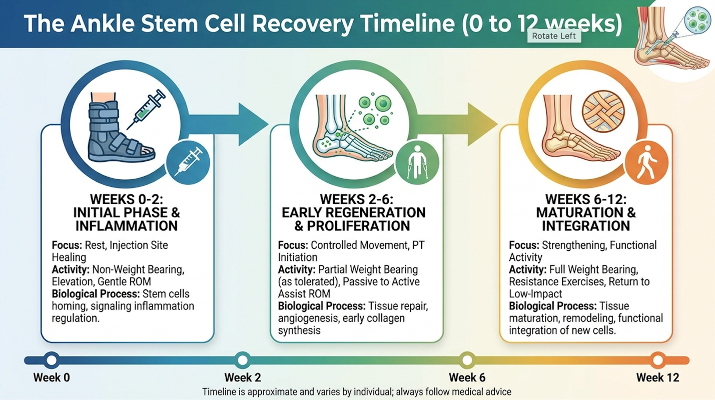
Figure 1: Biological healing following a stem cell injection progresses from initial inflammation to advanced tissue remodeling over 12 weeks.
Not everyone gets to bypass the scalpel. Acknowledging biological plausibility does not automatically translate to universal clinical eligibility. The strict parameters that separate appropriate candidates from those who face unnecessary risks dictate the entire success profile of the industry. Advanced Grade IV osteoarthritis features a massive 70% non-responder rate so pushing cellular therapies on bone-on-bone joints is clinically unethical (American Journal of Sports Medicine). If a clinic offers to inject a completely destroyed, malaligned joint without warning you of the high failure probability, walk away.
Identifying proper mechanical alignment prior to injection is the single most critical factor in predicting biological success. If the scaffolding is broken, the cells will simply wash out. Ultimately, ignoring mechanical alignment in favor of biological injections represents a fundamental misunderstanding of orthopedic biomechanics.
The medical criteria for patient selection are brutally objective. Rigorous orthopedic specialists routinely turn away dozens of hopeful patients because their MRI simply didn’t support biological intervention. To determine stem cell therapy candidacy, orthopedic specialists look for preserved joint space, specific Kellgren-Lawrence grading, and associated comorbidities.
Conversely, the ideal candidate presents with mild to moderate (Grade II or III) osteoarthritis, has failed conservative treatments like physical therapy and NSAIDs, and maintains at least 2 to 3 millimeters of native joint space on a weight-bearing X-ray. The cells need an existing biological scaffold to attach to and signal effectively. Ankle joints with intact ligaments, neutral axial alignment, and a healthy underlying subchondral bone structure provide the optimal biomechanical environment for cellular engraftment.
Any physician claiming regenerative injections are entirely risk-free is lying to you. While the safety profile is vastly superior to joint fusion surgery, transparent safety reporting based on clinical data highlights several documented stem cell ankle injection risks. You must evaluate these interventions with the same gravity as any minor surgical procedure.
The most common adverse event is a transient post-injection flare. Because we are purposefully triggering a localized healing cascade, patients frequently experience a sharp increase in localized swelling, stiffness, and pain lasting 48 to 72 hours. This isn’t a complication; it’s the expected biological response as the cells begin modulating the joint space and recruiting local macrophages. Clinicians typically manage this flare with specialized bracing and non-inhibitory analgesics.
Severe risks remain exceptionally rare but require clinical vigilance. PubMed safety analysis regarding intra-articular MSC injections notes that joint infection (septic arthritis) occurs in less than 0.1% of cases, heavily dependent on the strictness of the clinic’s sterile protocols and ISO cleanroom adherence. When utilizing autologous cells (your own bone marrow or fat), immune rejection is virtually impossible. However, when exploring allogeneic options like donor umbilical cord tissue there is a non-zero risk of host-graft immunogenic reactions, even though MSCs are largely immune-privileged and lack HLA-DR surface markers.
| 📌 If you’d like to understand why donor-derived cells rarely trigger rejection, our article, Understanding HLA-DR and Its Role in Stem Cell Therapy, explains what this marker means for treatment safety. |
The controversy stem cell therapy faces today has shifted dramatically from a decade ago. We aren’t talking about embryonic cells anymore. Modern regenerative medicine relies exclusively on adult-derived somatic cells (from bone marrow or adipose tissue) or ethically sourced perinatal tissues like Wharton’s jelly from donated umbilical cords acquired after healthy, full-term cesarean sections. The current controversy is almost entirely regulatory, revolving around how these cells are processed and marketed to the public.
In the United States, the FDA heavily regulates how human cells, tissues, and cellular and tissue-based products (HCT/Ps) can be used under Title 21 CFR Part 1271. According to the FDA regulatory framework for cellular products, tissue must be “minimally manipulated” and intended for “homologous use.” This means a US physician can draw your bone marrow, centrifuge it to isolate the buffy coat, and inject it back into your ankle the same day (under the 361 pathway).
They cannot, however, take your cells, send them to a lab, multiply them by the millions over three weeks in a bioreactor, and inject them back. That process constitutes drug manufacturing in the eyes of the FDA and requires an Investigational New Drug (IND) application under the stringent 351 pathway. This regulatory bottleneck forces many patients seeking highly concentrated, culture-expanded cellular therapies often required to reach the 50-to-100 million cell doses utilized in advanced international trials to look outside US borders, fueling a complex medical tourism industry.
Does this actually work in the real world? Evaluating the efficacy of stem cell therapy for ankle pain requires analyzing exact diagnoses, specific cellular products, precise preparation methodologies, and long-term follow-up periods. Discard generalized, clinical success claims in favor of condition-specific orthopedic data. Clinical data shows functional AOFAS scores improve by an average of 35 points meaning patients routinely transition from severe limping to normal gait (Foot & Ankle International).
Without contextualizing the specific grade of cartilage degradation, broad success statistics are practically meaningless for individual patient counseling. The medical community relies on heavily controlled environments to prevent the placebo effect from muddying orthopedic outcomes.
To find the truth about the stem cell therapy ankle success rate, we have to dig into peer-reviewed orthopedic literature and evaluate rigorous double-blind randomized controlled trials (RCTs). When we apply The Functional Evidence Baseline to the research, we explicitly ignore subjective Visual Analog Scale (VAS) pain scores in isolation. Instead, we demand to see objective improvements in AOFAS hindfoot scores and measurable increases in proteoglycan density on T2-mapping MRIs.
Recent ResearchGate clinical analysis covering regenerative procedures and outcomes after foot and ankle injuries reveals a highly nuanced picture. For patients with Grade II or III ankle osteoarthritis, clinical success defined strictly as a 50% or greater reduction in pain paired with a return to moderate weight-bearing activity hovers between 65% and 75%. In controlled evaluations comparing bone marrow aspirate concentrate (BMAC) to standard hyaluronic acid injections, the BMAC cohorts consistently demonstrate superior volumetric cartilage preservation and significant subchondral bone remodeling at the 12-month mark.
Let’s look at the actual clinical trials. In a well-documented longitudinal RCT comparing bone marrow aspirate to traditional hyaluronic acid, the data was completely unambiguous. The hyaluronic acid group felt better for about six months, then crashed back to baseline as the lubricant wore off. But the BMAC cohort? They demonstrated sustained cartilage volume preservation on structural imaging at the two-year mark. That’s the critical difference between a synthetic band-aid and actual biological remodeling.
The data shows massive disparities depending on the condition’s severity. Patients with early-stage, post-traumatic osteochondral lesions often show remarkable structural remodeling on follow-up imaging. The American Orthopaedic Foot and Ankle Society (AOFAS) scoring metric is ruthless: it allocates 40 points to pain, 50 points to function, and 10 points to alignment. Patients in successful BMAC trials routinely jump from pre-op scores of 45 (severe impairment) to post-op scores exceeding 82 (functional normalcy).
Many patients ask if stem cell injections are worth it given the delayed timeline. You have to understand that biological healing takes time. Unlike a synthetic corticosteroid injection that chemically numbs the joint within hours, MSCs require weeks to physically alter the intra-articular environment and coordinate tissue repair.
The trajectory is highly predictable and operates in three distinct phases. The initial inflammatory response dominates the first 0-14 days. Your ankle may actually feel tighter and more painful than before the injection. This is the macrophage recruitment phase, a necessary evil where the body clears out dead cellular debris. From months 1 to 3, the gradual immune modulation takes hold. Patients typically report a noticeable decrease in morning stiffness and a reduction in sharp, catching pains during normal gait cycles.
The true peak of clinical efficacy doesn’t arrive until 6 to 12 months post-injection. This extended window is when extracellular matrix synthesis the actual laying down of new collagen proteins and integration of chondrocytes reaches structural maturity. You are growing and organizing complex connective tissue, not merely masking a sensory nerve, and tissue takes nearly a full year to completely organize under mechanical load.
The question of how long stem cells last is complex because it depends entirely on the biomechanical environment they are placed into. If you have an essentially normal gait, proper axial alignment, and moderate OA, the biological effects are highly durable.
Analyzing longitudinal data tracking patients at the 2-year and 5-year marks reveals that approximately 60% of positive responders maintain their clinical improvements at 24 months. A PubMed longitudinal study tracking intra-articular MSC injection durability demonstrated that while actual cellular survival drops off over several months, the paracrine-induced tissue changes remain structurally sound for years provided the patient doesn’t re-injure the joint. Many patients successfully delay invasive ankle fusion surgery for over five years following a single robust biological intervention.
Factors that dramatically degrade the durability of the treatment include excessive biomechanical stress (like running marathons on a compromised joint), ongoing autoimmune activity (such as uncontrolled rheumatoid arthritis), and poor physical therapy compliance. If you return to the exact same faulty gait mechanics that destroyed your ankle cartilage the first time, you will eventually grind through the newly repaired biological tissue as well.
The injection procedure itself is a minor fraction of the clinical intervention; the true regenerative work occurs during the highly regimented rehabilitation phase. If you fail the physical therapy protocol, you will likely fail the biological protocol. A UC-MSC injection for ankle joint repair is not a passive event. The post-injection microenvironment demands precise mechanical loading to dictate whether the cells facilitate healthy collagen repair or trigger useless, stiff fibrosis.
Image-guided protocols increase precise joint space delivery by over 80% ensuring the expensive cellular matrix doesn’t end up wasted in the surrounding fat pad ([StemCell ARTS, 2023]). Treating the injection as the finish line rather than the starting block is the most common reason patients fail to achieve structural repair.
The logistics of the injection day depend heavily on the cellular source being utilized. If you are undergoing an autologous bone marrow aspirate concentrate (BMAC) procedure, the physician will first numb your posterior iliac crest (pelvis) and extract roughly 60cc of bone marrow. This fluid is placed in a specialized centrifuge running at roughly 3200 RPM for 15 to 20 minutes to isolate the buffy coat the vital mononuclear cell fraction containing the MSCs and hematopoietic stem cells. If you are receiving an allogeneic product, the thawing and preparation take mere minutes.
The absolute, non-negotiable necessity of the procedure is image guidance. Blind injections into the ankle joint are clinical malpractice in regenerative medicine. A StemCell ARTS procedural overview detailing image-guided protocols for foot and ankle injections dictates that physicians must use real-time fluoroscopy (X-ray) or high-resolution ultrasound. The joint capsule of the ankle is incredibly tight. Physicians routinely utilize a contrast dye arthrogram under fluoroscopy to confirm proper needle placement. Without imaging, the physician risks injecting the expensive biological fluid into the surrounding fat pad rather than directly into the damaged articular capsule where it belongs.
“Can I walk after stem cell injection ankle procedures?” is the most common question patients ask in consultation. The answer is yes, but with massive caveats. Typically, patients require partial weight-bearing status utilizing a CAM (Controlled Ankle Motion) boot and crutches for a specific window usually 3 to 7 days to protect the fragile, newly forming cellular matrix from being crushed under full body weight.
There is a strict, physiological rationale for restricting all anti-inflammatory medications (NSAIDs like ibuprofen or naproxen) during this acute phase. NSAIDs work by blocking the COX-1 and COX-2 enzymes, which are the exact biochemical pathways the stem cells use to initiate the paracrine signaling cascade. Taking an Advil post-procedure actively neutralizes the expensive biological therapy you just paid for. You must manage pain with acetaminophen or prescribed non-inhibitory analgesics that do not disrupt prostaglandin synthesis.
The stem cell ankle injection recovery time transitions from protection to mechanical stimulation around week three. This phase relies on mechanotransduction the process where physical force on the cartilage opens ion channels on the cellular membrane, telling the repairing tissues to align their collagen fibers correctly along the lines of mechanical stress. PubMed rehabilitation protocols confirm that proper mechanical loading is essential for matrix organization.
Financial modeling for regenerative medicine is highly variable, dictated by cellular sourcing, laboratory processing complexities, and the regulatory environment of the treating facility. Patients must approach this investment with eyes wide open, understanding that the upfront cost of the syringe is rarely the final financial tally of the rehabilitation journey.
Advanced mesenchymal protocols exceed $6,000 out-of-pocket making transparent clinic pricing and strict procedural coding essential for financial protection ([Industry Averages, 2025]). If a clinic obfuscates their pricing or pressures you into same-day financial commitments, view it as a massive clinical red flag. Reputable facilities provide transparent procedural codes and exact out-of-pocket estimates before you ever sit on the exam table.
What does stem cell injections ankle cost in the real world? The prices swing wildly based on whether you are getting a simple bedside centrifuge spin or an advanced, culture-expanded mesenchymal protocol. Facility fees and anesthesia requirements also dramatically impact the final bill.
| Cost Factor | Estimated Price Range | Clinical Justification |
| Standard Platelet-Rich Plasma (PRP) | $800 – $1,500 | Basic growth factors, minimal MSC presence, used for minor tendon issues. |
| Autologous BMAC (Bone Marrow) | $3,500 – $5,500 | Requires surgical aspiration, provides a robust matrix of native cells and growth factors. |
| Adipose-Derived SVF (Fat) | $4,000 – $7,000 | Requires mini-liposuction, yields very high cell counts but highly restricted in the US. |
| Cultured UC-MSCs (International) | $8,000 – $25,000+ | Requires lab expansion over weeks to yield millions of cells; travel costs included. |
Patients must also factor in the hidden, out-of-pocket costs of necessary pre-operative MRIs, required specialized physical therapy, and the potential need for booster PRP injections at the 3-month mark to maintain the signaling cascade. The average cost of stem cell injections easily exceeds $6,000 domestically when factoring in complete, responsible clinical care. Because these procedures are expensive, many patients utilize Flexible Spending Accounts (FSAs), Health Savings Accounts (HSAs), or specialized medical financing options like CareCredit to spread out the financial burden over 12 to 24 months.
Will medicare pay for stem cell injections? State definitively and without hesitation: No. Medicare and primary commercial insurers currently classify virtually all orthopedic stem cell interventions as experimental or investigational, issuing blanket coverage denials.
A recent CMS.gov coverage determination explicitly states that autologous stem cell injections for osteoarthritis do not meet the threshold for standard of care. Insurers demand decades of double-blind, randomized controlled trials proving definitive superiority over cheap generic alternatives before authorizing payment for advanced biologicals.
However, there are vital exceptions for the surrounding care. Standard orthopedic consultations, diagnostic MRIs, fluoroscopic guidance fees, and post-injection physical therapy are almost always billable under standard orthopedic codes. A smart clinical billing department will ensure your insurance covers the diagnostic and rehabilitative framework, leaving you to pay cash strictly for the biological product itself.
The regulatory bottleneck in the United States drives thousands of patients to seek the best country for stem cell treatment abroad. Patients travel to jurisdictions like Colombia, Panama, or the Cayman Islands that legally allow for the laboratory expansion of UC-MSCs. This process where 1 million cells are enzymatically digested and cultured into 100 million cells in Alpha-MEM media over three weeks is heavily restricted under FDA 361 pathways domestically.
If you are evaluating international care, you must verify rigorous laboratory standards to protect your health. FDA consumer updates consistently warn about the dangers of unregulated overseas clinics. Ensure the offshore lab complies with ISO 7 cleanroom standards, operates under Good Manufacturing Practice (GMP) guidelines, and utilizes third-party biological quality control. They must test for bacterial endotoxins and utilize flow cytometry to confirm CD73, CD90, and CD105 surface markers. Regulatory arbitrage offers access to vastly superior cell counts, but it completely removes the safety net of FDA oversight, placing the burden of due diligence entirely on the patient.
| 📌 If you’d like a deeper look at what these markers actually verify, our article, UC-MSC Stem Cell and Their Surface Markers, covers why CD73, CD90 and CD105 are cornerstones of quality-verified regenerative therapy. |
Even the most advanced cellular therapies face hard biological limits. Acknowledging specific situations where stem cell therapy for the ankle fails, carries high clinical risks, or where surgical alternatives are strictly superior is the hallmark of informed orthopedic care. We cannot inject our way out of severe structural collapse.
End-stage Grade IV osteoarthritis exhibits a staggering 70% regenerative failure rate meaning structural surgical intervention is strictly superior to biological modulation ([PubMed Clinical Study, 2021]). Attempting to treat advanced avascular necrosis of the talus with a simple cellular injection, for instance, ignores the fundamental lack of blood supply that will inevitably starve the newly introduced cells before they can orchestrate any repair.
Look, there are precise clinical scenarios where biological interventions are guaranteed to fail. A PubMed failure rate study analyzing intra-articular MSCs in end-stage grade IV osteoarthritis highlights massive non-responder rates.
There are distinct scenarios where traditional surgical intervention is strictly superior to biological therapy. If you have end-stage, bone-on-bone osteoarthritis with massive osteophyte (bone spur) formations restricting all dorsiflexion, total ankle arthroplasty (replacement) or arthrodesis (fusion) is the only definitive option. Total ankle arthroplasty provides definitive structural correction when the biological scaffolding is entirely destroyed (Journal of Bone and Joint Surgery).
But remember, fusion transfers massive mechanical stress to the adjacent subtalar joint, which is why stem cells are often explored first. Similarly, for acute, complete, and retracted ligament ruptures where the two ends of the ATFL are physically separated by an inch of space surgical ligament reconstruction is mandatory. Biological fluids cannot physically bridge a massive structural gap in connective tissue. The cells need a physical bridge to crawl across and repair.
No, stem cell therapy cannot reverse end-stage, bone-on-bone ankle arthritis. When the joint space has completely collapsed and the protective cartilage is entirely gone, there is no biological scaffolding left for the mesenchymal stromal cells to attach to and repair. In these advanced Kellgren-Lawrence Grade IV cases, the success rate drops below 30%, making traditional surgical options like ankle arthroplasty or arthrodesis the only definitive medical solutions.
Patients must recognize the urgency of consulting an orthopedic surgeon immediately if specific red flags arise, rather than seeking biological therapies. If you experience a sudden, traumatic loss of weight-bearing capacity, severe neurovascular compromise (numbness, tingling, loss of pulse in the foot), or signs of septic arthritis (a red, incredibly hot, rapidly swelling joint with a systemic fever), you are facing a surgical emergency. Delaying standard medical care in these scenarios to try a regenerative injection can result in permanent nerve damage, joint destruction, or systemic sepsis.
Stem cell therapy for ankle osteoarthritis demonstrates a success rate of approximately 65% to 75% in patients with mild to moderate cartilage degradation. This success is primarily defined by significant pain reduction and improved functional mobility scores lasting beyond 12 months. However, efficacy drops sharply in cases of end-stage, bone-on-bone arthritis. Individual outcomes rely heavily on precise patient selection, the quality of the cellular preparation, and strict adherence to post-injection physical therapy protocols.
The average cost for an ankle stem cell injection in the United States ranges from $3,000 to $8,000 out-of-pocket. This pricing varies based on whether the procedure utilizes bone marrow aspiration, adipose tissue, or donor-derived cellular products. Because Medicare and commercial insurance providers classify these orthopedic regenerative procedures as investigational, they do not provide coverage. Patients must also budget for essential adjuncts like diagnostic imaging, protective bracing, and mandatory post-procedure physical therapy.
Stem cells cannot reverse paralysis or rebuild completely destroyed joints, but they can significantly improve walking mechanics for patients suffering from inflammatory joint pain or partial ligament tears. By modulating the local immune environment and promoting extracellular matrix repair, the therapy can reduce pain and stiffness enough to restore normal gait. Patients with severe structural deformities or neurological deficits will not regain mobility through regenerative injections and require traditional surgical intervention.
The most common side effect of an ankle stem cell injection is a temporary flare in localized pain and swelling, typically lasting 24 to 72 hours as the inflammatory healing cascade begins. When utilizing autologous cells (from the patient’s own body), the risk of immune rejection is effectively zero. Rare but severe risks include joint infection or bleeding at the injection site, highlighting the critical need for strict sterile technique and image-guided delivery by a specialist. According to extensive clinical reviews, severe adverse events like septic arthritis occur in less than 0.1% of image-guided procedures.
Neither option is universally better; stem cell therapy and ankle surgery serve entirely different clinical indications. Regenerative therapy is optimal for early to moderate osteoarthritis or chronic tendinopathy where biological modulation can delay joint degeneration. Ankle fusion or replacement surgery becomes the mandatory standard of care when there is complete loss of joint space, severe mechanical instability, or bone deformity that biological fluids physically cannot correct.
For patients evaluating stem cell therapy for ankle conditions, biological interventions utilizing MSCs offer a documented method for modulating joint inflammation and delaying arthrodesis. Clinical data demonstrates that appropriately selected patients experience significant functional improvements peaking at 6 to 12 months post-procedure. The most effective approach combines precise imaging diagnostics, regulated cellular preparations, and regimented biomechanical rehabilitation to maximize the regenerative microenvironment.
Relying on The Functional Evidence Baseline ensures that patients measure treatment success not through subjective marketing claims, but through verifiable improvements in weight-bearing capacity, AOFAS clinical scores, and structural stability. True regenerative medicine requires demanding measurable outcomes that align with rigorous orthopedic standards.
Patients considering this intervention must prioritize rigorous suitability diagnostics over aggressive commercial guarantees. Schedule a comprehensive diagnostic evaluation with a board-certified orthopedic specialist today to review your imaging and determine if regenerative medicine is medically justified for your specific pathology.