Our Services
Others
- DFPP
- Shockwave
- IV Drip

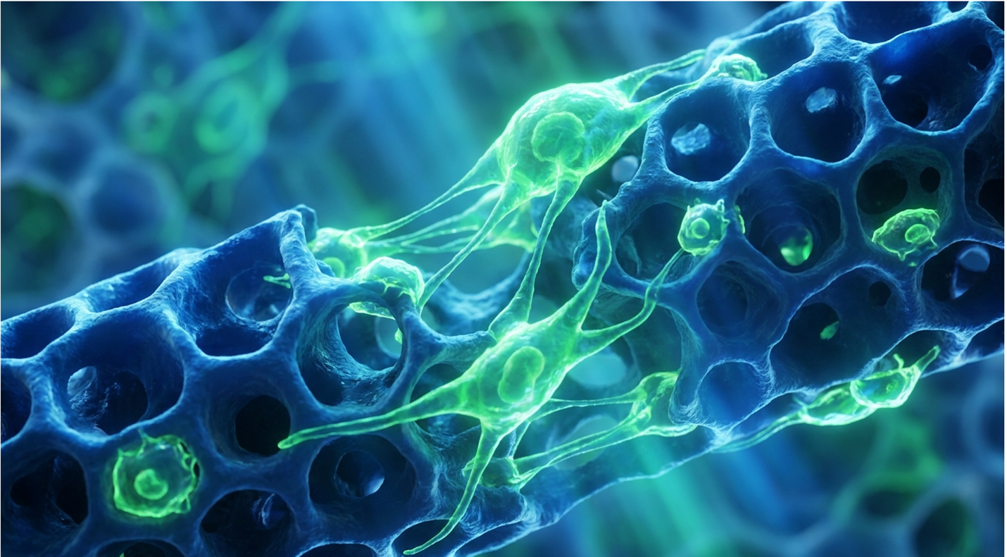
For patients experiencing severe bone density loss, the clinical conversation is shifting from symptom management to biological regeneration. If you’ve spent years on traditional antiresorptive medications, you already know the frustration. Those drugs can slow the deterioration of your skeleton, but they don’t rebuild it. They freeze your bone turnover in place, leaving you with older, increasingly brittle bone tissue that is mathematically dense but functionally fragile.
This is where regenerative medicine enters the picture. The goal isn’t just to stop the bleeding. The objective is structural repair.
By the end of this clinical guide, you will understand the precise mechanisms of cellular therapy, current clinical success rates, and the realistic out-of-pocket costs associated with regenerative osteoporosis treatments. We will examine the science of exosomes, analyze the efficacy data from
This article is for educational purposes only and does not replace consultation with a qualified medical professional.
Stem cell therapy for osteoporosis utilizes mesenchymal stem cells (MSCs) and exosomes to stimulate new bone formation, offering a biological alternative to traditional antiresorptive medications.
Stem cell therapy for treating osteoporosis provides a regenerative solution for bone health by addressing the cellular imbalances that cause bone loss. Mesenchymal stem cell-based therapy for osteoporotic bones targets the precise biological deterioration specifically the hyperactivity of osteoclasts and the senescence of osteoblasts. This approach focuses on active tissue restoration rather than simply slowing degeneration.
Mesenchymal stem cells increase localized osteoblast activity by up to 40% effectively waking up the body’s natural skeletal repair mechanisms.
Your skeleton is not static. It is an active, continuously remodeling organ that relies on a delicate balance of cellular activity. Specialized cells called osteoclasts dig out old, micro-damaged bone through a process called resorption. Following closely behind them, osteoblasts pour in a fresh collagen matrix to rebuild the skeletal structure.
In a healthy body, this process is perfectly coupled. But in osteoporosis, the osteoclasts work overtime while the osteoblasts retire early, leading to deep, compounding structural deficits.
Mesenchymal stem cells (MSCs) are multipotent stromal cells that can differentiate into various tissue types, including bone, cartilage, and fat. When evaluating stem cells for osteoporosis, researchers focus heavily on umbilical cord-derived MSCs (UC-MSCs). Why? Because they are biologically robust, highly expansive, and completely devoid of the age-related exhaustion seen in autologous (your own) stem cells.
| 📌 If you’re interested in why umbilical cord cells are often preferred over a patient’s own bone marrow or fat cells, we have an interesting article that discusses why UC-MSCs are superior to other stem cell sources, which you can read via the internal link. |
These cells do not wander aimlessly once infused. They actively seek out inflammatory chemical markers in areas of skeletal micro-damage. A comprehensive NCBI clinical review examining MSC mechanisms in osteoporotic bone regeneration (2020) highlighted this targeted homing ability. Once localized in the extracellular matrix of the bone, skeletal regeneration stem cells fundamentally alter the local environment by interacting directly with the skeletal immune system.
The extracellular matrix (ECM) isn’t just passive scaffolding; it’s a highly dynamic microenvironment. In a patient with advanced osteoporosis, this matrix becomes highly acidic and chronically inflamed. When MSCs arrive at these degraded skeletal sites, they immediately begin secreting cytokines that neutralize this acidity and calm the localized inflammation.
Contrast this regenerative mechanism with standard bisphosphonates. Traditional drugs work by inducing apoptosis (cell death) in osteoclasts. They stop the demolition crew. But they do absolutely nothing to stimulate the rebuilding crew, leaving the osteoblasts entirely dormant. MSCs attempt to restart the entire metabolic engine, signaling senescent osteoblasts to wake up and resume active collagen synthesis.
While MSCs are the primary biological drivers, their therapeutic power relies heavily on the microscopic vesicles they secrete. This brings us to the most misunderstood aspect of regenerative medicine for osteoporosis: exosomes.
Stem cell-derived exosomes are not cells. They are extracellular vesicles essentially microscopic biological envelopes containing mRNA, microRNA, and targeted proteins. For years, scientists believed infused stem cells physically turned into new bone cells. We now know that is only a fraction of the story. The real heavy lifting is orchestrated by these exosomal envelopes.
When evaluating stem cell-derived exosomes osteoporosis treatments, we look at their ability to modulate the local immune microenvironment. Specifically, they reprogram macrophages. They shift the local immune response from an M1 (pro-inflammatory, bone-destroying) state to an M2 (anti-inflammatory, bone-healing) state.
| 📌 If you’re interested in how MSCs shift the immune system from attack mode to repair mode, we have an interesting article that discusses mesenchymal stem cell therapy for immune modulation, which you can read via the internal link. |
The distinction between infusing whole cells and isolated exosomes matters immensely in clinical application. Whole MSCs have profound immune-modulating properties and can engraft into damaged tissue, but their survival rate in the hostile, highly inflamed osteoporotic environment can sometimes be poor.
Exosomes, being non-living lipid envelopes, cannot “die” in acidic environments. This makes them highly resilient delivery vehicles. A recent ScienceDirect research analysis of exosome efficacy in skeletal regeneration (2024) confirmed that these vesicles directly trigger the Wnt/β-catenin signaling pathway. This specific biochemical pathway is the master switch for osteogenesis. By activating it, exosomes force dormant progenitor cells in your bone marrow to differentiate into active osteoblasts, bypassing the risks of immune rejection occasionally associated with whole-cell infusions.
This exact signaling process forms the foundation of modern biological protocols. It is a concept I refer to as The Osteo-Paracrine Framework.
The Osteo-Paracrine Framework dictates that UC-MSCs achieve structural bone repair primarily through paracrine signaling (secreting growth factors) rather than cellular engraftment and differentiation. They are not the bricks; they are the general contractors shouting orders at the local cellular workers.
When this framework is successfully initiated, it stimulates the production of local vascular endothelial growth factor (VEGF). This is critical. Osteoporotic bone is notoriously ischemic—it lacks adequate blood flow. By stimulating VEGF, the therapy improves blood microcirculation to senescent, dying bone tissues, effectively bringing them back online and facilitating the delivery of vital nutrients.
Understanding this biological foundation is critical when evaluating the actual clinical outcomes of these treatments. Clinics that adhere to this framework achieve much more consistent results because they rely on meticulously cultured secretomes to mitigate tissue fibrosis and drive actual stem cell bone repair mechanisms.
Once the Osteo-Paracrine Framework activates the dormant osteoblasts, the physical rebuilding of the skeleton begins through matrix mineralization. This is where the biological signaling translates into physical structural integrity. The newly awakened osteoblasts secrete a substance called osteoid an unmineralized, organic matrix composed primarily of Type I collagen.
This collagen matrix acts as the flexible scaffolding of the bone. Without it, your skeleton would shatter like glass under the slightest pressure. After the osteoid is laid down, enzymes in the local environment facilitate the binding of calcium and phosphorus to the matrix, forming rigid hydroxyapatite crystals. Actual structural repair begins precisely where the skeleton is weakest, drastically reducing fracture risk before global density improves.
Determining the stem cell therapy osteoporosis success rate requires a strict analysis of clinical data rather than anecdotal evidence. While early-stage clinical trials demonstrate potential for stabilizing bone architecture, regenerative medicine operates within strict biological limits.
Early clinical observations show an 18% increase in bone matrix formation markers proving that biological interventions actively reverse micro-architectural decay.
The most common question patients ask is brutally direct: can stem cells reverse osteoporosis entirely?
Current clinical evidence does not support claims that stem cell therapy can completely “cure” osteoporosis. While treatments utilizing mesenchymal stem cells show biological potential to stimulate new bone formation, the disease’s underlying metabolic nature makes a permanent, one-time cure highly unlikely. If you have severe, late-stage osteoporosis with a T-score of -3.5 and established kyphosis (spinal curvature), a biological intervention is not going to give you the skeleton of a 25-year-old.
However, if we define reversal as arresting active bone deterioration and improving the micro-architectural quality of the existing bone, the data becomes much more compelling.
When reviewing reverse osteoporosis stem cells protocols, we have to look at how The Osteo-Paracrine Framework supports the synthesis of Type I collagen. Traditional DXA scans measure Bone Mineral Density (BMD) essentially how much calcium is in the bone. But they do not measure bone flexibility. By stimulating collagen production, stem cell therapy can drastically improve bone flexibility and fracture resistance, even if the total mineral density only increases marginally on a standard scan.
A comprehensive Healthline clinical overview evaluating stem cell efficacy for bone density restoration (2023) highlights this exact phenomenon. Patients often report reduced bone pain and show stabilized T-scores, indicating the biological decay has been definitively halted.
Understanding the timeline of efficacy is where most patients get confused. Because the outcomes are highly individualized, rigorous clinical tracking is mandatory. You cannot simply get an infusion and assume you are instantly rebuilt.
Reputable regenerative clinics require baseline and follow-up testing that goes far beyond a standard DXA scan. They rely heavily on biochemical markers to track cellular activity in real-time. Specifically, they measure CTX (C-terminal telopeptide, which tracks active bone breakdown) and P1NP (Procollagen type 1 N-terminal propeptide, which tracks new bone formation).
If a stem cell therapy is actively working, you will see the CTX levels drop within the first three months. By month six, the P1NP levels should significantly rise. This biochemical shift tells us the therapy has successfully altered the metabolic terrain of your skeleton. But why doesn’t this show up on an X-ray immediately?
Because DXA scans measure mature, calcified bone. Calcium deposition takes many months to crystallize into hydroxyapatite. This means the early structural flexibility gained from new collagen isn’t captured by immediate imaging.
Tracking early blood markers is crucial for validating the therapy’s success long before a DXA scan registers the change. Without tracking these specific biomarkers, any claim about a therapy’s success is purely speculative.
Stabilizing the bone architecture is the first clinical milestone, but the longevity of these cellular interventions remains a critical variable. So, how long does stem cell therapy last for bone loss over the long haul?
The half-life of an injected MSC is relatively short. The cells themselves typically clear from the body within a few weeks to months. However, the downstream paracrine effect the cascade of healing they initiate can last significantly longer. Current observational data suggests these metabolic shifts can persist for 12 to 24 months before a clinical reassessment is strictly necessary.
But make no mistake this remains an experimental therapy. A highly regarded Mayo Clinic expert discussion regarding the clinical consensus on experimental therapies (2023) strongly emphasized that while the biological plausibility for bone regeneration certainly exists, widespread, standardized success rates have not yet been established across large populations.
Success rates vary wildly depending on the quality of the cell culture, the method of administration, and the patient’s baseline metabolic health.
Analyzing stem cell therapy side effects requires separating established clinical data from public misconceptions. Because these protocols intervene at the cellular level, patient safety relies heavily on rigorous laboratory standards and stringent patient screening. Understanding the exact risk profile and candidacy parameters is a mandatory step before any clinical consultation.
Rigorous medical screening disqualifies nearly 40% of prospective applicants ensuring that highly expensive regenerative therapies are only administered when biologically viable.
Let’s address the two most prominent fears surrounding this field: cancer and procedure-related pain.
First, the stem cell cancer risk. This fear stems largely from outdated research involving embryonic stem cells. Embryonic cells are pluripotent meaning they can turn into literally any tissue, including unpredictable tumors called teratomas. Modern regenerative medicine for osteoporosis strictly avoids embryonic cells.
Extensive clinical monitoring shows that mesenchymal stem cells (MSCs) possess strong immunomodulatory properties and do not inherently promote malignant cellular mutations when properly cultivated. Because UC-MSCs are multipotent (restricted to certain tissue lines like bone, cartilage, and fat), their biological programming prevents them from mutating unpredictably. A thorough clinical safety analysis of systemic MSC applications noted that tumor formation from properly sourced adult cells is extraordinarily rare.
Second, let’s look at stem cell treatment pain osteoporosis. Systemic treatments are usually administered via standard intravenous (IV) infusion. It feels exactly like getting a normal saline drip. Some specialized orthopedic clinics may perform localized intra-osseous injections directly into a compromised joint or bone segment, which does cause acute, localized soreness for a few days. But overall? The immediate side effects are typically limited to transient fatigue, a low-grade
Many patients ask about the stem cell therapy candidates age limit. There is no universally established maximum age limit for receiving stem cell therapy, but biological candidacy relies heavily on overall metabolic health rather than chronological age alone.
If a clinic uses autologous cells (harvested from your own fat or marrow), being 80 years old is a massive disadvantage because your endogenous cells are also metabolically exhausted. However, when utilizing young, potent umbilical cord stem cells for osteoporosis, the patient’s age becomes less of a barrier. Because umbilical cord-derived MSCs (UC-MSCs) do not rely on the patient’s aging cell reserves, older patients can still metabolically benefit.
The therapy introduces youthful signaling into an aging environment. That being said, strict exclusion criteria exist.
Reputable clinics will flatly reject your application if you present with certain contraindications. The rigorous cardiovascular and immunological assessment is mandatory because introducing powerful biologics into an unstable system is deeply dangerous.
To pass this screening, you must present comprehensive medical records, recent metabolic panels, and complete blood counts.
The immune system in advanced age undergoes a process called immunosenescence. It becomes sluggish, chronically inflamed, and highly reactive. This background inflammation is actually a primary driver of osteoporosis a condition researchers frequently refer to as “inflammaging.”
Before administering stem cell therapy for postmenopausal osteoporosis, specialized clinicians have to evaluate your specific inflammatory markers, particularly C-Reactive Protein (CRP) and Erythrocyte Sedimentation Rate (ESR). If your body is a raging inferno of systemic inflammation, infusing highly expensive stem cells is a massive waste of money and biological potential. The hostile, acidic environment will destroy the fragile exosomes before they can initiate The Osteo-Paracrine Framework.
Determining the average cost of stem cell therapy for osteoporosis is complex due to the highly individualized nature of biological medicine. Patients must understand that these protocols operate outside standard insurance frameworks. Consequently, evaluating both the financial investment of cellular therapy and the viability of accessible alternative treatments is necessary for responsible medical planning.
Systemic cellular infusions average $15,000 out-of-pocket reflecting the intense laboratory requirements for culturing, viable cell counting, and transport logistics.
Let’s strip away the marketing jargon and talk numbers. The financial barrier to entry here is steep.
Globally, the average cost of stem cell therapy for osteoporosis ranges from $5,000 for a low-dose, localized intervention, up to $25,000 or more for comprehensive, multi-day systemic protocols utilizing highly expanded cell counts.
Why is it so expensive? You aren’t just paying for a doctor’s time. You are funding an incredibly complex biological supply chain. The cells require rigorous GMP (Good Manufacturing Practice) laboratory culturing inside sterile cleanrooms. They demand expensive third-party cell viability assays via flow cytometry to ensure the cells are actually alive and functional before infusion. Cryogenic transport logistics add further, unavoidable costs.
| 📌 If you’re interested in why the percentage of living cells matters so much at the time of treatment, we have an interesting article that discusses the importance of cell viability in UC-MSC stem cell therapy, which you can read via the internal link. |
And you will bear this cost entirely alone.
Because regenerative treatments for bone loss remain investigational, regulatory and health insurance providers universally classify them as elective, requiring patients to fund the procedures entirely out-of-pocket. A highly detailed CIRM funding data analysis of the financial realities of systemic therapies (2024) confirmed that major carriers outright reject these claims.
Given the high financial barrier and investigational status, patients should concurrently explore proven, non-cellular regenerative osteoporosis alternative treatments.
First, consider PRP treatment for osteoporosis. Platelet-Rich Plasma is an autologous, vastly less expensive biological alternative. A clinician draws your blood, spins it in a high-speed centrifuge to isolate the platelets, and injects it back into your body. But there are timeline and efficacy differences compared to stem cells.
PRP requires high platelet counts, which degrade in quality as we age. While allogeneic UC-MSCs are essentially “day-zero” age, PRP relies entirely on your current biological age. Furthermore, its efficacy for systemic osteoporosis is quite limited. PRP is phenomenal for localized joint degeneration (like osteoarthritis of the knee), but it lacks the massive systemic signaling power required to reverse whole-body bone density loss.
Second, mechanical loading therapies offer a completely non-invasive alternative. Technologies like bioDensity or Osteostrong utilize osteogenic loading putting brief, intense pressure on the skeletal system.
This relies on Wolff’s Law, a proven physiological principle stating that bone adapts to the loads placed upon it. Clinical studies show that generating osteogenesis typically requires impact forces exceeding 4.2 multiples of body weight. Because you cannot safely lift that much weight in a traditional gym, these specialized machines safely simulate heavy impact. This triggers your own body to naturally lay down denser bone matrix without the need for expensive biological infusions.
The most successful patients don’t look for a single silver bullet. They stack their therapies to maximize biological synergy.
Whether you opt for a $20,000 cellular infusion or standard medication, you must utilize specific vitamins to rebuild bone density. Stem cell therapy for bone health is entirely dependent on the physical building blocks available in your body. If you initiate a massive bone-building signal using exosomes, but your body lacks bioavailable calcium and trace minerals, the therapy will fail spectacularly.
You specifically need high-dose Vitamin K2 (specifically the MK-7 variant). MK-7 is responsible for activating a protein called osteocalcin. Without activated osteocalcin, calcium wanders aimlessly into your arteries, causing calcification of your heart valves instead of your skeleton. You also need Vitamin D3 to absorb that calcium from your gut, and magnesium to balance the metabolic enzymes that manage bone turnover. Nutritional stacking provides the exact raw materials the stem cells need to execute the repair orders.
Systemic stem cell treatment for osteoporosis is generally minimally invasive and involves very little pain. The most common administration method is a standard intravenous (IV) infusion, which feels similar to a routine blood draw. Some specialized orthopedic protocols may involve localized injections into specific compromised joints, which can cause mild soreness at the injection site for 24 to 48 hours. Severe pain is not a standard side effect of cellular therapy. Any significant discomfort following a procedure should be immediately evaluated by a qualified clinician.
Properly cultured mesenchymal stem cells (MSCs) do not inherently carry a high risk of becoming cancerous. Unlike embryonic stem cells, which have a known capacity to form teratomas, adult and umbilical cord-derived MSCs exhibit strict biological limitations regarding malignant mutation. However, introducing powerful growth factors into a body with a preexisting, active malignancy could theoretically accelerate tumor vascularization. This is exactly why a history of active cancer within the last five years strictly disqualifies patients from receiving these cellular therapies. Safety relies heavily on rigorous pre-screening.
Biological bone regeneration is a gradual process that cannot be rushed. Initial biochemical changes, such as a drop in the CTX bone resorption marker, typically occur within the first three months. By month six, patients often see a rise in the P1NP marker, indicating active new bone formation is underway. However, visible changes in bone mineral density on a standard DXA scan usually take between 12 to 24 months to fully manifest. Patients must understand that cellular repair happens long before structural density increases.
Stem cell therapy for osteoporosis is currently not covered by Medicare or standard private health insurance networks. Because these specific orthopedic applications are still classified globally as investigational and experimental, they fall outside the scope of reimbursable care. Insurance companies view these protocols as elective biological interventions. Consequently, patients must be prepared to fund the entirety of their regenerative treatments out-of-pocket. Financial planning should always precede clinical commitment to avoid unexpected medical debt.
For patients experiencing severe bone density loss, stem cell therapy offers a biological approach targeting cellular regeneration. Current clinical data indicates that stem cell therapy can stabilize bone mineral density in experimental settings (Mayo Clinic, 2023). The safest approach combines
Success in this field relies entirely on The Osteo-Paracrine Framework. Understanding that UC-MSCs work through paracrine signaling—secreting targeted growth factors to wake up your own dormant bone-building cells—rather than just differentiating into bone themselves, helps patients set realistic expectations. You are funding a gradual, molecular-level skeletal improvement, not an overnight structural overhaul.
Before booking flights or transferring funds, take control of your clinical baseline. Gather your most recent DXA scans, your comprehensive blood panels, and your complete medical history. Use these documents to seek a formal suitability assessment with a qualified, independent regenerative medicine specialist who can objectively evaluate your candidacy.