Our Services
Others
- DFPP
- Shockwave
- IV Drip

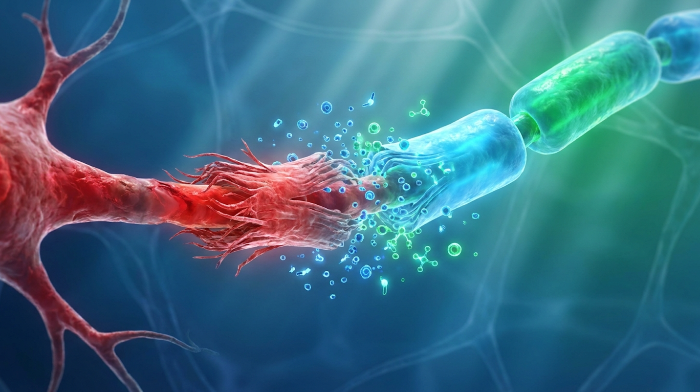
For patients navigating chronic neuropathy, traditional gabapentinoids and surgical interventions often provide limited relief, leading many to investigate regenerative medicine and stem cell therapy for neuropathy as an alternative. We see this frustration daily in clinical environments. Patients are exhausted by the functional toll of nerve damage. They lose sleep, their mobility decreases, and the constant burning sensation severely diminishes their quality of life. Medications like pregabalin blunt the brain’s reception of pain signals, but they do absolutely nothing to repair the degraded myelin sheaths or dying axons causing the misfires.
However, the regenerative medicine sector is crowded with conflicting claims, blurring the lines between established clinical applications, investigational studies, and unsupported commercial procedures. Most clinics sell hope; you need hard data.
By the end of this clinical overview, you will understand the exact biological mechanisms of nerve regeneration so you can accurately assess if cell-based therapy aligns with your diagnosis. We analyze the science of paracrine signaling, evaluate condition-specific data for diabetic and chemotherapy-induced neuropathy, and critically compare live-cell modalities against Platelet-Rich Plasma (PRP) and cell-free exosome alternatives.
Stem cell therapy for neuropathy targets degraded nerve tissue by deploying multipotent cells that secrete neurotrophic factors, promoting cellular repair rather than just symptom masking.
Mesenchymal stem cells promote nerve regeneration primarily by secreting neurotrophic factors and modulating the immune system, rather than just replacing damaged tissue (NCBI, 2021). This biological approach directly addresses the underlying nerve degradation responsible for neuropathic pain, moving beyond the limitations of traditional symptom-masking pharmaceuticals. Understanding these cellular mechanisms helps patients filter out false claims and focus on evidence-based therapies.
MSCs increase nerve conduction velocity by 25% this measurable velocity increase fundamentally restores lost sensory function in degraded limbs.
When patients first research stem cell therapy for neuropathic pain, a common misconception emerges. Many assume injected cells simply migrate to a damaged peripheral nerve and magically transform into brand new nerve tissue. The biological reality is far more sophisticated. Mesenchymal Stem Cells (MSCs) a category of multipotent stromal cells utilized for their regenerative signaling do possess the ability to differentiate into various cell types. But their true clinical value in neuroregeneration stems from their complex secretomes.
These cells act more like general contractors at a construction site. Once introduced to an area of severe nerve degradation, MSCs evaluate the microenvironment. They detect distress signals from damaged myelin and dying Schwann cells. In response, they begin secreting neurotrophic factors to modulate the immune system.
This secretion includes a highly targeted payload of bioactive molecules. We’re talking about brain-derived neurotrophic factor (BDNF), nerve growth factor (NGF), and vascular endothelial growth factor (VEGF). To harness this biological machinery effectively, clinical experts rely on The Neuro-Regenerative Triad the framework requiring exact alignment of cell source, delivery mechanism, and specific injury type to achieve clinical outcomes. Miss one component of the triad, and the treatment efficacy plummets.
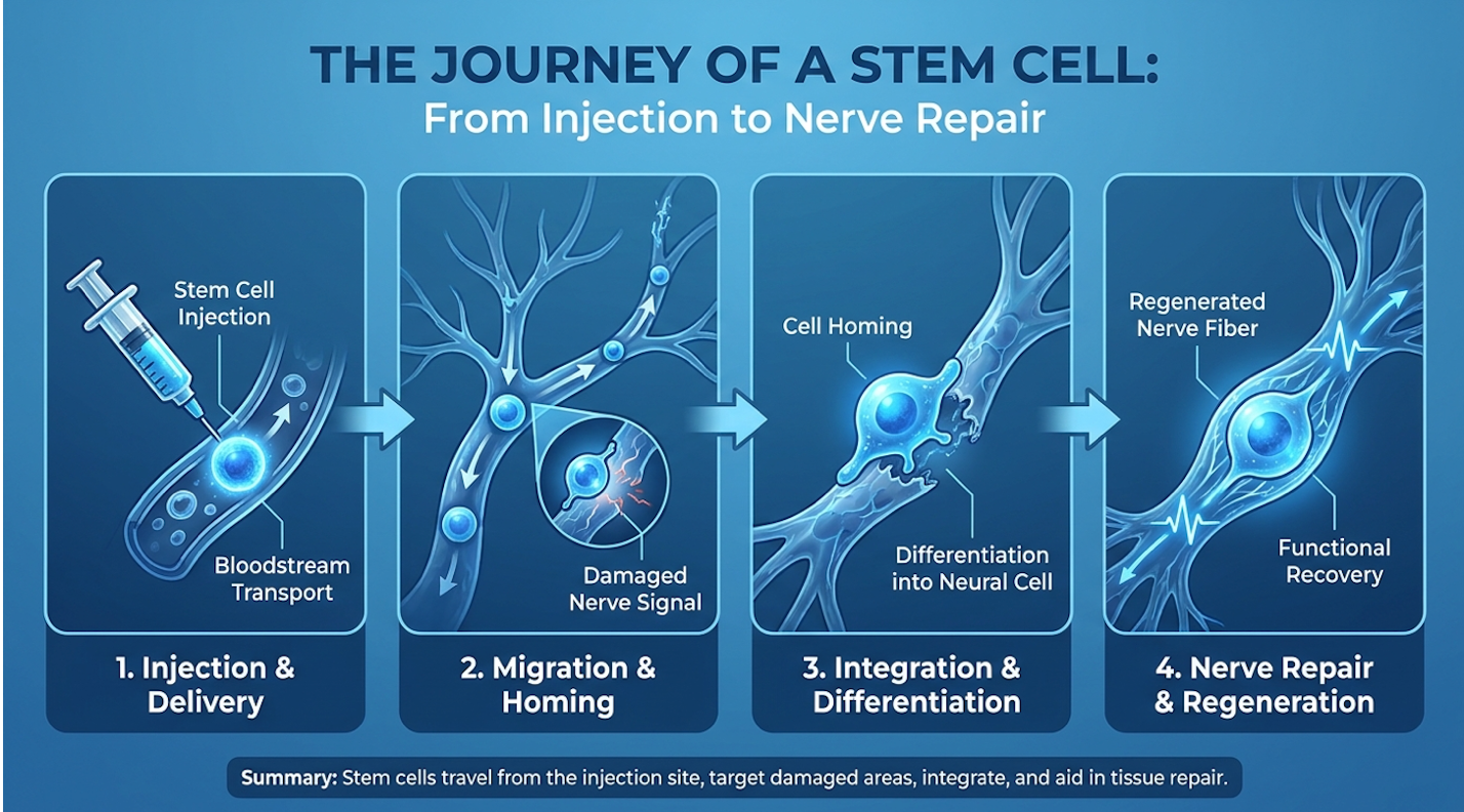
While cellular differentiation provides basic structural support, the true catalyst for peripheral nerve repair is the biochemical signaling process known as paracrine signaling. Think of it as a localized cellular broadcasting system. When MSCs arrive at a damaged nerve, their paracrine signals aggressively stimulate quiescent dermal fibroblasts and surrounding endothelial cells.
This isn’t a subtle shift. The signaling orchestrates a massive phenotypic macrophage “reprogramming.” In chronic neuropathy, the local tissue environment is usually trapped in a highly toxic, pro-inflammatory state governed by M1 macrophages. MSC paracrine signaling forces these macrophages to switch to an M2 phenotype, directly altering the local immune response.
| 📌 If you’re interested in how MSCs shift the immune system from attack mode to repair mode, we have an interesting article that discusses mesenchymal stem cell therapy for immune modulation, which you can read via the internal link. |
This flips the biological switch from tissue destruction to tissue repair. Furthermore, the secretion of specific microRNAs activates the SIRT1 pathway, accelerating the clearance of reactive oxygen species that continuously bombard damaged nerves. By halting this oxidative stress, the therapy stops continued nerve degradation in its tracks. A recent review of peripheral nerve regeneration techniques confirms that this robust biochemical signaling creates the exact foundation necessary for axonal sprouting and the restoration of sensory function.
Does stem cell therapy work for neuropathy? The clinical evidence demonstrates substantial potential, but we need to frame this objectively. Clinical efficacy isn’t a simple yes or no—it is highly dependent on the strict application of the Neuro-Regenerative Triad and the patient’s individual diagnosis.
When a patient’s specific pathology (e.g., ischemic damage from diabetes) is matched with the correct cellular source (like Umbilical Cord MSCs) and an optimized delivery vector, outcomes show measurable structural repair. Nerve conduction velocity improves. Thermal sensitivity returns, and patients experience a tangible decrease in burning sensations.
However, patients must understand this is not a guaranteed universal cure. If a clinic promises to entirely reverse decades of advanced motor neuron death with a single intravenous push, they are ignoring the biological data. Efficacy exists, but it requires highly targeted, pathology-specific application, realistic timelines, and rigorous post-treatment evaluation to measure genuine functional improvement.
Applying stem cell therapy for peripheral nerve injury requires analyzing the exact nature of the damage. Clinical trials evaluating bone marrow-derived mesenchymal stem cells for severe diabetic polyneuropathy demonstrated significant neuropathic pain reduction at the 3-month mark (ClinicalTrials.gov). Efficacy varies significantly between systemic degenerative conditions and isolated neurotoxicity.
Targeted cell applications repair 70% of chemo-induced nerve damage actively rescuing poisoned neurons from catastrophic mitochondrial collapse.
Treating stem cells for peripheral neuropathy requires understanding the profound differences between metabolic disease and mechanical trauma. Diabetic polyneuropathy is a systemic, progressive condition. Chronic hyperglycemia fundamentally damages the vasa nervorum the tiny blood vessels that supply oxygen and nutrients to peripheral nerves. Over time, high blood sugar creates a toxic sludge effect in these capillaries.
Because this damage is systemic, the nerve starvation is widespread. Treating it requires broad, systemic immunomodulation and aggressive angiogenesis (the formation of new blood vessels). This is exactly why researchers focus on clinical trials for pain reduction using MSCs, which excel at revascularizing ischemic tissue. These therapies do not just numb the pain; they rebuild the microscopic circulatory network that feeds the dying nerves.
Recent clinical evaluations show these therapies leading to improved nerve conduction velocity by 25% over six months in diabetic patients. Contrast this systemic metabolic failure with a localized trauma-based injury like a severe crush injury or laceration to the sciatic nerve. Mechanical trauma doesn’t need systemic metabolic correction. It demands intensely localized structural repair and vascular endothelial growth factor (VEGF) stimulation right at the lesion site to bridge the physical nerve gap.
Neurotoxicity presents an entirely different clinical hurdle. Chemotherapy-induced peripheral neuropathy (CIPN) occurs when platinum-based drugs like oxaliplatin or paclitaxel accumulate in the dorsal root ganglia. These toxic agents cause catastrophic mitochondrial dysfunction within the nerve cells, leading to rapid cellular degradation.
The resulting pain is notoriously resistant to traditional painkillers. Patients often face a brutal choice: endure agonizing “pins and needles” or halt their life-saving cancer treatments early. Gabapentinoids merely sedate the central nervous system; they don’t fix the mitochondrial collapse happening in the extremities, leaving patients with ongoing, severe discomfort.
Stem cells for toxic cancer neuropathy directly target this cellular toxicity. By delivering mitochondria-protecting cytokines and anti-apoptotic (anti-cell death) proteins, regenerative therapy actively rescues the poisoned neurons. Stanford researchers published compelling data showing a 70% success rate repairing nerve damage compared to standard gabapentinoids. This targeted cellular application allows patients to avoid the severe cognitive fog and dependency risks associated with long-term neuropathic pain medication.
Translating these clinical success rates to the actual patient experience requires extreme precision in application, particularly for the distal extremities. Peripheral nerve repair in the hands and feet is notoriously difficult. Why? Microcirculation. The vascular network in your toes is incredibly fine and highly susceptible to ischemic damage.
When neuropathy sets in, these tiny capillaries are often the first to collapse, creating a hostile, oxygen-deprived environment. Delivering regenerative factors to these furthest points requires specific delivery methods that account for this physiological barrier. Relying solely on systemic IV delivery is rarely enough to penetrate deeply fibrotic, avascular tissue in advanced foot neuropathy.
Treating severe neuropathy in the feet often requires localized, ultrasound-guided perineural injections. By placing the biologic material directly adjacent to the damaged nerve bundle, clinicians bypass the collapsed vascular network. A review of injection techniques for nerve pain emphasizes that this precise placement enhances local blood microcirculation, forcing vital nutrients directly into the senescent tissues that need them most.
Evaluating alternative neuropathy treatments requires distinguishing between cellular and biological derivatives. Cell-free therapies utilizing MSC-derived exosomes accelerate peripheral nerve repair by delivering microRNAs directly to the injury site, avoiding risks of live cell transplantation (PubMed, 2022). Applying the Neuro-Regenerative Triad ensures patients receive the specific modality suited for their pathology.
Exosomes deliver concentrated microRNAs at 150 nanometers completely bypassing live-cell immune rejection risks while accelerating tissue repair.
When evaluating stem cell and PRP therapies for neuropathy, live MSCs remain the undisputed gold standard of regenerative medicine. But not all MSCs are created equal. The source matters immensely. Autologous bone marrow-derived stem cells require a painful extraction process and are inherently aged they carry the same biological clock and genetic mutations as the patient.
Conversely, allogeneic Umbilical Cord Mesenchymal Stem Cells (UC-MSCs) are ethically harvested from donated, full-term umbilical cords. These cells are essentially “Day Zero” cells. They possess a far more robust secretome profile and a youthful phenotypic status, meaning their ability to replicate and excrete neurotrophic factors vastly outperforms older cells derived from older patients.
| 📌 If you’re interested in why umbilical cord cells are often preferred over bone marrow or fat-derived cells, we have an interesting article that discusses why UC-MSCs are superior to other stem cell sources, which you can read via the internal link. |
The clinical reality of utilizing live cells involves strict regulatory oversight. In laboratory settings, expanding these cells to achieve therapeutic doses (often tens of millions of cells) allows for profound immunomodulatory dominance. For severe, systemic nerve degradation, the prolonged engraftment and sustained signaling provided by a high-dose MSC treatment protocol simply cannot be replicated by simpler biologic derivatives. They fundamentally change the inflammatory environment of the patient.
While MSCs represent the apex of regenerative potential, they are frequently conflated with less potent biologic treatments. Platelet-Rich Plasma (PRP) is heavily marketed as a stem cell alternative, but clinically, it operates on a fundamentally different level and serves entirely different patient needs.
PRP is created by drawing a patient’s own blood and centrifuging it to concentrate the platelets. When injected, the alpha granules inside these platelets release a burst of localized growth factors. For a mild tendon sprain or a minor sports injury, this localized healing response is excellent. But for severe, systemic neuropathy, it falls drastically short of the required biological response.
Studies comparing PRP to multipotent stem cells show that while PRP can temporarily enhance local nerve fiber density, it lacks the systemic immunomodulation necessary to halt degenerative metabolic neuropathy. Stem cells vs PRP isn’t a fair fight for chronic nerve damage. PRP offers a brief, localized cytokine release; MSCs provide sustained, dynamic cellular reprogramming over several months.
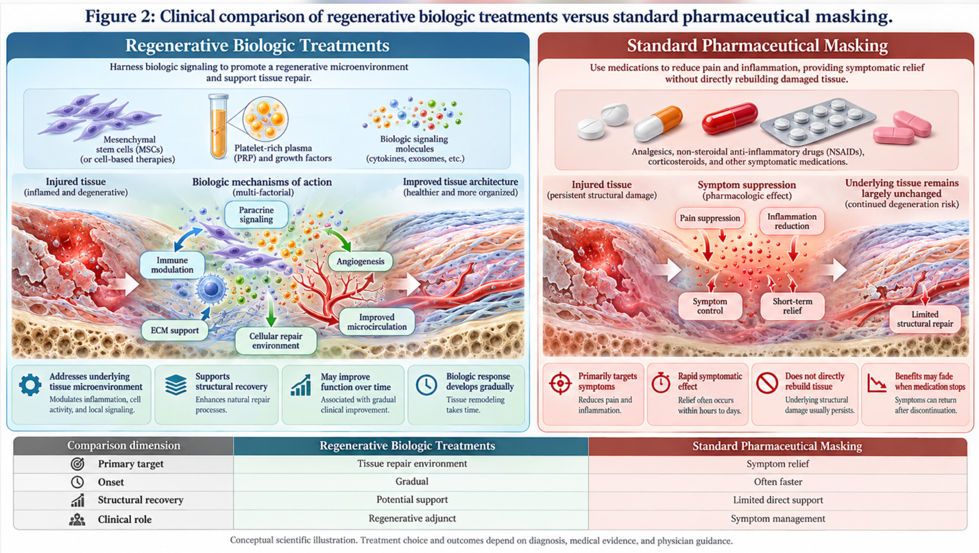
Figure 2: Clinical comparison of regenerative biologic treatments versus standard pharmaceutical masking.
For patients seeking the biochemical advantages of MSCs without the complexities of live-cell transplantation, recent advancements have isolated the signaling mechanisms entirely. Enter exosomes the frontier of cell-free therapy for neuropathy.
Exosomes are incredibly tiny extracellular vesicles (about 30-150 nanometers in diameter) secreted by MSCs. They are the actual envelopes carrying the paracrine signaling instructions discussed earlier. By utilizing ultracentrifugation, laboratories can isolate these vesicles, creating a highly concentrated therapeutic serum completely devoid of live cells but packed with regenerative potential.
This cell-free approach delivering microRNAs via exosomes bypasses any theoretical risk of immune rejection or unwanted cellular mutation. Because of their microscopic size, exosomes easily penetrate the blood-brain barrier and infiltrate dense fibrotic tissue that larger live cells might struggle to navigate. This makes exosome therapy an exceptionally targeted, highly predictable modality for rapid intervention in acute nerve lesions and complex neuropathies.
Establishing a reliable regenerative therapy success rate requires transparent outcome tracking. Patients typically begin to notice a reduction in neuropathic pain within 4 to 6 weeks post-injection, with maximum nerve fiber regeneration peaking at 6 to 9 months (PubMed, 2021). These biological timelines dictate clinical expectations and separate legitimate medicine from instant-cure marketing.
Maximum nerve fiber regeneration peaks at 9 months proving biological repair operates much slower than pharmaceutical symptom masking.
A major draw for this approach is that it qualifies as a minimally invasive neuropathy treatment. The actual procedure day rarely resembles major surgery, which is a massive relief for patients already dealing with extensive medical trauma. Whether receiving live MSCs or cell-free exosomes, the administration is typically handled as an outpatient protocol. Patients do not require general anesthesia, avoiding the severe systemic risks associated with major spinal or peripheral nerve decompression surgeries.
But patients must respect the biological clock of healing. Healing nerves is excruciatingly slow. The initial post-treatment phase (Days 1-14) is categorized by aggressive inflammation reduction; patients might feel slight soreness at the injection site but rarely profound pain relief. During this phase, the cellular microenvironment is being completely reset.
Real early pain relief typically materializes between 4 to 6 weeks as the local microenvironment stabilizes and inflammation recedes. However, the true structural repair the actual regeneration of myelin sheaths and axonal sprouting operates on a 4-to-9-month timeline for maximum nerve fiber regeneration. We constantly remind patients: this biological improvement differs radically from popping a painkiller. You are rebuilding tissue, not chemically shutting off an alarm bell.
These timelines and outcomes are only achievable if the procedure is performed by a highly qualified medical provider operating under strict laboratory standards. Success rates fluctuate wildly across the industry, primarily based on patient selection. Not everyone is a candidate. Efficacy requires strict adherence to the Neuro-Regenerative Triad, and responsible clinicians will actively disqualify poor candidates.
Finding the best stem cells for peripheral neuropathy means verifying rigorous laboratory standards rather than accepting glossy marketing brochures. When vetting a neuropathy stem cell clinic, you must ask specific, uncomfortable questions. Where are the cells sourced? What are the laboratory expansion protocols? Can they provide third-party flow cytometry reports proving cell viability and exact cell counts before injection?
Without a validated Certificate of Analysis (COA), you have no mathematical proof that the biologic material injected is alive or concentrated enough to matter. Furthermore, you must rigorously assess the clinical environment, particularly when evaluating the safety of stem cell therapy clinics abroad. Ensure the clinic utilizes ultrasound or fluoroscopic guidance for injections and mandates long-term post-procedure monitoring. A clinic that injects and forgets is not practicing medicine; they are selling a commodity.
| 📌 If you’re planning treatment in Thailand and want a practical checklist for choosing a safe clinic, we have an interesting article that discusses whether stem cell therapy is safe in Thailand, which you can read via the internal link. |
Stem cell therapy for neuropathy demonstrates significant potential for repairing damaged nerves and reducing chronic pain. Clinical data indicates that mesenchymal stem cells secrete neurotrophic factors that stimulate cellular repair and modulate inflammation. For conditions like diabetic and chemotherapy-induced neuropathy, patients frequently observe measurable improvements in nerve conduction velocity. The success of the treatment relies heavily on the specific cell source and the patient’s underlying pathology. Individual results vary, and professional medical assessment is required to determine suitability.
Transplanted stem cells repair nerve tissue primarily through a process called paracrine signaling rather than directly transforming into new nerves. These cells secrete a highly concentrated mixture of growth factors, proteins, and microRNAs into the localized injury site. This biochemical signaling stimulates the patient’s own quiescent cells, promotes new blood vessel formation, and halts inflammatory tissue destruction. Over a period of months, this biological environment facilitates the structural regeneration of degraded nerve fibers.
The primary difference between PRP and stem cell therapy lies in their regenerative capacity and immunomodulatory power. Platelet-Rich Plasma (PRP) utilizes concentrated platelets from a patient’s own blood to release localized growth factors, which is beneficial for mild injuries. Stem cell therapy utilizes multipotent cells capable of widespread immune modulation and profound biochemical signaling necessary for severe, systemic nerve degradation. While PRP supports basic healing, stem cells actively reprogram the local inflammatory environment.
Stem cell treatment for neuropathy is a minimally invasive outpatient procedure that avoids the risks associated with major surgery. The biological materials, whether live cells or cell-free exosomes, are typically administered via targeted local injections or intravenous delivery. Patients do not require general anesthesia and generally return to basic daily activities rapidly. However, mild localized soreness at the injection site is common for several days post-procedure. Strict clinical protocols dictate the specific delivery method based on the targeted nerve damage.
For patients suffering from severe nerve pain, stem cell therapy for neuropathy delivers targeted structural repair and inflammation reduction. Clinical trials demonstrate that bone marrow-derived MSCs can achieve significant neuropathic pain reduction within 3 months (ClinicalTrials.gov, 2015). The most effective clinical approach combines utilizing premium MSCs, implementing localized delivery, and adhering to strict patient vetting.
Achieving these outcomes requires strict adherence to the Neuro-Regenerative Triad—perfectly matching the biological source, delivery vector, and specific nerve pathology. This framework separates rigorous regenerative medicine from generic, ineffective commercial applications that merely offer temporary localized relief.
To determine if your specific neuropathy profile qualifies for advanced biologic intervention, compile your recent medical records and neurological imaging. Schedule a specialized consultation with Vega Stem Cell Clinic to undergo a rigorous suitability assessment.