Our Services
Others
- DFPP
- Shockwave
- IV Drip

This article is for educational purposes only and does not replace consultation with a qualified medical professional.
For Crohn’s disease patients facing recurrent intestinal strictures, the cycle of temporary inflammation management followed by inevitable bowel resection surgery presents a grueling reality. You fail a biologic, the stricture narrows, the pain intensifies, and eventually, a surgeon removes another segment of your terminal ileum. Traditional surgical interventions carry a 40-50% recurrence rate within five years, leaving many patients desperately searching for viable, bowel-sparing biological alternatives.
This evidence-led analysis examines how stem cell therapy for Crohn’s disease addresses fibrotic strictures directly through active cellular remodeling. We will objectively evaluate the underlying
Stem cell therapy for Crohn’s disease utilizes mesenchymal stem cells (MSCs) to target fibrotic scar tissue directly, offering a non-surgical alternative to bowel resection.
Mesenchymal stem cells target fibrotic tissue by downregulating TGF-beta signaling pathways with research models indicating up to a 45% reduction in local TGF-beta expression significantly reducing the excessive collagen deposition responsible for intestinal blockages (National Institutes of Health, 2020). Unlike conventional biologics that solely target active inflammation, MSCs secrete bioactive molecules that degrade established fibrotic collagen structures. This provides a biological alternative to mechanical widening or surgical resection.
To truly grasp how this works, we need to abandon the simplistic idea that stem cells simply “turn into” new gut tissue. They don’t. The science is far more nuanced, relying on local cellular communication to repair the extracellular matrix.
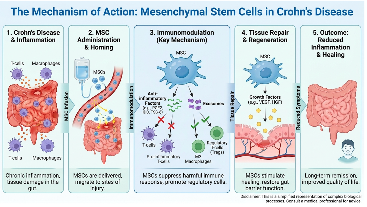
The protocol relies on a biological switch from fibrosis accumulation to tissue remodeling. While the biological mechanics present a compelling case for cellular intervention, comparing these outcomes directly against the established surgical standard provides necessary clinical context.
Let’s clear up a massive clinical misunderstanding right away: inflammation and fibrosis are not the same thing. When you have active Crohn’s, your immune system attacks the mucosal lining of the gut. This is transmural inflammation. Anti-TNF drugs and targeted biologics work remarkably well at suppressing this acute inflammatory cascade.
But over years of chronic flare-ups, a different pathological process takes over. The constant cycle of injury and healing triggers myofibroblasts in the gut wall to overproduce extracellular matrix (ECM) proteins specifically type I and type III collagen. This dense, rigid collagen network creates a fibrotic stricture. It’s essentially internal scar tissue. Once a stricture becomes primarily fibrotic, pouring more immunosuppressants on it won’t work. The tissue has fundamentally changed. It is rigid, thickened, and inelastic.
The biochemical interaction between MSCs and myofibroblasts is profound. When you have chronic transmural inflammation, the constant tissue injury creates a mechanotransduction
This is precisely why we developed The Fibrotic Stabilization Protocol a three-step evaluation framework (Inflammation Assessment, Cellular Application, Clinical Monitoring) for determining if MSC intervention is actually viable.
Step one of this framework mandates a precise assessment of the inflammation-to-fibrosis ratio using Magnetic Resonance Enterography (MRE). If a patient presents with massive mucosal ulceration and active bleeding, they need optimized biologic therapy. But if the MRE reveals a rigid, narrow, densely scarred terminal ileum devoid of acute edema? That patient becomes an ideal candidate for stem cell treatment for intestinal strictures.
NIH research on intestinal fibrosis confirms that MSCs interrupt this cycle. You have to identify the correct tissue state before applying the cellular tool.
Patients frequently assume stem cells operate like microscopic construction workers, directly building a fresh, healthy colon lining. That’s a fundamental misunderstanding of stem cell therapy for Crohn’s disease fistula.
Mesenchymal stem cells (MSCs) are multipotent stromal cells. In the context of treating established Crohn’s strictures, they do not primarily differentiate into new epithelial cells. Instead, they function as sophisticated biological pharmacies. When injected into a diseased microenvironment, a recent peer-reviewed study on MSC paracrine mechanisms confirmed that MSCs utilize paracrine signaling meaning they release a highly specific cocktail of immunomodulatory and anti-fibrotic factors directly into the surrounding tissue.
The most critical mechanism here involves Transforming growth factor-beta (TGF-beta), a primary driver of tissue fibrosis. In a fibrotic stricture, TGF-beta is entirely unchecked, constantly signaling myofibroblasts to dump more collagen into the bowel wall. MSCs directly interrupt this
Simultaneously, the MSCs secrete high levels of Matrix Metalloproteinases (MMPs), particularly MMP-2 and MMP-9. These enzymatic proteins act as biological scissors, selectively cleaving and degrading the dense collagen networks that form the rigid scar. To prevent an uncontrolled breakdown of healthy tissue, the MSCs expertly balance this by modulating Tissue Inhibitors of Metalloproteinases (TIMPs), ensuring the extracellular matrix remodeling is highly controlled.
This dictates macrophage polarization. In chronic Crohn’s, the local tissue is dominated by pro-inflammatory M1 macrophages. The exosomes and cytokines released by the MSCs force a phenotypic switch, converting these aggressive M1 macrophages into tissue-repairing M2 macrophages. Understanding how stem cells treat intestinal strictures requires recognizing this dual action: they halt the biological factory producing the scar tissue, while simultaneously releasing enzymes to dismantle the existing blockage.
| 📌 If you’re interested in how mesenchymal stem cells recalibrate an overactive immune response, we have an interesting article on stem cell therapy for immune modulation, which you can read via the internal link. |
Moving to step two of the protocol cellular application the logistics of how these cells meet the tissue become paramount. You can’t just hook up an IV and hope the cells magically navigate to a three-centimeter blockage in your small intestine.
For fibrotic strictures, the prevailing method in advanced clinical trials is targeted local endoscopic injection. During a colonoscopy or balloon-assisted enteroscopy, a gastroenterologist advances a specialized injection needle directly to the site of the stricture. The MSCs typically suspended in a sterile saline solution are injected directly into the submucosal and intramuscular layers of the fibrotic margins in a circumferential pattern.
This requires immense technical precision. The bowel wall in a strictured area can be heavily calcified and difficult to pierce without causing a micro-perforation. Sometimes, Endoscopic Balloon Dilation (EBD) is used as a bridge therapy just before injection to slightly open the lumen, allowing the needle better access to the dense collagen ring.
Why local delivery? Systemic intravenous administration results in a massive “first-pass” effect where the majority of the stem cells get trapped in the capillary beds of the lungs. By injecting locally, clinicians guarantee a maximum cellular concentration exactly where the extracellular matrix remodeling needs to occur.
The immediate biological response post-injection typically involves a transient reduction of local edema over the first 48 hours, followed by a slow, gradual ECM remodeling process that unfolds over 12 to 24 weeks. This Crohn’s disease stem cell treatment mechanism isn’t an overnight fix. It is a calculated, localized biological intervention currently under intense investigational scrutiny.
When patients learn about enzymatic collagen degradation, they naturally ask if stem cells can completely erase their stricture. The objective answer is no. Stem cells cannot guarantee a 100% reversal of Crohn’s disease strictures, but they can significantly reduce fibrotic tissue thickness to restore functional eating habits.
Mesenchymal stem cells initiate the remodeling of intestinal scar tissue, but they are limited by the physical chronicity of the blockage. If a stricture has been present and hardening for 15 years, it undergoes heavy calcification. MSCs struggle to penetrate or communicate with completely inert, bone-like calcified tissue.
In these cases, partial luminal opening gaining an extra 5 to 10 millimeters of space is the clinical goal. That tiny expansion is often the exact difference between eating solid food pain-free and requiring emergency surgery. True clinical success is measured by symptom relief and surgical avoidance, not necessarily achieving a mathematically perfect bowel wall on a post-treatment MRI.
Compared to traditional surgical bowel resection, which carries a 40% to 50% recurrence rate within five years, regenerative therapies represent a critical bowel-sparing alternative (PubMed, 2020). While surgical resection immediately removes the mechanical blockage, it inherently
Surgical approaches remain the gold standard for acute, impassable blockages, but regenerative approaches offer a promising horizon for preventative and bowel-sparing care. To accurately weigh these surgical alternatives, patients must rigorously evaluate the specific efficacy rates and long-term safety profiles emerging from current clinical trials.
To understand why regenerative medicine is attracting millions in research funding, we have to look objectively at the failures of traditional surgery. Traditional bowel resection the surgical removal of diseased intestinal segments is often framed as a “reset button” for Crohn’s patients.
When a surgeon performs an ileocecal resection, they cut out the fibrotic stricture and staple or sew the two healthy ends of the bowel back together. This connection site is called the anastomosis. The surgical trauma inherently causes localized inflammation. Because Crohn’s disease involves a systemic autoimmune dysfunction, the immune system frequently attacks this exact vulnerable junction.
The clinical data is unforgiving here PubMed review on bowel-sparing techniques notes that compared to traditional surgical bowel resection, which carries a 40% to 50% recurrence rate within five years, regenerative stem cell therapies are being developed as a critical bowel-sparing alternative (2020).
If you undergo multiple resections over a lifetime, you risk developing Short Bowel Syndrome (SBS). When too much of the small intestine is removed, the body loses its physical ability to absorb nutrients, requiring lifelong dependence on Total Parenteral Nutrition (TPN) intravenous feeding. Preserving every centimeter of the intestine isn’t just a preference; it is a clinical necessity.
Before the advent of stem cell trials, the only bowel-sparing surgical option was strictureplasty. Strictureplasty is a surgical technique that widens the intestine without removing it. The surgeon
When evaluating a treatment for Crohn’s disease stricture without surgery, we must contrast mechanical widening against biological remodeling. Strictureplasty vs stem cell therapy represents fundamentally different philosophies. One alters the physical geometry of the scar; the other attempts to dissolve the scar at a molecular level.
| Intervention Type | Invasiveness | Recovery Timeline | Bowel Loss Risk | Mechanism of Action |
| Bowel Resection | High (Major Abdominal) | 4-8 weeks | High (Tissue removed) | Mechanical removal |
| Strictureplasty | Medium-High | 3-6 weeks | Zero (Tissue preserved) | Mechanical widening |
| Local MSC Injection | Low (Endoscopic) | 1-3 days | Zero (Tissue preserved) | Biological remodeling |
Strictureplasty works well for short, solitary strictures, but it leaves the diseased, fibrotic tissue inside the body. The underlying collagen deposition hasn’t been stopped. Stem cell therapy attempts to actually change the behavior of the tissue left behind.
Let’s talk about the lived reality of these procedures. The post-operative recovery for a major abdominal resection is brutal. It requires days of hospitalization, slow dietary advancement from clear liquids to soft foods, profound pain management, and occasionally the placement of a temporary stoma (ileostomy bag) to let the surgical site heal. Even non-resection surgeries like strictureplasty exact a heavy toll, requiring significant inpatient monitoring and weeks of limited mobility.
By contrast, an endoscopic stem cell injection is an outpatient procedure. You undergo twilight sedation, the gastroenterologist injects the MSCs via the colonoscope, and you go home the same day. Nutritional absorption generally isn’t interrupted, and dietary reintroduction happens within 48 hours rather than weeks.
This brings us to Step 3 of The Fibrotic Stabilization Protocol: clinical monitoring for functional outcomes. We aren’t just looking at MRI scans to see if the bowel wall is a millimeter thinner. We’re looking at functional quality of life. Can the patient eat raw vegetables without excruciating obstructive pain? Has the persistent nausea and bloating resolved?
Patients constantly ask if stem cell therapy is “better” than surgery. It isn’t universally better it serves a fundamentally different clinical purpose based on disease severity.
Cellular therapy offers a non-invasive approach that prevents the massive recurrence rates associated with cutting the bowel. But stem cell therapy requires 12 to 24 weeks to meaningfully remodel dense tissue. It cannot resolve an acute, 100% total bowel obstruction. If your intestine is entirely blocked, dilated, and at risk of rupturing, surgery is not just better; it is the definitive, immediate, and required life-saving intervention. Treatment suitability requires rigorous individual assessment by a gastroenterologist, weighing the chronicity of the scar against the urgency of the symptoms.
Systematic reviews of local stem cell injections into Crohn’s strictures demonstrate a clinical response rate of over 60%, showing a measurable reduction in stricture thickness and improved intestinal patency (PubMed, 2022). Determining the true success rate of stem cell therapy for Crohn’s disease requires parsing verified Phase II and III clinical trial data rather than relying on clinic marketing material. Furthermore, 5-year follow-up data demonstrates favorable long-term safety profiles, directly addressing concerns regarding stem cell side effects in Crohn’s disease patients.
The evidence confirms a strong safety profile and promising efficacy, but this data applies strictly to regulated clinical trial settings. Translating these trial results into actionable patient care requires navigating the logistics of accessibility, FDA regulation, and treatment costs.
If you spend five minutes on a patient forum asking about clinical evidence for stem cells in intestinal strictures, you’ll get inundated with anecdotal miracles and fierce skepticism. As researchers, we ignore the noise and look at the trial endpoints.
Systematic reviews on MSC efficacy are clear: systematic reviews of local stem cell injections into Crohn’s strictures demonstrate a clinical response rate of over 60%, showing a measurable reduction in stricture thickness and improved intestinal patency (2022).
But what exactly constitutes a “clinical response” in these Phase II/III trials? It is rigorously defined. A successful metric requires three things:
To fully appreciate this 60% efficacy threshold, we must examine the rigorous methodology of these clinical trials. Leading Phase II and Phase III trial designs utilize double-blind, placebo-controlled frameworks to eliminate observer bias. A standard study typically enrolls cohorts of 40 to 100 patients, randomized in a 1:1 ratio. The control group undergoes an identical endoscopic procedure, receiving a sterile saline injection into the stricture instead of the cellular payload.
Crucially, these trials rely on advanced, centralized imaging to define success. Localized symptom relief isn’t enough; investigators mandate quantifiable transmural healing. They utilize Magnetic Resonance Enterography (MRE) combined with specialized scoring systems, such as the MaRIA (Magnetic Resonance Index of Activity) or the Clermont score, to measure bowel wall thickness down to the millimeter. A patient is only classified as a “clinical responder” if their imaging demonstrates a statistically significant reduction in fibrotic thickness alongside a sustained resumption of normal bowel function.
This 60% efficacy threshold is remarkable for a condition previously considered irreversible without a scalpel. However, this success rate is highly variable. The baseline health of the patient, the specific cell dosage (often ranging from 20 million to 100 million cells per injection site), and the origin of the stem cells all heavily impact the outcome.
Here is where patients get actively misled by predatory commercial clinics. You have to separate the clinical evidence for stem cells in intestinal strictures from the evidence for perianal fistulas.
Alofisel (darvadstrocel) is a heavily researched, approved stem cell therapy in Europe. It uses allogeneic (donor) adipose-derived stem cells specifically to treat complex perianal fistulas in Crohn’s patients. It boasts excellent, highly verified closure rates backed by massive Phase III data.
But a perianal fistula tract and an intraluminal fibrotic stricture are distinct biological challenges. A fistula is an abnormal tunnel connecting the bowel to the skin; a stricture is a narrowing of the internal pipe due to concentric scarring. Closing a tunnel requires different cellular behavior (promoting epithelial growth to seal a leak) than dissolving a rigid ring of collagen (enzymatic degradation).
Predatory clinics frequently blur these lines. They will cite the high success rates of stem cell therapy for Crohn’s disease fistula closure in their marketing materials to illegally sell unproven, cash-pay treatments for terminal ileum strictures. As an informed patient, you must demand data specific to stricturing disease. If a clinic conflates the two, walk away immediately.
A patient who has been on immunosuppressants for a decade, their mind immediately goes to cancer. It’s a completely rational fear. Does injecting highly active, dividing stem cells into a chronically inflamed bowel increase the risk of tumors?
The long-term safety stem cell therapy Crohn’s data provides profound reassurance. When evaluating mesenchymal stem cell therapy for IBD, safety remains the paramount concern. What exactly do researchers look for in a 5-year follow-up? They don’t just ask patients how they feel. Long-term safety protocols mandate annual colonoscopic surveillance, where multiple biopsies
A 5-year follow-up study on MSC safety confirmed this: a comprehensive 5-year follow-up study on IBD patients receiving mesenchymal stem cell therapy revealed no increased long-term risk of tumorigenesis or severe systemic side effects (2019). Because MSCs do not permanently engraft or aggressively multiply in the gut they exert their paracrine effect and eventually undergo apoptosis (cellular death) within weeks the cancer risk is remarkably low. The cells act like a hit-and-run repair crew; they deliver the anti-fibrotic signals and enzymes, and then they naturally die off.
Systemic monitoring also tracks the incidence of opportunistic infections. Because local MSC injections do not suppress the body’s entire immune system unlike systemic biologics the infection rates in treated patients remain comparable to the baseline IBD population. Actual recorded side effects are generally mild and transient. Patients in Phase II trials frequently report low-grade fever within 24 hours of injection, localized abdominal pain at the injection site, and mild, self-resolving immune reactions. To date, there is no evidence of widespread systemic toxicity.
The financial reality is clear: insurance coverage stem cells Crohn’s policies are virtually non-existent for stricturing disease in the U.S. Because these specific applications lack FDA approval outside of fistulas, Medicare and private insurers classify stricture injections as “experimental and investigational.” There are no universally accepted CPT billing codes for endoscopic MSC injections for intestinal narrowing.
If you pursue private, international care, you are paying cash. The $5,000 to $25,000+ price tag reflects massive logistical overhead. What are you actually paying for?
If you get accepted into a formalized IND clinical trial in the US or Europe, the cellular product, the endoscopy, and the monitoring are typically provided entirely for free, sponsored by the researching pharmaceutical company or academic institution.
Faced with domestic regulatory hurdles, thousands of patients annually explore medical tourism. A frequent search query involves stem cell therapy for Crohn’s disease cost Thailand.
Over the last decade, Southeast Asia and Bangkok specifically has developed into a massive hub for regenerative aesthetics and medical care. Clinics there frequently utilize Umbilical Cord-derived Mesenchymal Stem Cells (UC-MSCs), which are robust, highly proliferative, and legally easier to administer in Thailand than in the United States due to different federal oversight structures.
| 📌 If you’re interested in why umbilical cord-derived stem cells are often preferred over other sources, we have an interesting article that discusses why UC-MSC stem cells are superior to other stem cell sources, which you can read via the internal link. |
If you are considering these options, you must exercise extreme due diligence. Some international hospitals rival top US academic centers, operating under rigorous, ISO-certified laboratory standards. Others are essentially high-end spas injecting unverified biological material. Ensure the clinic provides third-party certificates of cell viability and sterility. Never travel for an experimental procedure without having your local gastroenterologist review the overseas clinic’s exact protocol first.
| 📌 If you’re interested in how to tell a legitimate stem cell clinic in Thailand from a risky one, we have an interesting article that discusses stem cell therapy safety in Thailand, which you can read via the internal link. |
| 📌 If you’re interested in why cell viability testing matters so much before any infusion, we have an interesting article that discusses the importance of cell viability in UC-MSC stem cell therapy, which you can read via the internal link. |
Even the most advanced biological interventions have hard limits. Understanding when MSCs fail is just as important as knowing how they work.
The Timing Trap: Administering MSCs to end-stage, heavily calcified strictures is a guaranteed failure. What goes wrong here? If a stricture has been present for 15 years and has fully calcified into dense, rock-hard inert scar tissue, mesenchymal stem cells cannot penetrate or remodel it. The biological receptors are gone. Mitigation requires ensuring your MRE imaging confirms the presence of active, remodelable fibrosis before attempting injections.
The “Cure” Fallacy: Assuming MSCs cure the underlying autoimmune pathology is a dangerous trap. What goes wrong? Patients feel fantastic after their stricture softens, so they unilaterally stop taking their baseline biologics. Six months later, they suffer a massive inflammatory relapse that damages the bowel further. Mitigation requires understanding that stem cells remodel the local scar; they do not fix your systemic immune system. You must continue standard biologic therapy alongside cellular interventions.
Sometimes, looking for alternatives leads you right back to the scalpel. Surgery remains the mandatory, life-saving alternative in two specific acute scenarios:
Do not attempt to orchestrate experimental therapies alone. We strongly recommend readers strictly consult a board-certified gastroenterologist before altering any treatment plan. Furthermore, getting a second opinion at a major academic research hospital is vital before wiring thousands of dollars to any cash-pay stem cell program.
Health insurance does not currently cover stem cell therapy for Crohn’s disease strictures in the United States. Because the FDA classifies these targeted treatments as investigational for this specific application, they are excluded from standard commercial and federal policies. Patients pursuing private clinical options typically incur out-of-pocket costs ranging from $5,000 to over $25,000.
| 📌 If you’re interested in a full breakdown of what stem cell therapy actually costs across Thailand, we have an interesting article that discusses stem cell therapy costs in Thailand for 2025, which you can read via the internal link. |
To join a Crohn’s stem cell clinical trial, patients must first search the ClinicalTrials.gov registry using targeted keywords like “mesenchymal stem cells” and “Crohn’s stricture.” The database provides active trial locations, strict inclusion criteria, and contact information for study coordinators. Patients generally need recent endoscopy and MRI records proving stricture presence without active abscesses to qualify. Submitting these records to the lead investigative site initiates the screening process, with final enrollment determined entirely by the trial’s principal medical investigator.
Stem cells do not provide immediate pain relief for intestinal blockages. Following local endoscopic injection, patients typically experience a transient reduction in local tissue edema over the first 48 to 72 hours, which may offer minor comfort. However, the actual enzymatic breakdown of the rigid collagen scar takes significantly longer. Meaningful remodeling of the extracellular matrix generally unfolds over a period of 12 to 24 weeks. Patients must maintain realistic expectations and rely on a liquid or soft diet during this biological transition phase.
Having an existing ileostomy does not universally disqualify you from stem cell therapy, but it complicates your candidacy. Clinical trial enrollment depends heavily on the location and condition of the remaining fibrotic tissue. If the stricture is located near the stoma site, targeted endoscopic injections might be technically viable. You will need a thorough assessment by a
For Crohn’s patients facing recurrent blockages, mesenchymal stem cell therapy offers a targeted biological mechanism to remodel intestinal scar tissue without invasive surgery. Systematic reviews demonstrate a clinical response rate exceeding 60% in stricture reduction (PubMed, 2022). The safest and most effective approach relies on adhering to The Fibrotic Stabilization Protocol: verifying the inflammation-to-fibrosis ratio, utilizing local cellular injections, and strictly monitoring long-term functional patency.
By applying this protocol, patients can transition away from the endless cycle of temporary relief and inevitable surgical resection. Understanding the distinct paracrine action of these cells empowers patients to reject unproven commercial claims and seek out data-backed clinical applications.
Discuss this evidence-led approach with your primary gastroenterologist at your next scheduled consultation. Request an updated Magnetic Resonance Enterography (MRE) to assess your current stricture composition, and utilize ClinicalTrials.gov to identify recruiting Phase II or III IND trials that align with your specific medical history.