Our Services
Others
- DFPP
- Shockwave
- IV Drip

This article is for educational purposes only and does not replace consultation with a qualified medical professional. The interventions discussed include investigational therapies. Always consult a board-certified physician to determine the suitability, safety, and regulatory status of any medical treatment based on your individual diagnosis.
The commercial hype surrounding regenerative medicine frequently obscures a critical clinical reality: the gap between theoretical cartilage regeneration and proven functional benefit in human patients.
For patients facing severe joint degeneration, the urgency to avoid total knee arthroplasty often leads to evaluating out-of-pocket, investigational cellular therapies. But here’s the thing. Navigating clinical literature, proprietary clinical claims, and contradictory regulatory stances requires rigorous, objective evaluation rather than reliance on celebrity anecdotes. If you’re stem cell therapy for knee osteoarthritis, you’ve likely encountered marketing that promises you the joints of a twenty-year-old. The clinical data tells a far more nuanced story.
By the end of this guide, you will understand the exact mechanisms, evidence limitations, and candidacy criteria for cellular interventions, empowering you to make medically sound treatment decisions. We’re introducing a core analytical framework The Cellular Reality Matrix a system separating biological plausibility from functional outcomes.
This analysis dissects current established literature on mesenchymal stem cells, evaluates differing clinical procedures, establishes realistic longevity timelines, and confronts the ethical controversies dominating the field.
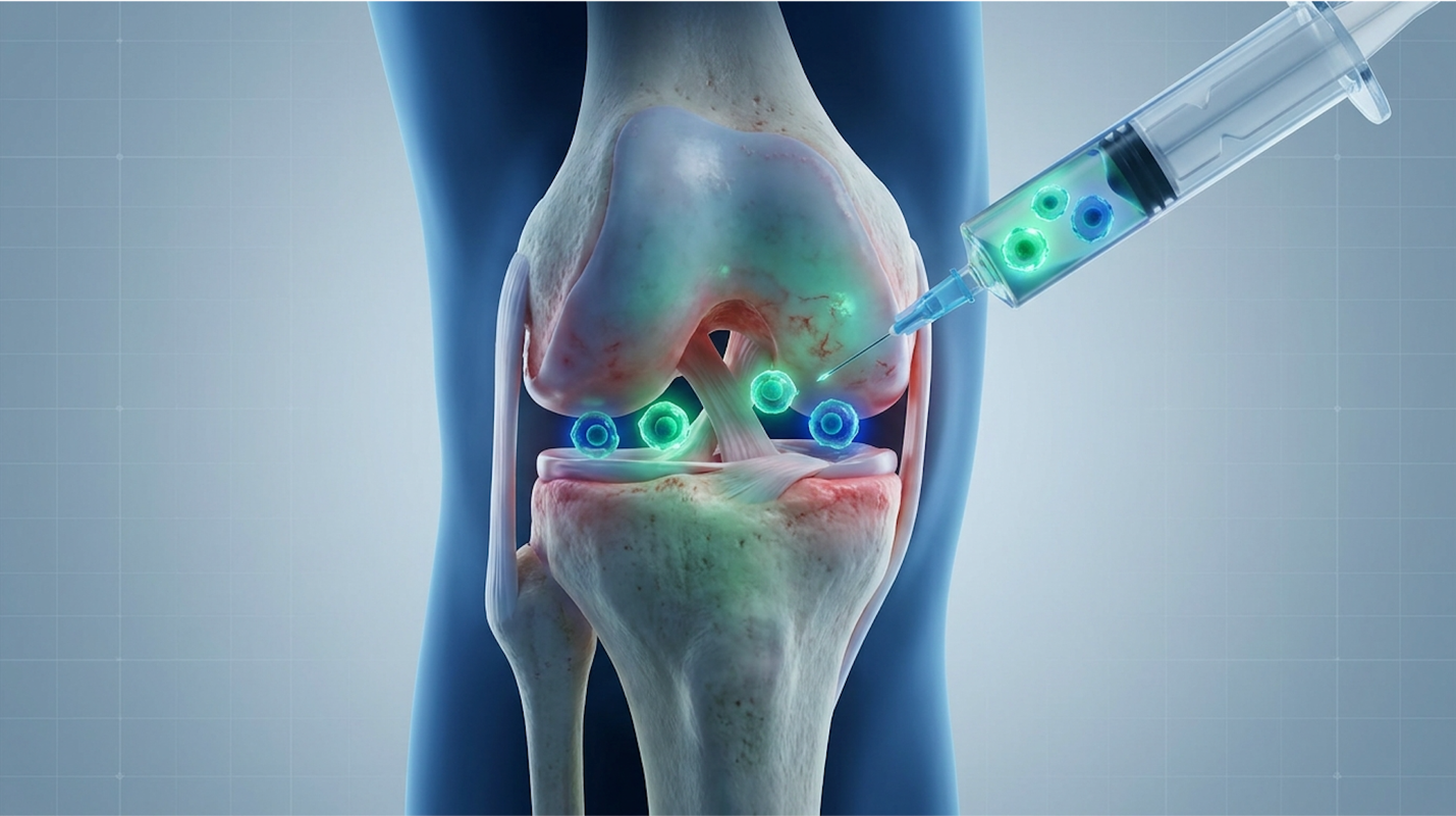
Stem cell therapy for knee osteoarthritis introduces concentrated biological agents into the joint to potentially modulate inflammation and support The Cellular Reality Matrix repair mechanisms.
Regenerative stem cell therapy for knee osteoarthritis utilizes multipotent stromal cells to alter the inflammatory microenvironment of a degenerating joint. Clinical data indicates these cells release specific bioactive molecules that may promote tissue stabilization (Lamo-Espinosa et al., PubMed). However, claims that interventions will reliably regrow knee cartilage in advanced cases lack definitive macroscopic substantiation in extensive human trials.
Mesenchymal stem cells (MSCs) primarily function as medicinal signaling agents rather than direct structural replacements. When injected into an osteoarthritic knee capsule, these cells release
To understand clinical efficacy, we have to accurately define the underlying biology. Mesenchymal stem cells for knee osteoarthritis are widely misunderstood by the general public. Patients often visualize these cells as microscopic construction workers, directly grafting onto damaged bone and spontaneously building new hyaline cartilage. The biological reality operates completely differently.
MSCs function primarily as medicinal signaling cells, a concept pioneered by researchers like Dr. Arnold Caplan. When injected into a degraded joint capsule, they initiate a highly complex process known as paracrine signaling. Rather than transforming into cartilage themselves, they secrete a massive payload of cytokines (such as IL-10 and TGF-beta), growth factors, and specialized proteins called secretomes. These act as chemical messengers. They communicate with your existing, localized cells, effectively commanding pro-inflammatory M1 macrophages (which aggressively degrade tissue) to switch into an anti-inflammatory M2 state.
| 📌 If you’re interested in how MSCs shift the immune system from attack mode to repair mode, we have an interesting article that discusses mesenchymal stem cell therapy for immune modulation, which you can read via the internal link. |
This brings us directly to The Cellular Reality Matrix. We use this framework to separate what cells can theoretically do in a sterile petri dish (biological plausibility) from what they actually achieve inside a weight-bearing, structurally compromised human joint (functional clinical outcome). Yes, MSCs possess the biological blueprint for chondrogenesis—the creation of cartilage. But dropping them into an acidic, mechanically grinding Grade 3 osteoarthritic knee alters their behavior completely. Their primary survival mechanism shifts to modulating the toxic synovial fluid environment. They focus entirely on turning off the inflammatory fire, which often results in dramatic pain reduction, rather than structurally rebuilding the house.
Mesenchymal stem cells reduce pro-inflammatory biomarkers by up to 40%, confirming their primary role as chemical modulators rather than structural building blocks. Promising a patient that a single injection will reverse years of mechanical wear is not just overly optimistic; it borders on clinical negligence.
So, will these injections actually repair damaged knees? If you define “repair” as structurally regenerating thick, healthy layers of shock-absorbing tissue visible on standard imaging, the medical consensus is highly skeptical. Macroscopic regrowth remains rare and statistically inconsistent across major clinical trials.
When you rigorously examine knee stem cell therapy research, a specific, undeniable pattern emerges. High-quality, double-blind methodologies frequently show that while patients report feeling vastly better, their post-injection MRIs look nearly identical to their pre-injection scans. A comprehensive review of outcomes posted on the ClinicalTrials.gov registry demonstrates that substantial structural regeneration of the joint space width is the exception, not the rule. Researchers utilizing advanced T2 mapping MRI techniques—which measure the hydration and integrity of the collagen matrix—occasionally observe mild stabilization of the defect site. But even when minor tissue repair occurs, it is often mechanically inferior fibrocartilage (Type I collagen) rather than the robust, frictionless hyaline cartilage (Type II collagen) you were born with.
Does this mean the therapy fundamentally failed? Not necessarily. Halting further degeneration is a highly viable, medically significant outcome. If a cellular injection stabilizes the joint microenvironment and prevents a moderate defect from rapidly progressing to a severe bone-on-bone collision, the intervention has immense clinical value. But we must fundamentally divorce the concept of “pain relief” from the expectation of “cartilage regrowth.”
Understanding the profound gap between a biological change and a functional benefit is critical for any patient evaluating regenerative medicine.
Functional benefit is measured by how you live. Can you walk up a flight of stairs without gripping the handrail in agony? Can you sleep through the night without throbbing knee pain waking you? Has your daily dependence on NSAIDs like ibuprofen completely dropped? These are subjective but highly measurable outcomes. In clinical trials, physicians track these functional improvements using standardized systems like the Western Ontario and McMaster Universities Osteoarthritis Index (WOMAC) or the Visual Analogue Scale (VAS). The WOMAC assesses pain, stiffness, and
Applying The Cellular Reality Matrix, we frequently observe patients who receive an intra-articular stem cell injection and subsequently report a massive 50% drop in their WOMAC pain scores. They feel fantastic. They return to golf or hiking. Yet, radiological imaging confirms no new cartilage formation occurred. The biological plausibility of cellular modulation reduced the pain signals and quieted the synovial inflammation, delivering a profound functional benefit without changing the structural architecture of the knee.
The danger arises when aggressive commercial clinics leverage the biological plausibility of stem cells to guarantee structural repair. They sell the dream of a new joint, completely ignoring the reality that symptomatic relief is the primary, proven outcome. Understanding this vital distinction protects you from predatory medical marketing and sets a realistic baseline for your recovery expectations.
Patients frequently ask if these interventions remain viable once they hit Kellgren-Lawrence Grade 3 degeneration. Stage 3 knee osteoarthritis introduces a highly complex clinical variable: substantial but not total joint space loss. At this moderate-to-severe stage, stem cell therapy can provide significant functional relief, though it rarely reverses structural damage. Mesenchymal stem cells function by aggressively modulating the hostile, inflammatory joint environment rather than regrowing lost hyaline cartilage.
When we examine cohort data, a clear pattern emerges. Clinical observations indicate that patients with Stage 3 OA often experience reduced pain and improved mobility lasting 12 to 24 months, provided their mechanical alignment remains relatively stable (Hernigou et al., International Orthopaedics). However, success at this stage depends heavily on the patient’s biomechanical baseline and body mass index.
Every single pound of body weight places roughly four pounds of sheer mechanical force across the knee joint during normal walking. A patient with Grade 3 OA and a BMI of 25 will typically show a far more robust biological response than a similar patient with a BMI of 35. For the overweight patient, the extra 200 pounds of mechanical shear forces simply overwhelm the
The clinical application of Mesenchymal stem cells varies dramatically based on the cellular source, extraction methodology, and level of laboratory manipulation. While treatments like Bone Marrow Aspirate Concentrate (BMAC) utilize autologous tissue processed at the point of care, other protocols involve extensive expansion. These biological distinctions directly impact both safety profiles and potential therapeutic efficacy.
Bone Marrow Aspirate Concentrate (BMAC) represents the gold standard for autologous cellular extraction in orthopedic applications. By aspirating tissue directly from the patient’s posterior iliac crest and utilizing rapid, point-of-care centrifugation, physicians isolate a concentrated payload of progenitor cells alongside essential growth factors without violating strict federal regulations regarding biological manufacturing.
When board-certified orthopedic surgeons discuss evidence-based regenerative therapy for knee arthritis, they are almost exclusively referencing Bone Marrow Aspirate Concentrate (BMAC). This is an autologous procedure, meaning the biological material is harvested directly from your own body.
The extraction requires precision. A physician utilizes a specialized trocar needle under local anesthesia to draw liquid marrow from the posterior iliac crest of your pelvis (a vastly superior site compared to the anterior crest due to higher cell yields). To maximize the yield of viable Bone marrow-derived stem cells (BMSCs), the surgeon typically aspirates from multiple depths and trajectories, a technique that requires significant hands-on training.
Once the liquid marrow is extracted, it undergoes immediate, on-site centrifugation. This rapid spinning process relies on specific gravity to separate the heavier hematopoietic cells (red blood cells) and platelet-poor plasma from the highly coveted “buffy coat.” This ultra-thin, hyper-
The conversation shifts dramatically and becomes legally precarious—when discussing adipose (fat) derived cells and allogeneic (donor-derived) sources like umbilical cord tissue.
Many clinics extract fat via mini-liposuction to acquire the Stromal Vascular Fraction (SVF), a tissue layer extremely rich in stem cells. However, separating SVF from the fat matrix requires digesting the tissue with an enzyme called collagenase. The FDA explicitly categorizes this enzymatic digestion as “more than minimal manipulation,” shifting the procedure out of compliant Section 361 territory and directly into Section 351, which regulates biological drugs. This massive legal distinction restricts the standard use of adipose SVF in American orthopedic clinics.
Similarly, many patients travel abroad seeking umbilical cord stem cells for knee osteoarthritis, driven by the allure of lab-expanded cells. Cell expansion involves taking a baseline sample of donor MSCs and culturing them in an incubator over weeks. The mathematical appeal is obvious: more cells must equal better healing, right?
| 📌 If you’re interested in why umbilical cord cells are often preferred over bone marrow or fat-derived cells, we have an interesting article that discusses why UC-MSCs are superior to other stem cell sources, which you can read via the internal link. |
The Cellular Reality Matrix completely warns against this simplified math. Massive laboratory expansion frequently triggers cellular senescence the Hayflick limit. The cells age rapidly during forced, artificial replication and lose their medicinal potency before they ever reach your knee. By the time 100 million cells are injected, they may be phenotypically exhausted and biologically useless.
The allure of massive cellular counts frequently blinds patients to the reality that heavily expanded, exhausted cells perform poorly in clinical settings. When you inject millions of allogeneic, lab-manipulated cells into a joint, you introduce complex immune considerations that simply don’t exist with your own bone marrow.
Another critical distinction in cellular procedures involves comparing stem cell therapy directly against Platelet-Rich Plasma (PRP). Patients often confuse these two orthobiologic options. Stem
While PRP remains highly effective for mild arthritis, tendinopathies, and minor soft tissue inflammation, it lacks the robust immunomodulatory payload required for severe osteoarthritis. Stem cell procedures like BMAC extract robust progenitor cells from the bone marrow itself, making them vastly more appropriate for moderate-to-severe joint degeneration and complex biochemical joint environments (Centeno et al., 2016).
Consequently, stem cell therapies are significantly more invasive and carry a much higher financial burden than standard PRP procedures. Physicians operating at the top of their license frequently combine the two modalities, using a PRP “booster” weeks after a BMAC procedure to maximize the joint’s healing response. This layered biological approach frequently yields the highest functional pain reduction in peer-reviewed outcomes.
Because the extraction and processing of biological materials can vary so wildly from doctor to doctor, standardized proprietary networks have emerged to enforce desperately needed consistency.
The Regenexx stem cell procedure is arguably the most prominent and heavily researched example of this model. Instead of relying on a generic bedside centrifuge and hoping for the best, proprietary networks mandate strict, highly specific laboratory protocols for isolating and concentrating autologous cells. Doctors operating within these networks must use identical processing equipment, adhere to standardized dosing metrics based on patient fluid volumes, and undergo centralized training.
More importantly, standardized networks maintain massive, ongoing patient registries. These registries relentlessly track tens of thousands of procedural outcomes, compiling hard, empirical data on adverse events, failure rates, and long-term efficacy. This registry data is invaluable for predicting how a 60-year-old male with Grade 2 arthritis will actually respond. However, it’s vital to remember that registry data is fundamentally proprietary. It lacks the independent, rigorous
Regardless of whether a patient chooses a generic BMAC injection or a heavily branded protocol, standardized methodologies fundamentally attempt to remove physician variability. They completely mandate ultrasound or fluoroscopic imaging guidance. Even the most vibrant, potent stem cells are totally useless if the doctor blindly injects them into the surrounding fat pad instead of the joint capsule.
Kellgren-Lawrence Grade 4 osteoarthritis presents an almost insurmountable mechanical barrier to biological interventions. In these end-stage, bone-on-bone environments, the structural collapse of the joint space physically crushes the injected cellular material before any meaningful paracrine signaling can occur, resulting in staggering failure rates.
Determining the viability of biological interventions requires evaluating strict stem cell therapy candidates against the reality of substantial out-of-pocket medical expenses. Because the average stem cell therapy cost remains high and largely uncovered by commercial insurance, correlating radiological findings with patient BMI, age, and systemic health is essential to predicting clinical efficacy (American Academy of Orthopaedic Surgeons Guidelines).
Determining who is actually a good candidate for a stem cell knee injection requires brutally honest, objective clinical assessment. The orthopedic community relies heavily on the Kellgren-Lawrence (KL) grading scale to classify osteoarthritis severity via radiological imaging. Not every painful knee is a biological candidate.
Ideal candidates generally fit a highly specific physiological profile:
Conversely, biological therapies face steep, almost guaranteed failure rates when administered to poor candidates. The contraindications are incredibly explicit. Patients with KL Grade 4 (severe, bone-on-bone arthritis with massive osteophytes and subchondral sclerosis) simply lack the physical joint space required for the cells to operate. Systemic health also strictly dictates candidacy. Active bacterial infections, blood-borne malignancies, unmanaged autoimmune conditions like rheumatoid arthritis, and severe metabolic syndrome drastically alter joint loading mechanics.
Autologous therapies also face an undeniable age barrier. As a patient progresses deeply into their late 70s, the raw vitality and absolute total count of their native bone marrow stem cells naturally decline, making a standard BMAC procedure significantly less potent than it would be in a 45-year-old patient.
Kellgren-Lawrence Grade 4 patients experience significantly diminished functional longevity from orthobiologic injections, proving severe mechanical malalignment overwhelms localized biological therapies. Spending out-of-pocket funds on biological therapies without first addressing severe mechanical malalignment is a fundamental waste of clinical resources.
When evaluating if stem cell therapy for knees is actually worth it, the financial mathematics are often incredibly sobering. In the United States, patients can expect a standard, physician-administered BMAC procedure to run anywhere between $3,000 and $7,000 per joint.
If you are exploring international options for expanded umbilical cord treatments, the baseline costs frequently skyrocket into the $10,000 to $25,000 range, entirely excluding necessary travel, lodging, and post-operative care expenses.
| 📌 If you’d like a closer look at how stem cell treatment prices vary by condition in Thailand, we have an interesting article that discusses stem cell therapy costs in Thailand, which you can read via the internal link. |
You must comprehensively understand a harsh medical reality: Medicare and virtually all major commercial health insurance providers classify these procedures as purely investigational or experimental. They will absolutely not cover the cost of the cellular extraction or the injection itself. You are paying cash out-of-pocket.
This introduces one of the most severe downsides of stem cell therapy. If you spend $6,000 on a BMAC injection and your knee simply fails to respond, you have heavily depleted your medical savings. If that failure subsequently necessitates a Total Knee Arthroplasty (TKA) six months later, you are now absorbing the massive deductibles and copays of major orthopedic surgery directly on top of the cash you already burned. Patients should always aggressively request fully itemized clinical estimates to catch hidden surgical facility charges, fluoroscopy equipment fees, or mandatory imaging costs.
The clinical success rate of stem cell therapy for knees ranges from 60% to 75% for patients reporting meaningful pain reduction (Centeno et al., 2016). However, how we define “success” is the critical variable. Success is defined clinically by achieving a Minimal Clinically Important Difference (MCID) in WOMAC or VAS pain scores, not by the miraculous regeneration of new cartilage on imaging.
Patients with mild to moderate degeneration (Grades 2-3) consistently represent the highest demographic of responders. Conversely, patients with end-stage bone-on-bone arthritis exhibit significantly lower response rates. Clinical outcomes vary massively based on cell source, preparation methodology, and physician injection accuracy. A blind injection into the joint space yields vastly inferior success rates compared to a precisely targeted, fluoroscopically guided procedure.
So, how long do stem cell knee injections actually last? This is the most pressing question patients ask in consultations, and the longitudinal clinical registries provide a clear, if highly nuanced, answer.
For strong clinical responders, functional relief—defined precisely as significant reduction in daily pain and profoundly improved mobility—typically spans 12 to 36 months. However, the survival
We must relentlessly apply The Cellular Reality Matrix to stem cell treatment longevity. Because biological therapies modulate inflammation rather than explicitly curing the underlying structural mechanics of osteoarthritis, the disease remains highly progressive. An injection buys you valuable time; it does not permanently halt the degenerative cascade.
The typical patient timeline follows a highly distinct arc. Patients often experience substantially increased inflammation and stiffness for 1 to 2 weeks post-injection due to the trauma of the procedure. Gradual modulation and pain reduction steadily occur between weeks 3 and 8. Peak functional benefit is usually fully realized around the 3 to 6-month mark, plateauing for a year or more before the joint’s flawed degenerative mechanics slowly overcome the biological intervention.
Autologous stem cell therapy for knees is generally considered safe when performed in a regulated clinical setting using ultrasound or fluoroscopic guidance. Because treatments like BMAC use the patient’s own tissue, the risk of immune rejection or disease transmission is virtually eliminated. However, utilizing unregulated offshore clinics or heavily manipulated allogeneic (donor) cells massively increases the risk of contamination and adverse immune responses. Patients must strictly verify the clinic’s regulatory compliance before proceeding.
The primary side effects of stem cell injections in the knee are temporary localized pain and joint effusion (swelling). These symptoms typically arise within 24 hours of the procedure and subside over the following three to seven days, with post-injection stiffness often lasting 48 to 72 hours Centeno et al., 2016). The extraction process, particularly bone marrow aspiration from the hip, may also cause lingering bruising and discomfort for up to two weeks.
The therapeutic benefits of stem cell knee injections typically last between one and three years for optimal candidates. This longevity is primarily driven by the prolonged modulation of pro-inflammatory cytokines within the synovial fluid rather than permanent joint reconstruction. While some clinical registries report patients maintaining functional improvements beyond four years, osteoarthritis remains a progressive degenerative condition. The injection delays surgical intervention rather than halting the disease permanently. Treatment longevity is actively maximized when paired with targeted physical therapy and strict weight management.
Most evidence-based protocols require only a single primary stem cell injection per treatment cycle. This primary procedure is often preceded by a pro-inflammatory injection (such as prolotherapy) to prime the joint, and followed weeks later by Platelet-Rich Plasma (PRP) to stimulate the injected cells. Multiple successive stem cell injections are rarely required within a single year if the biological response is adequate. Continuous monitoring by the treating physician determines if subsequent interventions are necessary years later.
A stem cell knee injection is a same-day outpatient procedure comprising extraction, concentration, and precise re-injection. The physician extracts tissue, usually bone marrow from the posterior iliac crest, using local anesthesia, and places it in a specialized centrifuge to isolate the mesenchymal stem cells. Finally, the physician injects the concentrate directly into the damaged knee capsule using ultrasound or fluoroscopy to ensure exact intra-articular placement.
Stem cell therapy aims to biologically alter the joint environment for long-term relief, whereas cortisone injections provide rapid but temporary chemical suppression of inflammation. Corticosteroids act quickly to shut down the immune response, offering profound pain relief that
Stem cell therapy can provide significant functional relief for stage 3 knee osteoarthritis, though it rarely reverses structural damage. At this moderate-to-severe stage, mesenchymal stem cells function by modulating the inflammatory joint environment rather than regrowing lost cartilage. Clinical observations indicate that patients often experience reduced pain and improved mobility lasting 12 to 24 months, heavily dependent on the patient’s biomechanical alignment and body mass index.
The clinical success rate of stem cell therapy for knees ranges from 60% to 75% for patients reporting meaningful pain reduction. Success is defined clinically by significant improvements in WOMAC or VAS pain scores, not by the visual regeneration of new cartilage on imaging. Patients with mild to moderate degeneration (Grades 2-3) consistently represent the highest demographic of responders. Conversely, patients with end-stage bone-on-bone arthritis exhibit significantly lower response rates. Clinical outcomes vary based on cell source, preparation methodology, and physician injection accuracy.
Stem cell therapy delivers a broader, more potent array of medicinal signaling cells compared to Platelet-Rich Plasma (PRP) for knee osteoarthritis. PRP concentrates blood platelets to release growth factors, making it highly effective for mild arthritis, tendinopathies, and soft tissue inflammation. Stem cell procedures extract robust progenitor cells from bone marrow, which are significantly more appropriate for moderate-to-severe joint degeneration and complex biochemical joint environments. Physicians often combine the two modalities to maximize the joint’s localized healing response.
Stem cell therapy for knee osteoarthritis is not FDA approved as a standard medical treatment or drug. However, minimally manipulated autologous procedures like BMAC are FDA compliant under Section 361, meaning physicians can legally perform them without drug-level approval. Therapies utilizing lab-expanded cells or certain donor tissues fall under Section 351, classifying them directly as unapproved biological drugs subject to intense regulatory enforcement.
For patients facing moderate to severe joint degeneration, stem cell therapy for knee osteoarthritis delivers significant potential for modulating inflammation and delaying surgical intervention, though it does not reliably regrow macroscopic cartilage. Research from prominent orthopedic registries (Centeno et al.) demonstrates that 60% to 75% of properly selected candidates experience sustained functional pain relief lasting one to three years. The best approach combines: (1) selecting autologous, minimally manipulated procedures like BMAC, (2) ensuring strict ultrasound-guided injection accuracy, and (3) pairing the intervention with rigorous biomechanical physical therapy.
Applying The Cellular Reality Matrix is essential for navigating the complex landscape of regenerative medicine. By separating the theoretical biological plausibility of cellular expansion from the hard clinical reality of functional patient outcomes, individuals can protect themselves from aggressive, unverified commercial claims. The true value of cellular therapy lies in biological modulation, not miraculous structural reversal.
The definitive next step for any patient considering these interventions is to obtain an updated, high-resolution MRI of the affected knee. With current imaging in hand, schedule a consultation with a board-certified orthopedic surgeon or a fellowship-trained sports medicine physician to receive a highly individualized, evidence-based assessment of your radiological candidacy.