Our Services
Others
- DFPP
- Shockwave
- IV Drip

Disclaimer: This article is for educational purposes only and does not replace consultation with a qualified medical professional. Suitability for any cellular therapy requires rigorous individual clinical assessment.
For patients navigating advanced heart failure, conventional pharmacology often reaches a ceiling of efficacy, prompting serious investigation into cellular medicine. Clinical experience across advanced cardiology settings demonstrates this trajectory frequently. You optimize the beta-blockers, dial in the ACE inhibitors, and yet the ejection fraction refuses to budge. At that point, stem cell therapy for heart disease stops being a sci-fi concept and becomes a highly relevant clinical consideration.
But here’s the reality. The regenerative medicine sector is heavily saturated with commercial clinics making wildly unsubstantiated claims. This severely complicates the ability to separate biological plausibility from established clinical outcomes. Are these cells actually rebuilding heart muscle, or just temporarily masking symptoms?
By the end of this guide, you’ll understand exactly how stem cell therapy supports heart disease treatment based exclusively on peer-reviewed trial data, FDA guidance, and documented mortality risks. This analysis dissects the biological mechanisms of cardiac repair, the clinical outcomes of major trials like DREAM-HF, exact eligibility requirements, and the brutal reality of the 100-day recovery timeline. We aren’t glossing over the dangers here.
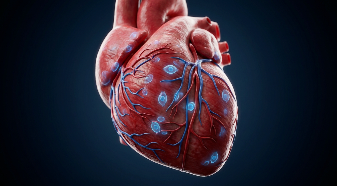
Stem cell therapy for heart disease operates primarily through paracrine signaling to reduce inflammation, rather than directly replacing dead cardiac muscle tissue.
Stem cell therapy for heart disease operates by modifying the localized cardiac microenvironment rather than simply replacing necrotic tissue. Current scientific models indicate that cardiac stem cell therapy primarily utilizes paracrine signaling to reduce chronic inflammation and stimulate native tissue repair. This precise biological mechanism aggressively differentiates authentic regenerative protocols from standard pharmacological symptom management, which only masks progressive deterioration.
Let’s dismantle the oldest myth in regenerative cardiology. If you think injecting stem cells means those cells instantly transform into beating heart muscle, you’re operating on outdated science. The actual mechanism is far more elegant and heavily reliant on molecular communication networks.
The paracrine effect is a form of cell-to-cell communication where administered stem cells secrete localized signaling molecules directly into the damaged myocardium. After a major cardiac event, the heart becomes a hostile, highly inflammatory environment. Pro-inflammatory macrophages (M1) swarm the damaged tissue, leading to aggressive fibrosis, cellular apoptosis (programmed cell death), and extensive scarring.
Mesenchymal stem cells (MSCs) operate as biological catalysts when introduced to this toxic zone. They release exosomes, cytokines, and microRNAs that effectively “talk” the local immune system down. They force a critical phenotypic transition from destructive M1 macrophages to reparative M2 (anti-inflammatory) macrophages. This localized cytokine storm downregulation is the exact mechanism that makes people wonder if stem cells reverse heart disease. They don’t magically erase a scar. Instead, they halt the pathological remodeling process by halting the overactive immune response, which in turn limits the structural damage that leads to chronic heart failure.
| 📌 If you’re interested in how these signaling molecules calm a damaged organ, we have an interesting article that discusses mesenchymal stem cell therapy for immune modulation, which you can read via the internal link. |
If you’re looking at regenerative therapy for heart failure, this localized immune modulation is the biological foundation. By downregulating the inflammation at the cellular level, the heart is spared from the vicious cycle of continuous cellular degradation. Exosomes released by these cells carry genetic material that blocks further cardiomyocyte death, essentially freezing the disease progression in its tracks.
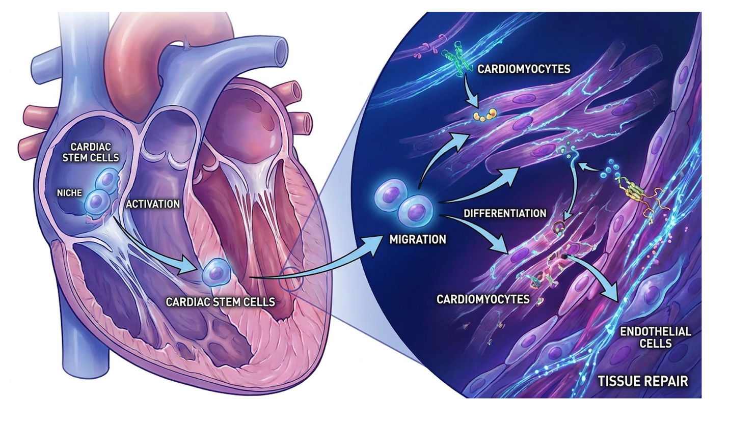
To understand the sheer biological horsepower of this process, look at the exosomal RNA transfer. These exosomes aren’t just passive bubbles; they carry microRNAs that penetrate dying cardiomyocytes and literally reprogram their genetic expression to resist apoptosis. This molecular reprogramming drastically reduces left ventricular wall stress. It stops the physical stretching and thinning of the heart muscle that inevitably leads to terminal dilation. By halting this structural dilation at the cellular level, mesenchymal stem cells actively prevent the heart from ballooning into a weak, inefficient pump.
Stem cells have the capacity to regenerate cardiovascular tissue and significantly reduce inflammation by promoting localized angiogenesis (NHLBI).
NHLBI research on stem cell heart repair mechanisms confirms that these stem cells have the capacity to regenerate cardiovascular tissue and significantly reduce inflammation, thereby repairing damaged heart muscle and restoring better blood flow.
While this profound inflammation reduction stabilizes the cardiac environment, the secondary phase of repair involves the actual structural regeneration of the vascular network.
Reducing inflammation is critical, but surviving severe heart failure requires robust, continuous blood flow. This is where repairing the heart with stem cells transitions from immune modulation to structural biological rebuilding.
When cardiac tissue is starved of oxygen a state known clinically as ischemia the cells begin to die rapidly. To combat this hypoxic environment, stem cells secrete massive amounts of vascular endothelial growth factor (VEGF) and hypoxia-inducible factor (HIF-1 alpha). These specific proteins act as biological catalysts for angiogenesis, which is the formation of entirely new microvascular networks. Think of it as the heart growing its own microscopic bypass grafts to route blood around the dead zones.
For patients seeking stem cell treatment after a myocardial infarction, this micro-perfusion is absolutely critical. Standard bypass surgery or coronary stenting fixes the massive coronary arteries on the surface of the heart. But what about the millions of microscopic capillaries deep within the muscle wall that actually feed the tissue? Surgery can’t reach those microscopic depths.
However and this is a massive caveat there are hard biological limitations to tissue regeneration. Stem cell therapy after a myocardial infarction works best on the “border zone” (or penumbra) of the damage. If a patient has a massive area of entirely necrotic, dead tissue, stem cells cannot bring it back to life. Dead is dead. What they can do is rescue the struggling, oxygen-starved tissue surrounding the dead zone, preventing the infarct from expanding.
Clinical evaluations frequently reveal patients misunderstanding this critical distinction. They expect a brand-new heart with flawless function. Instead, they get a slightly more efficient, significantly better-perfused older heart. The clinical evaluation of these mesenchymal precursor cellular pathways confirms their role in chronic heart failure models, demonstrating exactly how these microvascular networks improve over time, providing necessary oxygenation to the surviving myocardial border zones.
Understanding these biological mechanisms is paramount, but patients and clinicians must ultimately rely on large-scale, peer-reviewed clinical trials to prove that these microscopic changes translate to actual human survival.
When evaluating stem cell therapy for heart failure, clinical efficacy must be measured through structured, randomized trials rather than anecdotal reports or clinic marketing materials. The survival rate after stem cell transplant depends heavily on the specific cellular product, the processing methodology, and the delivery mechanism. Recent phase 3 trials have established concrete statistical baselines for evaluating cardiovascular mortality and major adverse cardiac events.
If you want to understand real-world efficacy and stringent medical science, look closely at the parameters of the DREAM-HF trial. This wasn’t a small-scale, unregulated study from a commercial strip-mall clinic. This was a massive, Phase 3, sham-controlled trial evaluating allogeneic (donor) mesenchymal precursor cells for chronic heart failure in over 500 patients.
The methodology utilized here matters intensely for setting expectations. Commercial clinics often offer a simple, basic IV stem cell infusion for heart disease. The DREAM-HF trial intentionally avoided that route. Why? Because injecting cells directly into a peripheral vein means the vast majority of them end up trapped in the microvasculature of the lungs a phenomenon known clinically as the pulmonary first-pass effect. Very few cells actually make it to the heart, rendering the intervention practically useless for targeted cardiovascular regeneration.
Instead, DREAM-HF utilized precise transendocardial delivery. Using a specialized NOGA electromechanical mapping catheter, interventional cardiologists mapped the electrical voltage of the beating heart from the inside. They identified the exact borders of the dead tissue distinguishing between viable muscle and non-viable scar tissue and directly injected 150 million cells straight into the struggling myocardium.
The rigorous sham-controlled nature of this study is exactly what separates legitimate clinical science from placebo-driven marketing. In the control group, patients underwent the exact same grueling catheterization procedure, complete with the NOGA electromechanical mapping, but received a sterile saline injection instead of cells. This completely eliminated the psychological placebo effect from the efficacy data, ensuring that any functional improvements recorded were purely biological.
Furthermore, understanding how the NOGA system maps the heart is critical. Dead scar tissue does not conduct electricity. Viable, healthy tissue conducts robustly. The border zone the struggling, hypoxic tissue shows specific low-voltage electrical signatures. The interventional cardiologists used this real-time electrical mapping to navigate the catheter tip, firing the 150 million cells exclusively into that highly vulnerable border zone where angiogenesis could actually make a difference. If you blindly inject cells into the dead center of a massive scar, they simply die. Precision electromechanical targeting was the defining variable that made this trial successful.
When evaluating stem cell treatment clinical trials, precise physiological delivery accuracy dictates the entire outcome. The parameters of the DREAM-HF clinical trial were structured specifically to evaluate how these allogeneic mesenchymal precursor cells impact major adverse cardiovascular events in chronic heart failure patients. Unregulated IV infusions simply cannot replicate this targeted physiological delivery, rendering their outcomes vastly inferior to trial data.
The rigorous, electromechanically guided methodology of DREAM-HF yielded specific data regarding patient survival that fundamentally challenges previous assumptions about cellular therapy capabilities.
So, what did injecting 150 million targeted, allogeneic cells actually accomplish in a Phase 3 setting? The statistical results were incredibly nuanced, completely shattering the simplistic binary “it works” or “it doesn’t work” narrative.
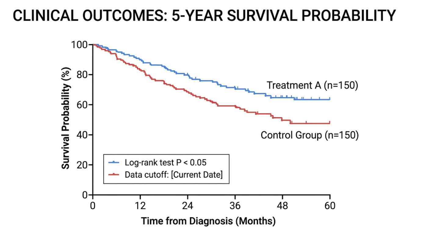
First, the therapy did not significantly reduce the overall rate of non-fatal heart failure hospitalizations across the general cohort. If a patient was prone to fluid overload and required diuresis, they still occasionally needed hospitalization. But here is the massive breakthrough that stunned the cardiological community: the data demonstrated a stunning 58% reduction in cardiovascular mortality.
Read that exact statistic again. The therapy didn’t stop all hospital visits, but it fundamentally stopped patients from dying of cardiovascular collapse.
This cardiac death reduction stem cells statistic is vital for patients and caregivers managing severe outcome expectations. Data shows a 58% reduction in mortality so patients with high baseline systemic inflammation derive the most significant survival benefits. Specifically, patients presenting with elevated C-reactive protein (CRP > 2 mg/L) prior to the procedure saw the most profound benefits. The cells targeted their systemic inflammation, shut down the pathological cardiac remodeling, and kept the patients alive significantly longer than the placebo group. It proved that while cellular therapies might not immediately resolve daily fluid retention symptoms, they forcefully interrupt the terminal progression of the disease.
Targeted stem cell delivery reduces cardiac mortality by 58% proving localized biological repair outweighs systemic symptom management (JACC).
According to published, peer-reviewed literature detailing the 58% reduction in cardiovascular mortality, clinical outcomes demonstrated a notable decrease in fatal heart attacks for patients who received this targeted mesenchymal stem cell therapy.
However, impressive mortality statistics must always be contextualized within the broader scope of patient quality of life. Being alive is the first metric. Living well is the second.
Understanding stem cell transplant risks is paramount for patient safety and informed consent. While trial data shows incredible promise for survival, determining who is a candidate for stem cell therapy requires rigorous immunological and cardiovascular screening. The medical community acknowledges that adverse events, ranging from localized injection site arrhythmias to systemic, fatal immune rejection, represent a significant barrier to widespread clinical adoption.
Let’s address the most terrifying complication in the transplant world: profound immunological rejection. When you introduce biological material from a donor (allogeneic cells) into a host, the host’s immune system naturally views those foreign cells as an invading threat.
The most severe manifestation of this biological warfare is Graft-Versus-Host Disease (GVHD). GVHD occurs when the donor’s immune T-cells hitching a ride with the stem cell infusion recognize the recipient’s body as foreign and actively begin attacking the patient’s native organs. It is a brutal, agonizing, and frequently fatal complication. Acute GVHD typically ravages the skin (causing severe blistering rashes and desquamation), the liver (causing dangerous spikes in bilirubin and hepatic failure), and the gastrointestinal tract (causing severe, uncontrollable diarrhea and mucosal sloughing).
The pathophysiology of this rejection is complex. When allogeneic mesenchymal cells are introduced, they are generally considered immune-privileged because they lack Class II human leukocyte antigens (HLA) on their surface. However, processing methodologies vary, and some cellular surface markers can still trigger host immune surveillance. If donor T-cells contaminate the infusion and proliferate, they immediately recognize the recipient’s major histocompatibility complex (MHC) as a foreign invader. This triggers a massive cascade of cytotoxic T-cell activation. The donor cells effectively mount an insurgency against the host.
| 📌 If you’re interested in why donor-derived cells carry less rejection risk than you’d expect, we have an interesting article that discusses why umbilical cord-derived UC-MSC stem cells are superior to other stem cell sources, which you can read via the internal link. |
| 📌 If you’re interested in how clinicians verify a cell batch’s identity and quality before it’s ever infused, we have an interesting article that discusses the CD73 marker and how important it is for stem cell therapy, which you can read via the internal link. |
While highly purified mesenchymal stem cells carry a lower risk than traditional bone marrow transplants, the risk of severe immune reactions never fully drops to absolute zero. Patients navigating stem cell therapy long-term side effects often deal with prolonged, medically induced immunosuppression. To prevent the body from destroying the newly engrafted cells, doctors must aggressively suppress the patient’s native immune system using potent drugs.
This regimen leaves the patient incredibly vulnerable. A standard viral cold can rapidly escalate into fatal, bilateral pneumonia. The body’s native T-cell responses are completely blunted. Fungal infections, normally fought off effortlessly, become a genuine daily threat to survival. The biological tightrope between allowing the stem cells to successfully graft and keeping the patient alive through the immunosuppressed window is incredibly thin and requires expert hematological management.
Managing this tightrope relies heavily on drugs like calcineurin inhibitors (such as tacrolimus or cyclosporine), combined with anti-metabolites like mycophenolate mofetil. These medications carry their own devastating toxicity profiles. Calcineurin inhibitors are notoriously nephrotoxic. A patient’s kidneys, already struggling from low cardiac output due to their pre-existing heart failure, are suddenly forced to filter highly toxic immunosuppressants, frequently pushing them toward acute renal injury.
Additionally, the prophylactic pill burden is staggering. Patients must consume potent broad-spectrum azole antifungals and antivirals daily just to survive their own home environments. The constant blood draws required to monitor drug trough levels become a logistical nightmare, ensuring the medication is suppressing the immune system enough to prevent rejection, but not enough to trigger complete organ failure.
Beyond these inherent immunological risks, patients face massive, preventable dangers from unregulated, non-compliant medical providers operating outside federal guidelines.
The regulatory environment surrounding stem cells is a complex legal minefield. The FDA Center for Biologics Evaluation and Research explicitly regulates highly processed cellular products as 351 biological drugs, requiring extensive, multi-phase clinical trials. Yet, hundreds of commercial clinics bypass these strict regulations by exploiting the 361 exemption pathway. They extract adipose (fat) tissue, spin it in a basic centrifuge to isolate the stromal vascular fraction, and inject it back via IV, illegally claiming it will cure severe heart failure.
These unapproved procedures are not just medically ineffective; they are actively lethal. What are the actual causes of death after stem cell transplant at these rogue clinics? Irreversible organ damage, fatal bacterial sepsis from contaminated laboratory equipment, and terrifyingly, uncontrolled tumor growth.
| 📌 If you’re interested in what actually separates a legitimate clinic from a dangerous one, we have an interesting article that discusses whether stem cell therapy is safe in Thailand, which you can read via the internal link. |
Stem cells are highly proliferative by nature. If they are not strictly controlled, cultured, and processed under rigorous IND (Investigational New Drug) protocols, they can form teratomas aggressive tumors made of multiple tissue types (hair, bone, muscle) directly inside the delicate heart muscle. Because these fat-derived cells have zero biological business being inside cardiac tissue, they simply proliferate out of control, obstructing coronary flow and triggering lethal ventricular arrhythmias. Cardiological assessments often reveal patients researching overseas medical tourism options, completely unaware they are risking catastrophic tumor growth.
The stem cell treatment recovery time is extraordinarily arduous and requires immense patient discipline and caregiver support. The initial acute phase involves managing severe systemic fatigue, intense pharmacological side effects, and strict isolation. But the critical milestone universally monitored by all transplant clinicians is the 100 days after stem cell transplant. This extended period dictates the long-term viability of the cellular integration and patient survival. It’s a grueling marathon, not a sprint.
Why exactly is the 100-day mark so critical? In the complex transplant world, this is the universally recognized window for initial immune reconstitution. When a patient undergoes conditioning regimens to prepare their body to accept donor cells, their native immune system is effectively zeroed out through intense chemotherapy. They have virtually no white blood cells. They exist in a terrifying state of absolute neutropenia.
During these first 100 days (specifically Weeks 4 through 12), the donor cells are desperately attempting to engraft in the tissue, while the body slowly attempts to rebuild a brand-new immune defense. This constitutes the highest vulnerability window for lethal opportunistic infections like Cytomegalovirus (CMV) or Epstein-Barr Virus (EBV) reactivation. A simple, unrecognized fever during this phase constitutes an absolute medical emergency requiring immediate hospitalization and broad-spectrum intravenous antibiotics.
The absolute worst days after transplant typically hit between Day +5 and Day +15. This is the physiological nadir, where absolute neutrophil counts (ANC) drop below 500. Fatigue during this window isn’t just tiredness; it’s a profound, crushing cellular exhaustion. The isolation protocols are incredibly strict. Patients cannot have fresh flowers in the room due to fungal spores. They face rigorous dietary restrictions, avoiding all unpasteurized dairy, raw fruits, and undercooked meats to prevent gastrointestinal sepsis. Continuous HEPA air filtration is mandatory, and the psychological toll of this absolute physical isolation is frequently underestimated by patients. The burden on caregivers during this specific window is immense, as the patient requires 24/7 dedicated support simply to navigate daily hygiene and medication adherence.
As detailed in clinical guidelines regarding the critical first 100 days of recovery, the patient requires constant, exhaustive hematological monitoring as their immune system rebuilds entirely from zero, making this the most perilous phase of the entire medical journey.
Navigating the severe, life-threatening immunosuppression of the first 100 days slowly gives way to a longer, chronic phase of rigorous physical rehabilitation.
Once a patient successfully clears Day +100 and initial engraftment is confirmed, the medical focus shifts toward long-term monitoring and aggressively managing the residual physical trauma. And let’s be entirely honest about the profound pain after stem cell transplant.
If transendocardial catheter delivery is used, the direct cardiac pain is usually minimal often just some arterial insertion site soreness or mild chest pressure. But the systemic side effects are undeniably brutal. Mucositis an agonizing, blistering inflammation of the mucous membranes can make simply swallowing water feel like swallowing glass, frequently requiring IV PCA (patient-controlled analgesia) opioid pumps. As the marrow begins rapidly producing new cellular lines, patients frequently report deep, intense aching bone pain in their sternum and pelvis.
The realistic timeline for returning to a baseline “normal” life is 6 to 12 months. During this extended period, patients are subjected to continuous echocardiograms to check ejection fraction, regular Holter monitoring to watch for fatal arrhythmias, and extensive biomarker blood panels to track systemic inflammation.
This rehabilitation isn’t a casual walk around the block. It involves intensive, telemetry-monitored cardiopulmonary exercise testing (CPET) to safely push the newly vascularized heart muscle to adapt. Physical recovery is frequently derailed by the profound psychological toll of the transplant process. Patients often experience severe medical PTSD, terrified that every minor chest twinge signals catastrophic rejection or a fatal arrhythmia. You are navigating intense survivorship anxiety while your body is still fundamentally broken.
Rebuilding skeletal muscle mass lost during the acute isolation phase requires months of specialized hypertrophic training. Therefore, setting the expectation that you will ‘feel completely normal’ at month three is not just overly optimistic it’s clinically irresponsible.
Clinical overviews detailing the realistic timeline for pain management and rehabilitation confirm that patients must expect severe, debilitating fatigue and painful side effects during the acute window, with full functional recovery lagging significantly behind the initial biological healing.
Given the intense severity of this timeline and the inherent medical risks, patients must carefully weigh this extreme intervention against standardized, proven alternatives.
Not every patient suffering from severe cardiomyopathy is an appropriate candidate for stem cell intervention. In fact, pursuing experimental cellular therapy at the wrong time or in the wrong, unregulated clinical setting can rapidly accelerate heart failure and cause premature death. Risk mitigation requires ruthless objectivity from both the patient and their primary cardiological team.
The most dangerous pitfall in modern cardiology is medical tourism. A massive trend involves patients actively researching stem cell therapy heart disease Thailand medical tourism packages, hoping to find a quick, luxurious fix. These offshore clinics frequently market themselves as high-end getaways where patients can receive unapproved infusions while recovering on a beach. This is an incredibly dangerous illusion.
When patients travel internationally for these therapies, they completely forfeit the rigorous FDA safety oversight required to prevent catastrophic infections. Commercial clinics intentionally bypass FDA Investigational New Drug (IND) protocols entirely to maximize profit. They frequently extract fat tissue, spin it in a basic, non-sterile centrifuge, and inject it back via an IV, outrageously claiming it will magically cure severe heart failure.
Furthermore, the sheer cardiac stem cell therapy cost at these offshore facilities is staggering. Commercial clinics frequently charge upwards of $30,000 to $50,000 out-of-pocket for entirely unproven, highly dangerous intravenous fat-derived cells. In stark contrast, legitimate Phase 3 clinical trials in the United States and Europe are sponsored by research institutions, meaning the experimental therapy is often provided at reduced or zero cost to enrolled participants. The authoritative ISSCR guidelines on unproven stem cell interventions aggressively warn that these unapproved, non-homologous cellular infusions carry extreme infection risks and zero verified cardiac efficacy.
| 📌 If you’re interested in what realistic treatment pricing looks like, we have an interesting article that discusses stem cell therapy costs in Thailand for 2025, which you can read via the internal link. |
Patients must always demand proof of active ClinicalTrials.gov registration. If a clinic guarantees a cure, requires a massive upfront cash payment, or promises immediate results without explicitly outlining GVHD risks, walk away immediately.
There are stark, undeniable clinical scenarios where standard cardiological care utterly outperforms experimental stem cells. If a patient has progressed to end-stage heart failure (NYHA Class IV) with severe mechanical deterioration and resting breathlessness, stem cells cannot save them.
In these advanced cases, debating stem cell therapy versus a heart transplant is a dangerous waste of critical time. A patient actively in cardiogenic shock requires immediate mechanical circulatory support, such as a Left Ventricular Assist Device (LVAD), or an immediate orthotopic heart transplant evaluation. Cellular therapy takes months to mature; it is entirely too slow to rescue a patient experiencing acute hemodynamic collapse.
Furthermore, according to the major AHA guidelines on standard heart failure therapy, patients who have not fully exhausted maximized doses of ACE inhibitors, beta-blockers, and SGLT2 inhibitors should never abandon proven pharmacology for experimental cells. Standard medications have decades of proven mortality reduction data backing them up. Maximize the established standard of care first. Always.
Stem cells cannot magically erase existing scar tissue or completely reverse heart disease, but they can effectively halt its rapid progression. The therapy works by reducing localized cardiac inflammation and promoting the biological growth of new micro-blood vessels around damaged areas, stabilizing the heart’s existing structure.
The failure rate of a stem cell transplant varies significantly based on the delivery method, the specific cell type utilized, and the patient’s baseline immunological health. In late-stage clinical trial cohorts, data shows up to a 40% non-response rate where functional capacity does not measurably improve. Engraftment failure frequently occurs when the patient’s native immune
Yes, patients can absolutely live 20 years or more after a stem cell transplant, provided they successfully overcome the highly critical acute recovery phases without fatal complications. Long-term survival relies entirely on surviving the initial 100-day immune reconstitution period without acquiring a fatal systemic infection or developing severe Graft-Versus-Host Disease (GVHD). Ongoing trial data for cardiac-specific applications continues to track longitudinal survival, with early clinical indicators showing sustained mortality reduction over 5 to 10 years. Continuous cardiological monitoring and strict, lifelong adherence to prescribed immunosuppressive protocols remain mandatory for decades.
The cellular infusion itself is generally not painful, but the intense preparative chemotherapy regimens and subsequent immunological side effects cause massive systemic discomfort. While transendocardial catheter delivery typically only causes mild chest pressure or transient arrhythmias, the most severe pain arises post-procedure from acute mucositis or intense bone aching as the marrow produces entirely new cell lines. Clinical transplant teams consistently utilize aggressive pharmacological pain management protocols, including PCA opioid pumps, to navigate these excruciating acute recovery windows.
For patients battling advanced heart failure, targeted stem cell therapy offers a mechanism to reduce cardiovascular mortality by up to 58% through localized inflammation reduction and angiogenesis. The safest and most effective approach requires strict, unwavering adherence to FDA-regulated clinical trials, rigorous patient eligibility screening to prevent GVHD, and a comprehensive understanding of the severe immunological risks inherent in the 100-day recovery
Navigating this reality requires returning frequently to The Biological-Functional Recovery Framework. Even when laboratory markers of cardiac inflammation decrease rapidly post-transplant, true functional recovery the ability to walk upstairs without breathlessness, exercise comfortably, and live entirely independently requires months of patience and intense, supervised cardiac rehabilitation. Expectations must remain firmly grounded in physiological reality, not offshore marketing promises.
Collect your complete medical records, compile your most recent echocardiogram imaging, and thoroughly document your current cardiovascular medication list. Schedule a focused consultation with a board-certified transplant cardiologist to discuss whether pursuing an FDA-approved clinical trial is a medically sound option for your specific cardiac baseline.