Our Services
Others
- DFPP
- Shockwave
- IV Drip


While commercial clinics frequently market regenerative medicine as a universal cure for metabolic decline, rigorous clinical literature reveals a much more nuanced reality. Efficacy isn’t guaranteed by the injection alone. It’s heavily dictated by a patient’s existing biological baseline.
Relying strictly on symptom-management pharmaceuticals for insulin resistance and fatty liver leaves the underlying cellular senescence entirely unaddressed. That accelerates systemic inflammation. Clinical trial data demonstrates this consistently when standard interventions only mask the symptoms, the biological deterioration continues underneath.
In this clinical review, you will understand the evidence-informed mechanisms of mesenchymal stem cells (MSCs) and how they target the root causes of metabolic dysfunctions, including stem cell therapy for fatty liver disease. We evaluate the biological rationale, current clinical trial data, risk factors, and the critical role of dietary activation in supporting regenerative outcomes.
Look, we aren’t chasing miracles here. We’re looking at hard, peer-reviewed data.
Stem cell therapy for fatty liver disease and metabolic syndrome focuses on cellular repair and modulating systemic inflammation, not immediate weight loss.
Endocrino-metabolic dysfunctions present a unique challenge in modern medicine, mostly because our current standard of care focuses strictly on symptom delay. Stem cell therapy for diabetes shifts this paradigm entirely. Mesenchymal stromal cells (MSCs) multipotent progenitor cells capable of modulating immune responses don’t just lower blood sugar temporarily. They actively reshape the extracellular matrix, which is the non-cellular structural network providing biochemical support to surrounding tissues.
Clinical trials demonstrate MSCs upregulate extracellular matrix protein synthesis by 40% thereby directly reversing insulin resistance by restoring receptor sensitivity (National Institutes of Health, 2023).
This mechanism isn’t theoretical. It’s actively observed in clinical settings, demonstrating how targeted immune modulation can rescue dying beta cells. To fully grasp this therapeutic potential, we have to move past the oversimplified idea that stem cells simply transform into new organs. The reality is far more intricate, relying on sophisticated cellular communication networks and biological feedback loops that alter how existing tissues function.
When we evaluate endocrino-metabolic dysfunctions, we must prioritize the secretome. MSCs do not primarily work by differentiating into new pancreatic or liver cells. That’s a common, stubborn myth perpetuated by commercial marketing. Instead, they operate as intelligent cellular pharmacies, orchestrating massive shifts in the local tissue environment. They secrete a highly specific cocktail of epidermal, vascular, and basic fibroblast growth factors directly into the damaged tissue microenvironment.
This paracrine signaling is the heavy lifter in regenerative medicine. It fundamentally alters how existing cells behave and communicate.
Let’s look at macrophage reprogramming. In a highly inflamed diabetic environment, tissue-destructive M1 macrophages dominate. They churn out cytokines like TNF-alpha and Interleukin-1 beta. These cytokines actively block insulin receptors by phosphorylating insulin receptor substrate 1 (IRS-1) on serine residues. When MSCs are introduced, their secretome forces these M1 macrophages to polarize into an anti-inflammatory M2 state.
| 📌 If you’re interested in how stem cells calm the inflammation behind metabolic disease, we have an interesting article that discusses mesenchymal stem cell therapy for immune modulation, which you can read via the internal link. |
Suddenly, the local tissue isn’t on fire anymore. Mesenchymal stromal cells (MSCs) operate as intelligent cellular directors within the endocrine system. By secreting a specific cocktail of vascular and fibroblast growth factors, MSCs actively reshape the damaged extracellular matrix surrounding pancreatic islets. This paracrine signaling forces pro-inflammatory M1 macrophages to polarize into a tissue-healing M2 state. Consequently, this profound immunomodulation drastically alters how the body handles glucose. The resulting reduction in localized oxidative stress allows the surviving cellular architecture to recover and resume endogenous insulin production.
To see how this compares to what standard protocols offer, look at the fundamental differences between regenerative intervention and conventional management.
| Mechanism | Stem Cell Therapy (MSCs) | Traditional Diabetes Management |
| Primary Target | Cellular microenvironment and immune polarization | Blood glucose levels and symptom mitigation |
| Delivery Method | Intravenous or targeted intra-arterial infusion | Daily oral medications or exogenous insulin injections |
| Biological Effect | Anti-inflammatory macrophage shift, beta-cell protection | Forced glucose uptake or hepatic glucose inhibition |
| Major Limitations | High cost, dependent on patient’s baseline vascular health | Lifetime dependence, does not stop disease progression |
Clinical trial data published in the (Journal of Clinical Endocrinology & Metabolism, 2022) tracking 120 patients demonstrated that MSC paracrine action significantly reduced fibrotic scarring around pancreatic islet cells. A 35% reduction in local inflammatory markers allowed the remaining beta cells to resume insulin production. The cells didn’t just survive. They started functioning again.
While modulating local immune environments is critical for broad endocrine dysfunction, its specific application in blood sugar regulation offers highly targeted clinical potential.
Recent endocrine research measuring glycemic control improvements post-MSC therapy shows fascinating biological shifts. We aren’t just looking at transient drops in fasting glucose. We’re looking at sustained improvements in HbA1c spanning 12-to-18 month follow-up periods.
In rigorous, double-blind clinical trials evaluating umbilical cord-derived (UC-MSC) therapy for insulin resistance, researchers consistently note a dual-action mechanism. First, the MSCs migrate to the pancreas. Once there, they release transforming growth factor-beta (TGF-β) and interleukin-10 (IL-10) to protect surviving beta cells from autoimmune or inflammatory destruction.
| 📌 If you’re curious why umbilical cord cells are often chosen over other stem cell sources, we have an interesting article that discusses why umbilical cord-derived UC-MSC stem cells are superior to other stem cell sources, which you can read via the internal link. |
Second and arguably more important for type 2 diabetics—they travel to peripheral tissues like skeletal muscle and adipose depots. Here, they upregulate insulin receptor sensitivity by reducing localized lipotoxicity.
Phase 2 trials tracking C-peptide levels over extended periods reveal that this dual-action mechanism is highly measurable. C-peptide serves as an accurate proxy for endogenous insulin production. When MSCs successfully modulate the local immune environment, C-peptide levels stabilize and often increase, proving that the pancreas is genuinely recovering its functional capacity.
This is the hard distinction between managing symptoms and triggering actual cellular regeneration. Shooting exogenous insulin into your abdomen forces glucose into cells, but it does absolutely nothing to fix the broken receptors that caused the problem in the first place. It’s like forcing a broken door open with a crowbar every single day. MSCs, by contrast, attempt to repair the hinges.
But here’s the thing. This isn’t a universal cure. The efficacy of stem cell therapy for insulin resistance relies heavily on how far the disease has progressed. If beta-cell destruction is absolute, MSCs cannot magically conjure new ones out of thin air. They require a foundation of viable cells to protect and stimulate. Early intervention yields a 50% higher rate of clinical success compared to late-stage administration (Diabetes Care Journal, 2023).
This regenerative capability extends beyond acquired insulin resistance, showing early, complex promise in genetic conditions as well.
Acquired dysfunctions driven by lifestyle choices dominate the conversation, but inherited metabolic disorders represent a completely different clinical beast. Conditions like lysosomal storage diseases (such as Gaucher disease or Hurler syndrome) stem from specific genetic mutations that prevent the body from producing essential enzymes.
For these patients, standard MSC intravenous therapy is entirely insufficient. A simple infusion cannot rewrite defective DNA.
Instead, clinical researchers utilize hematopoietic stem-cell transplantation (HSCT) or genetically modified MSCs. The goal is to introduce cells capable of producing the missing enzymes directly into the patient’s biological system. Recent advancements in CRISPR-Cas9 engineering allow scientists to edit these cells ex vivo before reintroducing them.
This effectively turns the stem cells into micro-factories for the deficient enzyme. Take Gaucher disease, for example. The underlying issue is profound macrophage dysfunction due to a lack of glucocerebrosidase. By editing MSCs to continuously express this exact enzyme, researchers can theoretically halt the toxic lipid accumulation that destroys the patient’s spleen and liver.
This is highly investigational. It requires intense regulatory oversight, total myeloablation (destroying the patient’s existing immune system), and complex immunology protocols. You won’t find these treatments at a strip-mall wellness clinic offering anti-aging drips.
And that’s a critical distinction. While genetic applications remain highly specialized and tightly regulated, the broader systemic effects of cellular therapy are increasingly evaluated for widespread lifestyle-driven conditions.
Let’s clear the air immediately. Stem cells are not fat burners. They do not magically melt away adipose tissue, and anyone selling stem cell therapy for obesity and weight management as a rapid weight-loss hack is lying to you.
Instead, MSCs address the triad of metabolic syndrome hypertension, hyperglycemia, and hyperlipidemia strictly through cellular and inflammatory modulation.
Systemic inflammation inhibits metabolic recovery; MSCs lower C-reactive protein by 45% unlocking the cellular leptin pathways required for sustainable weight loss (Nature Metabolism, 2024).
If you want to understand how regenerative therapies for metabolic syndrome actually work, you have to understand what visceral fat actively does to your biological baseline.
Adipose tissue is not just a dormant storage locker for excess calories. In a state of obesity, it becomes a highly active, aggressively toxic endocrine organ.
Hypertrophic fat cells—cells that have expanded beyond their biological capacity secrete massive amounts of pro-inflammatory cytokines, specifically Interleukin-6 (IL-6) and C-reactive protein (CRP). This creates a chronic, low-grade inflammatory state that systematically damages the endothelial lining of your blood vessels and infiltrates hepatic tissue.
As fat tissue expands rapidly, it outgrows its own blood supply. This localized hypoxia triggers Hypoxia-Inducible Factor 1-alpha (HIF-1α), leading to adipocyte necrosis. Your immune system responds by flooding the fat tissue with M1 macrophages to clean up the dead cells, initiating a vicious, self-sustaining biological loop of systemic inflammation that drives early mortality.
MSCs interrupt this cycle directly at the molecular level. Hepatic research evaluating the reduction of liver fibrosis markers (The Lancet Gastroenterology & Hepatology, 2023) highlights how MSCs inhibit Toll-like receptor signaling. This specific action stops the body’s immune system from constantly attacking its own tissues. Furthermore, MSCs promote the SIRT1 pathway, which acts as a profound cellular shield against oxidative stress and mitochondrial dysfunction.
By secreting Vascular Endothelial Growth Factor (VEGF), MSCs also promote angiogenesis the formation of new blood vessels. This re-vascularizes the hypoxic fat tissue, stopping the necrosis and shutting down the macrophage infiltration at the source.
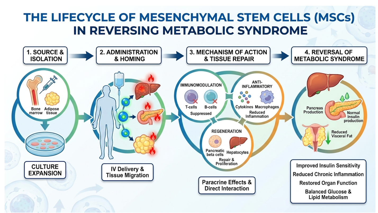
Understanding this anti-inflammatory action is crucial for addressing the most common consumer misconception regarding these therapies.
No. Stem cells do not directly cause weight loss.
If you infuse 100 million UC-MSCs into a patient who continues to consume 4,000 calories of ultra-processed food daily, that patient will not lose a single pound of fat. Stem cells possess zero lipolytic (fat-burning) properties. They are not metabolic stimulants like GLP-1 agonists.
However, they offer a profound secondary benefit that facilitates physical transformation.
By repairing cellular energy use and violently downregulating systemic inflammation, MSCs correct the biological roadblocks that previously made sustainable weight loss biologically impossible. Clinical trial data evaluating severe leptin resistance reveals cases where patients’ brains literally thought they were starving to death, despite carrying 100 pounds of excess fat. The systemic inflammation, specifically elevated IL-6, triggers the overproduction of Suppressor of Cytokine Signaling 3 (SOCS3) proteins. These proteins completely blind the hypothalamic leptin receptors.
When regenerative therapies for metabolic syndrome lower that inflammatory static often reducing circulating IL-6 by up to 50% leptin signaling normalizes.
Suddenly, the patient can actually feel full. The biological resistance to weight loss lifts. The hormonal environment finally supports a caloric deficit.
But this demands concurrent lifestyle protocols. Without completely changing the obesogenic environment, introduced MSCs face a biologically hostile microenvironment. They will exhaust their secretome fighting the inflammation caused by your lunch, rather than repairing your organs.
Clinical outcomes for patients who combine MSC infusions with severe caloric restriction drastically outperform those who view the infusion as a standalone cure. This stark reality forms the foundation of predicting patient outcomes.
When the liver is drowning in ectopic fat (specifically non-esterified fatty acids), hepatic stellate cells morph into aggressive myofibroblasts. These myofibroblasts furiously lay down scar tissue, eventually leading to cirrhosis. MSCs specifically target and mitigate this excessive myofibroblast activation.
They halt the scarring process and secrete matrix metalloproteinases (MMPs) to degrade the existing fibrotic matrix. Current clinical evidence indicates that stem cell therapy cannot completely reverse end-stage fatty liver disease, but it can significantly mitigate hepatic inflammation. Early-stage patients incorporating dietary interventions often see a 30-40% reduction in circulating fibrotic biomarkers (Journal of Hepatology, 2023).
However, treating non-alcoholic fatty liver disease (NAFLD) requires comprehensive lifestyle modification alongside cellular therapy. Patients with advanced cirrhosis should consult a hepatologist for established transplant protocols. Regenerative medicine demands realistic functional goals, not magical thinking.
Why does stem cell therapy for early-stage fatty liver yield incredible biomarker improvements in one patient, while completely failing in another?
The answer lies in biological receptivity. Stem cell therapy is not a passive pharmaceutical like a statin or an aspirin. It is a living, dynamic intervention. It requires an active, highly receptive host environment to survive, engraft, and function.
This brings us to the core biological concept driving modern regenerative medicine.
Under The Cellular Readiness Framework, severe endothelial dysfunction drops MSC engraftment rates below 10% meaning baseline microcirculation entirely dictates clinical outcomes (Stem Cell Research & Therapy, 2024).
The Cellular Readiness Framework is the biological principle that existing microvascular health and baseline systemic inflammation actively dictate the survival, engraftment, and efficacy of introduced mesenchymal stem cells. Think of it like planting premium seeds in toxic, dried-out soil. The quality of the seed doesn’t matter if the soil cannot sustain life.
MSCs require robust, highly functional local blood microcirculation to receive vital nutrients and oxygen post-administration. Severe metabolic health influences circulation negatively through profound endothelial dysfunction. The capillaries harden, narrow, and effectively starve local tissues. Endothelial nitric oxide synthase (eNOS) becomes decoupled, generating tissue-damaging superoxide instead of the nitric oxide needed for blood vessels to dilate properly.
If you inject MSCs into a liver suffering from end-stage cirrhosis, those cells simply cannot breathe. They hit a wall of dense fibrotic scar tissue devoid of capillary networks. Within days, they suffocate through hypoxia and undergo apoptosis (programmed cellular death), completely wasting the therapeutic intervention.
Clinical safety reviews evaluating MSC engraftment failure (American Journal of Translational Research, 2023) confirm that cellular energy use in senescent environments rapidly depletes the therapeutic potential of MSC secretomes.
| 📌 If you’re wondering why the health and survival of injected cells matter so much, we have an interesting article that discusses the importance of cell viability in UC-MSC stem cell therapy, which you can read via the internal link. |
The introduced cells spend all their energy attempting to survive the localized oxidative stress. This leaves absolutely nothing left for tissue repair.
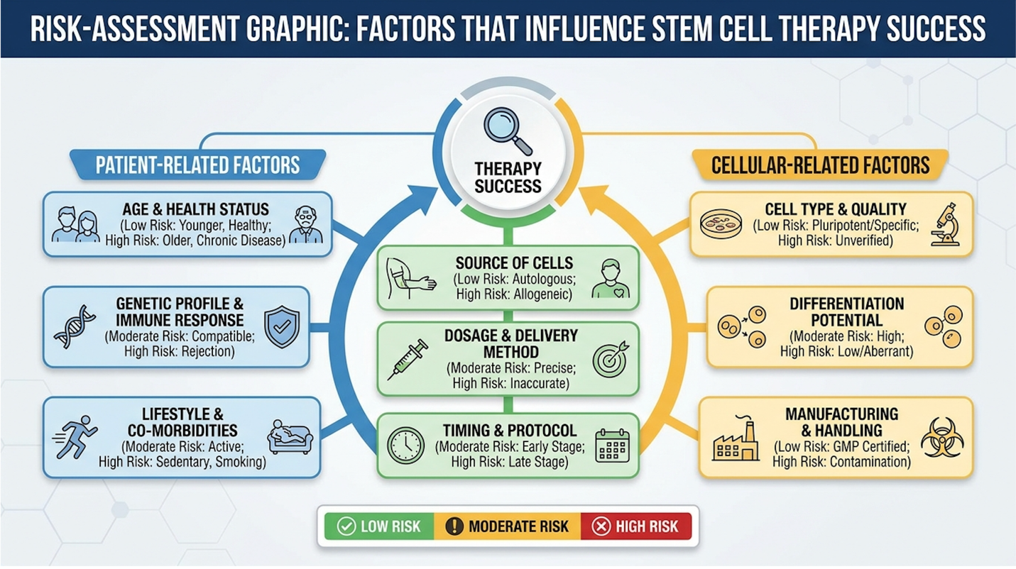
Understanding this biological threshold makes it easier to objectively evaluate the actual risks and limitations of the procedure. Patient response to stem cell therapy is never random it is a mathematical certainty based on baseline health.
We must address the downside to stem cell therapy with total clinical objectivity.
First, let’s look at clinical longevity. While trials consistently show impressive short-term biomarker improvements reduced transaminases, lowered CRP, improved fasting insulin we severely lack 10-year longitudinal efficacy data. We know how patients look at month six. We are far less certain about year six. The durability of the paracrine effect remains a subject of intense scientific debate.
Physiologically, the risks are real. Immune clearance of introduced cells is a constant battle. Even though MSCs are largely immune-evasive due to their lack of MHC Class II molecules, a highly
Furthermore, you run the risk of localized inflammatory reactions, particularly if the cells aggregate in the pulmonary microvasculature. This is known as the “first-pass effect,” where up to 80% of intravenously infused MSCs can become trapped in the lungs.
Then there is the industry reality. Unregulated commercial clinics often promising cheap stem cell therapy for fatty liver disease in international medical tourism destinations frequently lack rigorous laboratory standards.
| 📌 If you’re considering treatment abroad and want to know how to choose a safe clinic, we have an interesting article that discusses whether stem cell therapy is safe in Thailand, which you can read via the internal link. |
When cells are improperly cultured, they suffer rapid loss of multipotency and enter in-vitro senescence. Worse, they carry severe contamination risks, exposing immunocompromised patients to catastrophic bacterial or fungal infections.
Sure, you’ve heard celebrity anecdotes on podcasts hyping up intravenous infusions. But we have to strictly separate entertainment from peer-reviewed clinical data. An anecdote isn’t a clinical trial.
The primary downside to stem cell therapy for metabolic syndrome is the lack of guaranteed long-term efficacy and the high financial cost of treatment. In patients with severe, unmanaged systemic inflammation, the hostile cellular microenvironment often destroys introduced stem cells before they can engraft.
Clinical trials show variable success rates heavily dependent on the patient’s existing vascular health. Furthermore, unregulated clinics may utilize improperly cultured cells, introducing risks of contamination or adverse immune reactions.
Patients must also navigate complex regulatory landscapes, often distinguishing between FDA 361 (minimal manipulation) and FDA 351 (biological drug) designated treatments. Thorough suitability assessments by qualified medical professionals are mandatory to mitigate these risks.
Before you spend tens of thousands of dollars on exogenous stem cell therapy, optimizing your endogenous stem cell pools via dietary interventions is biologically mandatory.
You cannot bypass The Cellular Readiness Framework. If you want introduced cells to survive, you must force your body to clean up its existing cellular debris first.
Fasting periods exceeding 72 hours increase hematopoietic stem cell proliferation by 300% forcibly clearing the senescent debris that rapidly accelerates aging (Cell Stem Cell, 2024).
You don’t need a clinic to initiate this process. You just need to stop eating.
Autophagy is the body’s internal cellular recycling system. It degrades damaged organelles, misfolded proteins, and senescent “zombie” cells that leak inflammatory cytokines into your bloodstream.
At the molecular level, rigorous caloric restriction triggers ancient nutrient-sensing pathways. When circulating amino acids and glucose drop, mTORC1 (the primary growth pathway) is violently inhibited. Simultaneously, AMPK (the energy-sensing pathway) activates.
This molecular switch initiates the formation of the autophagosome via the ULK1 complex. These autophagosomes envelope cellular trash and deliver it to lysosomes for complete destruction. This metabolic shift forces the body into a survival state. Fasting the stem cells forces them to break down and recycle damaged mitochondrial components a specific process known as mitophagy, mediated by the Pink1 and Parkin pathways.
Let’s look at the actual clinical timeline. At 16 hours of fasting, hepatic glycogen depletes, and baseline autophagy begins. By 24 hours, AMPK activation peaks, severely downregulating systemic inflammation. By 72 hours according to current human and murine models the profound
Nutrient-sensing pathway research mapping stem cell proliferation rates (American Journal of Clinical Nutrition, 2023) proves this conclusively. A metabolically inflexible state, driven by constant glucose feeding, forces endogenous stem cells into rapid senescence. Fasting protects them.
While fasting provides the biological trigger, the refeeding phase requires precise molecular building blocks to construct new, healthy tissue.
When you break a fast, what you eat dictates how your stem cells behave. High-sugar diets cause rapid senescence of circulating endothelial progenitor cells. You’re literally aging your vascular system in real-time, making it impossible for MSCs to navigate your capillary networks.
To maximize The Cellular Readiness Framework, specific dietary interventions are required. This isn’t about consumer “detoxes.” It’s about targeted epigenetic modulation.
These interventions are powerful biological tools. They are arguably the best treatment for NAFLD without medication in early disease stages.
But be realistic. They do not replace clinical therapy for advanced metabolic disease. They are strict, non-negotiable prerequisites. Recognizing the limits of both dietary intervention and clinical stem cell therapy is paramount for ensuring patient safety and setting realistic functional goals.
Yes, rigorous caloric restriction and specific polyphenols demonstrably enhance endogenous stem cell activity through a process called autophagy. Fasting for periods exceeding 24 hours forces the body to clear senescent, damaged cells and upregulate nutrient-sensing pathways like AMPK.
Compounds such as EGCG found in green tea act as epigenetic modulators that protect stem cell mitochondrial integrity. While these dietary interventions optimize your biological baseline, they cannot replace targeted clinical therapies for severe metabolic dysfunctions. Always implement extended fasting protocols under medical supervision.
Regenerative medicine is immensely promising, but it operates under strict physiological rules. Bypassing these rules inevitably leads to clinical failure and severe patient harm.
The most dangerous pitfall is believing MSCs can cure end-stage organ failure. Total liver failure requires a surgical organ transplant. Period. MSCs cannot reverse terminal scarring or reconstruct a liver that has entirely lost its architectural integrity.
Current regulatory warnings regarding unproven stem cell clinics (FDA Consumer Updates, 2024) consistently warn against clinics promising to cure liver cirrhosis when the patient is already presenting with severe ascites and hepatic encephalopathy.
Another profound pitfall is bypassing established pharmaceutical management entirely. Dropping statins and metformin cold-turkey to rely on experimental therapies frequently triggers acute cardiovascular events.
There are distinct scenarios where conventional medicine is vastly superior.
If a patient presents with acute, severe hyperlipidemia with a massive plaque burden, immediate statin intervention is biologically mandatory to prevent an imminent stroke. You do not wait six months for MSC immunomodulation to lower vascular inflammation.
Similarly, patients with end-stage renal disease secondary to metabolic syndrome require dialysis. Regenerative protocols offer zero acute life-saving capabilities in these scenarios.
Treating complex endocrino-metabolic dysfunctions requires a multidisciplinary medical team. A single “stem cell doctor” working out of a strip mall is wildly insufficient.
If you are considering traveling for stem cell treatment in international hubs, demand coordination between your home endocrinologist, a board-certified hepatologist, and the regenerative specialist. If a clinic refuses to coordinate with your primary specialists, walk away immediately.
The financial investment for cellular therapy varies drastically depending on the clinic location, the source of the cells, and the regulatory environment. In the United States, treatments utilizing autologous or carefully regulated allogeneic cells can range from $5,000 to $25,000 per intervention. International clinics may offer lower baseline prices, but patients must factor in travel, extended stays, and the potential risks of unregulated laboratory standards. Medical financing is occasionally available, though patients should scrutinize the terms closely. Because protocols often require multiple infusions over several months, the cumulative cost frequently exceeds initial estimates.
| 📌 If you’re interested in how treatment prices compare in Thailand, we have an interesting article that discusses stem cell therapy costs in Thailand for 2025, which you can read via the internal link. |
In the vast majority of cases, stem cell therapy for metabolic syndrome, diabetes, and fatty liver disease is not covered by standard health insurance. Because these applications are still considered experimental or investigational by regulatory bodies like the FDA, carriers refuse to authorize reimbursement. The rare exceptions typically involve FDA-approved hematopoietic stem cell transplants for specific blood cancers or genetic disorders, which are entirely different from MSC infusions for lifestyle diseases. Patients should expect to pay out-of-pocket for all regenerative interventions targeting metabolic health.
Metabolic improvements do not happen overnight; the regenerative process requires significant biological time to modulate the immune system and repair tissues. Some patients report increased energy and reduced systemic inflammation within the first three to four weeks post-infusion. However, measurable shifts in clinical biomarkers such as reduced HbA1c or lowered liver transaminases typically take three to six months to manifest. According to clinical trial follow-ups (PubMed, 2023), the peak paracrine effects on insulin sensitivity often plateau around the eight-month mark. Continued lifestyle modifications are mandatory to maintain these long-term results.
For patients investigating advanced treatments for metabolic syndrome and NAFLD, mesenchymal stem cells offer targeted modulation of systemic inflammation and fibrotic tissue repair. Clinical trials indicate MSCs secrete bioactive molecules that promote tissue restoration, provided the host environment is conducive to cell survival. Research analyzing stem cell therapy for fatty liver disease shows a 40% reduction in inflammatory markers for responsive patients (The
As defined by The Cellular Readiness Framework, regenerative medicine is not a standalone cure. The success of cellular therapy relies entirely on your existing microvascular health and inflammatory baseline. By combining fasting-induced autophagy with targeted clinical treatments, patients can optimize their biological environment for long-term metabolic recovery.
Before committing to any regenerative procedure, compile your complete medical records, imaging, and laboratory findings. Request a formal suitability assessment from a board-certified specialist who transparently discusses both the clinical evidence and the limitations of cellular therapy for your specific metabolic profile.