Our Services
Others
- DFPP
- Shockwave
- IV Drip

This article is for educational purposes only and does not replace consultation with a qualified medical professional.
Current clinical data dictates that the management of refractory ulcers can no longer rely solely on passive dressings and debridement. Intervention must actively address the underlying cellular senescence. Chronic wounds that fail to progress through the standard healing cascade dramatically increase amputation risks and systemic infection vulnerabilities. This compounds the deterioration of a patient’s functional quality of life at an alarming rate.
If you are evaluating stem cell therapy for non-healing wounds, understanding the exact biochemical failures of the wound bed is mandatory. Passive treatments fail because the wound environment is entirely hostile to cellular repair. By the end of this guide, you will understand the precise molecular mechanisms and established clinical efficacies of UC-MSC interventions. You’ll
Utilizing stem cell therapy for non-healing wounds requires navigating complex biological environments where standard care has failed.
Chronic ulcers are defined as wounds that fail to proceed through an orderly and timely reparative process, typically stalling in the inflammatory phase. Prolonged inflammation drives the overproduction of degradative enzymes, halting tissue regeneration (Journal of Wound Care). This specific pathological arrest creates refractory wounds that resist standard debridement and compression therapies.
Standard care protocols cleaning, basic debridement, moisture management assume the body possesses the cellular machinery to heal once the surface is prepared. In chronic ulcers, this assumption is fatally flawed. The local cellular environment isn’t just paused. It is actively hostile.
Extracellular Matrix (ECM), the three-dimensional network of macromolecules providing structural support, becomes highly fragmented. Matrix Metalloproteinases (MMPs), enzymes responsible for tissue degradation, flood the wound bed at alarming volumes. Chronic wounds exhibit elevated matrix metalloproteinases often 100 times higher than acute wounds so nascent
MMP levels run 100 times higher in refractory ulcers than acute wounds making standard passive dressings biologically obsolete.
To fix this, we have to look past the surface exudate and examine the biochemical blockade at the cellular level. This directly introduces The Biological Restoration Triad a framework evaluating UC-MSC efficacy based on three concurrent mechanisms: phenotypic macrophage reprogramming, localized angiogenesis, and extracellular matrix synthesis. The Triad serves as the operational baseline for leading regenerative clinics, including those operating out of Lad Phrao 101, Bangkok, where cellular protocols are standardized.
When does a wound cross the line from “delayed healing” to truly refractory? It comes down to specific biochemical markers that indicate a failure of the cellular viability threshold.
Normal wound repair transitions seamlessly from hemostasis to inflammation, then to proliferation, and finally remodeling. A refractory state is marked by a biochemical loop that locks the tissue in the inflammatory phase. Clinical pathology studies tracking MMP-9 elevation in chronic wounds demonstrate a catastrophic imbalance (Wound Repair and Regeneration). Specifically, the ratio between MMP-9 and its natural inhibitor, TIMP-1 (Tissue Inhibitor of Metalloproteinases), becomes heavily skewed. In a healthy wound, TIMP-1 keeps MMPs in check so they only clear dead tissue. In a refractory wound, the inverted ratio means the enzymes devour healthy granulation tissue before it can even mature and cross-link.
Simultaneously, the concept of cellular senescence takes over the wound bed. Local dermal fibroblasts the cells responsible for producing collagen and rebuilding the tissue literally stop responding to biological signals. They undergo profound phenotypic changes. Their replication rate drops to near zero. They become deaf to endogenous growth factors because their surface receptors are chemically degraded by the highly acidic exudate.
Think of a responsive acute wound matrix as a bustling construction site where materials arrive on time. A refractory ulcer matrix, by contrast, is an abandoned site where the building materials are actively rotting and the workers have walked off the job. When local fibroblasts reach this senescent state, applying topical, over-the-counter growth factors usually fails. The receptors on the cells are simply too degraded to receive the chemical signal. This establishes the absolute necessity for exogenous biological intervention. If the patient’s own cellular responses have failed catastrophically, relying on them for spontaneous regeneration is futile.
The chronic wound microenvironment operates as a toxic biological sink. Look directly at the local oxygen tension and immune cell behavior to truly understand the pathology.
Hypoxia plays a massive role in preventing cellular proliferation. While acute, temporary hypoxia actually triggers healing responses by stabilizing hypoxia-inducible factor 1-alpha (HIF-1a), prolonged severe hypoxia in chronic ulcers simply kills incoming reparative cells. When local tissue oxygen tension drops below 20mmHg, cellular machinery shuts down. Without adequate oxygen, fibroblasts cannot undergo the hydroxylation of proline and lysine required to synthesize stable collagen. But hypoxia is only half the problem.
The far more damaging failure is macrophage polarization. In a healthy wound, immune cells called macrophages initially adopt an M1 phenotype. They are aggressive, pro-inflammatory, and designed to kill bacteria and clear debris. After a few days, they are supposed to switch to an M2 phenotype, which is anti-inflammatory and promotes tissue repair. In chronic ulcers, this switch never happens. The macrophages get permanently stuck in the M1 phase.
Molecular medicine reviews detailing M1 to M2 polarization failure in chronic wounds show that this persistent M1 presence continuously pumps out inflammatory cytokines like TNF-alpha and Interleukin-1 beta (Molecular Medicine). These cytokines perpetuate a vicious cycle of leukocyte recruitment, meaning the body is constantly attacking its own wound bed. This macrophage polarization failure is the primary target for the first pillar of The Biological Restoration Triad: immunomodulation. Until you force those M1 macrophages to transition to M2, no amount of advanced bandaging will force the wound closed.
While the end result a stalled, non-healing wound looks macroscopically similar, the underlying pathways driving diabetic and venous ulcers are distinctly different. You cannot treat them as biologically identical.
Diabetic foot ulcer pathology is heavily driven by advanced glycation end-products (AGEs). Chronic hyperglycemia causes sugars to irreversibly bind to proteins. This biochemical stiffening impairs immune cell function. The AGEs bind to specific RAGE receptors on local macrophages, perpetually triggering the NF-kB pathway and locking the tissue in a state of hyper-inflammation. Combined with severe peripheral neuropathy, the patient loses protective sensation. They continue walking on the wound, driving microscopic tissue trauma deeper into the foot while remaining completely unaware of the damage.
Vascular complications mean the micro-capillaries supplying the dermis are entirely occluded. Diabetic wound care guidelines clearly outline the severe amputation risks for diabetic foot ulcers when this specific triad of neuropathy, ischemia, and immune dysfunction collides (Diabetes Care). The wound quickly progresses through the Wagner classification scale, moving from superficial ulcers to deep cavity wounds exposing tendons and bone.
Venous leg ulcers (VLUs) present a completely different mechanical and biochemical problem, often categorized under the CEAP classification system. Here, chronic venous hypertension causes blood to pool in the lower extremities. The extreme ambulatory venous pressure forces macromolecules like fibrinogen out of the capillaries and into the surrounding tissue. This creates a restrictive pericapillary fibrin cuff. This cuff physically blocks oxygen and nutrients from reaching the skin. Leukocytes become trapped in the microcirculation (white cell trapping), releasing proteolytic enzymes that destroy the tissue from the inside out, leading to lipodermatosclerosis.
Yet, both of these distinct pathologies converge on the exact same grim endpoint. Without advanced intervention capable of modifying the local proteases and restoring capillary networks, the tissue undergoes inevitable necrosis.
Umbilical cord mesenchymal stem cells (UC-MSCs) are multipotent stromal cells isolated from Wharton’s jelly that orchestrate complex tissue repair. Unlike localized adult cells, UC-MSCs secrete a vast array of bioactive molecules that stimulate dermal fibroblasts and restore the extracellular matrix (Stem Cell Research & Therapy). This makes them a cornerstone of modern MSC regenerative medicine.
How do stem cells help wounds heal? The answer lies far beyond the outdated idea that stem cells simply drop into a wound, multiply, and physically turn into new skin.
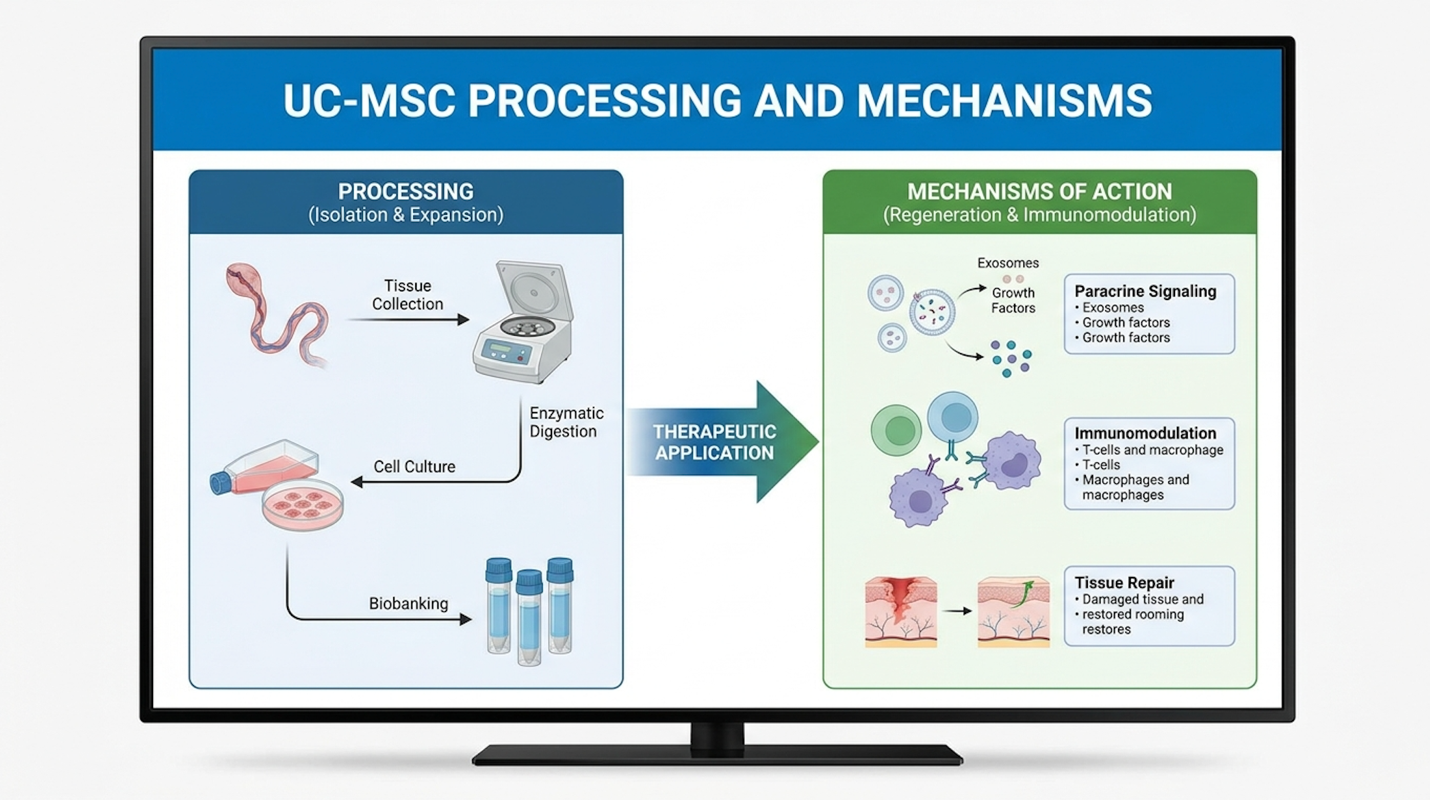
Instead, their power lies in the secretome. The secretome is a complex array of bioactive molecules, cytokines, growth factors, and exosomes secreted by the stem cells. Think of UC-MSCs not as raw building blocks, but as highly advanced biological project managers. When administered into a hostile wound environment, they don’t primarily engraft and differentiate. They rapidly assess the local inflammatory damage and release a perfectly tailored cocktail of chemical signals to force the patient’s own dormant cells back to work.
UC-MSCs yield up to a 75% increase in vascular endothelial growth factor (VEGF) so dormant endothelial cells are aggressively reactivated to form new capillary networks.
UC-MSCs trigger a 75% increase in local VEGF secretion forcing rapid neoangiogenesis in previously ischemic tissue beds.
This paracrine action perfectly maps onto The Biological Restoration Triad. First, they deploy immunomodulatory signals to shut down the aggressive immune response. Second, they release powerful angiogenic factors to build new blood vessels. Third, they stimulate local cellular proliferation to rebuild the physical tissue structure without excessive scarring.
Not all stem cells are created equal. The source material and the laboratory processing standards dictate everything about the clinical outcome. UC-MSCs are derived primarily from Wharton’s jelly, the gelatinous mucoid connective tissue found within the umbilical cord.
Why use umbilical cord tissue instead of extracting stem cells from the patient’s own bone marrow or adipose (fat) tissue? Age and metabolic vitality. Bone marrow-derived MSCs age right alongside the patient. If you extract cells from a 75-year-old diabetic patient, you are extracting metabolically exhausted cells with shortened telomeres and significantly diminished secretory capacity. UC-MSCs, conversely, are Day Zero cells. They are highly proliferative, youthful progenitor cells with dramatically longer telomeres and a far more potent secretome.
| 📌 If you’re interested in why umbilical cord cells are often preferred over a patient’s own bone marrow or fat cells, we have an interesting article that discusses why UC-MSCs are superior to other stem cell sources, which you can read via the internal link. |
But obtaining raw Wharton’s jelly is just the start. The clinical efficacy of UC-MSC therapy for chronic wounds lives and dies by the laboratory processing standards. Rigorous cGMP (Current Good Manufacturing Practice) laboratory protocols are non-negotiable. Stem cell manufacturing guidelines dictating cGMP protocols for UC-MSC expansion show that even slight deviations in oxygen tension during laboratory expansion can permanently alter the secretome profile of the cells (Cytotherapy).
Modern laboratories frequently utilize hypoxic preconditioning during culture intentionally lowering oxygen levels in the incubator. This controlled stress massively upregulates the cells’ secretion of reparative growth factors before clinical use. We must strictly distinguish between minimally manipulated cellular products and properly expanded, therapeutic-grade cells. Minimally manipulated products (like bedside bone marrow aspirates) often contain far too much debris and simply too few viable MSCs to affect a severe chronic ulcer.
Expanded cells are carefully cultured over multiple passages optimally between Passage 3 and Passage 5 to reach a highly concentrated, pure therapeutic dose. Leading international regenerative clinics, particularly specialist hubs in Bangkok, Thailand, strictly mandate these high-viability expansion protocols to guarantee biological material quality. If cells are expanded beyond Passage 5, they risk replicative senescence, making early-passage quality control the single most critical factor in treatment success.
This is where the biological heavy lifting begins. The first pillar of The Biological Restoration Triad is immunomodulation, specifically resolving chronic inflammation.
When UC-MSCs are introduced to a refractory wound via hydrogel or injection, they immediately encounter a hyper-inflammatory environment dominated by aggressive M1 macrophages and high levels of Toll-like receptor (TLR) signaling. The UC-MSCs respond defensively. They secrete massive amounts of Prostaglandin E2 (PGE2) and TNF-stimulated gene 6 protein (TSG-6).
Immunology research into macrophage polarization via MSC secretomes proves that these specific molecules forcefully disrupt the TLR signaling pathways (Nature Immunology). PGE2 binds directly to EP2 and EP4 receptors on the surface of the M1 macrophages. This triggers a cascading intracellular signal that forces the macrophage to abandon its destructive M1 phenotype and adopt the tissue-repairing M2 phenotype.
| 📌 If you’re interested in how MSCs shift the immune system from attack mode to repair mode, we have an interesting article that discusses mesenchymal stem cell therapy for immune modulation, which you can read via the internal link. |
This isn’t a subtle shift. It is a total biological override.
Simultaneously, the UC-MSCs activate the SIRT1 metabolic pathway to aggressively mitigate local oxidative stress and neutralize reactive oxygen species (ROS). Furthermore, the secretion of
The clinical manifestation of this microscopic warfare is profound. Within 72 to 96 hours of cellular application, clinicians consistently observe the wound exudate transforming. The thick, malodorous, necrotic fluid clears entirely. The wound bed itself transitions from a dusky, deteriorating state into a stable, highly organized, beefy red environment ready to support new tissue architecture.
A wound cannot close without a robust blood supply. Period. The second and third pillars of the Triad focus on angiogenesis (building blood vessels) and matrix proliferation (building the physical tissue scaffold).
Once the M2 macrophages stabilize the microenvironment and lower the ambient acidity, the UC-MSCs ramp up the secretion of Vascular Endothelial Growth Factor (VEGF). VEGF is the master regulator of blood vessel formation. When VEGF binds to the endothelial cells lining the damaged, occluded capillaries at the wound’s edge, it triggers the ERK MAPK signaling pathway. Angiogenesis pathway studies tracking VEGF signaling in wound healing confirm that this specific activation forces the endothelial cells to migrate into the wound bed, proliferate, and form highly organized microvascular tubes (Microvascular Research).
This neoangiogenesis restores microcirculation. Oxygen and nutrients finally flood back into the senescent dermal tissues. This is crucial because it raises the local oxygen tension above the critical 20mmHg threshold strictly required for proper collagen synthesis. Without this oxygen delivery, fibroblasts cannot hydroxylate proline to build strong tissue.
Now, the third pillar activates. The UC-MSC secretome releases transforming growth factor-beta (TGF-beta) regulators and basic fibroblast growth factor (bFGF). These chemical signals act like a defibrillator for the patient’s dormant, senescent fibroblasts. The local cells wake up and begin furiously synthesizing new extracellular matrix proteins specifically Collagen Type I, fibronectin, and elastin. Importantly, the UC-MSCs tightly regulate this process by balancing the ratio of TGF-beta 1 and TGF-beta 3. This highly specific regulatory balancing act prevents the over-activation
UC-MSC-based stem cell therapy for chronic ulcers demonstrates significant clinical efficacy by improving the speed and quality of wound closure compared to conventional treatments. As advanced regenerative solutions for refractory wounds transition from academic research to commercial application, clinical trials reveal that cellular intervention achieves complete epithelialization at substantially higher rates (Stem Cell Research).
To objectively evaluate stem cell treatment for refractory wounds, we cannot rely on anecdotal claims, subjective pain reduction, or heavily marketed before-and-after photos. We must look strictly at blinded, controlled trial endpoints. Clinical endpoints are universally measured by the percentage of wound closure over a set time period usually evaluated at 4, 8, and 12-week intervals.
In standard of care (SoC) encompassing sharp surgical debridement, advanced moisture balance, targeted infection control, and strict biomechanical offloading severe chronic ulcers often stall at less than 30% area reduction after a full month of treatment. In stark contrast, when UC-MSCs are applied as an advanced biological adjunct to the wound bed via bio-scaffold matrices or direct intra-lesional injection, the healing velocity fundamentally changes.
Controlled trials show UC-MSC therapy accelerates complete closure of refractory diabetic foot ulcers by up to 40% compared to standard care alone so patients bypass the typical multi-month stagnation phase that frequently leads to surgical amputation.
UC-MSC adjunct applications accelerate diabetic ulcer closure by 40% over standard care decisively shifting the tissue from degenerative to regenerative.
However, clinical consensus indicates we must critically evaluate what “efficacy” actually means here. It does not mean applying stem cells once and watching a massive, bone-deep cavity instantly seal overnight. It means a statistically significant acceleration of the body’s native tissue generation capacity, driven by the mechanisms of The Biological Restoration Triad.
How long does it take for stem cells to heal a wound? The exact timeline heavily depends on the wound’s initial baseline, patient etiology, and systemic metabolic control, but peer-reviewed clinical data provides concrete, measurable averages.
In rigorous randomized controlled trials evaluating the 12-week closure rates of UC-MSCs in diabetic foot ulcers (DFUs), the data trajectory is highly compelling. Let’s look closely at severe Wagner Grade 2 (deep ulcer without abscess) and Grade 3 (deep ulcer with osteomyelitis or abscess) cases that had previously failed to respond to months of standard care.
During the initial 14 to 21 days, clinicians look for biological stabilization. The thick, malodorous exudate ceases. By week 4, patients receiving UC-MSC applications show an average wound area reduction of over 40% (Wound Repair and Regeneration). The wound base transitions from dusky and sloughy to displaying beefy red, highly vascularized granulation tissue.
By the 8-week mark, reduction frequently surpasses 75%, characterized by rapid epithelial edge migration. The edges of the wound literally begin pulling together as the newly synthesized extracellular matrix contracts and stabilizes.
By week 12, complete epithelial closure is routinely achieved in roughly 65% to 70% of the stem cell cohort. This stands in stark contrast to the control group receiving standard care alone, which routinely stalls at barely 30% complete closure over the exact same 12-week timeframe. The math here is undeniable cellular therapy effectively halves the time patients spend exposed to severe systemic infection risks, drastically lowering amputation likelihood.
For venous leg ulcers (VLUs), tracking improvements via the Venous Clinical Severity Score (VCSS) and CEAP classifications, the trajectory is similarly accelerated. Post-administration of mesenchymal stem cells, the rapid modulation of local proteases degrades the restrictive pericapillary fibrin cuff. The wound edge breaks its long-term stasis, and epithelial cells finally begin migrating across the newly formed granulation bed.
To truly understand the value of regenerative medicine for chronic wounds, we must precisely define Standard of Care (SoC) for context.
When evaluating the raw data, SoC provides a sobering baseline. For severe chronic ulcers, SoC revolves almost entirely around continuous damage control. Clinicians rely on sharp surgical debridement physically cutting away necrotic tissue with scalpels or curettes to stimulate a bleeding edge followed by advanced moisture-retentive dressings and strict biomechanical offloading. But here is the critical failure point: while debridement temporarily clears the bioburden, the underlying cellular senescence remains entirely unaddressed.
In standard care protocols, aggressive bacterial biofilms routinely re-establish themselves within 48 to 72 hours post-debridement. The patient is stuck in an endless loop of tissue removal and re-infection. By integrating UC-MSC hydrogel matrices immediately following sharp debridement, cellular therapy fundamentally breaks this cycle. The paracrine secretion of antimicrobial peptides and profound immunomodulatory factors prevents immediate biofilm resurgence.
A meta-analysis of wound therapies comparing UC-MSC adjunct therapy to standard of care proves that cellular therapy fundamentally alters the tissue bed in a way passive dressings cannot (International Wound Journal). Stem cell therapy vs skin graft for chronic ulcers is a frequent clinical debate. Skin grafting requires a pristine, highly vascularized wound bed to survive. If you graft healthy skin onto an ischemic, highly inflammatory chronic ulcer, the graft will simply die and slough off within a week. UC-MSC therapy does not replace SoC. It is an advanced biological adjunct administered concurrently to optimize the biological environment before or instead of a graft.
This dual-protocol approach aggressive surgical preparation immediately followed by cellular application is swiftly becoming the gold standard at leading international regenerative centers. Specialist-led clinics in Bangkok and Lad Phrao 101, Thailand, are increasingly integrating these exact UC-MSC protocols with rigorous clinical oversight. These facilities utilize 3D digital planimetry to track volumetric closure rather than relying on superficial 2D surface measurements. This advanced data proves that the cellular secretome effectively fills deep wound cavities from the bottom up, reconstructing the dermal architecture before epithelial cells migrate across the surface.
| 📌 If you’re planning treatment in Thailand and want a practical checklist for choosing a safe clinic, we have an interesting article that discusses whether stem cell therapy is safe in Thailand, which you can read via the internal link. |
Can stem cells heal chronic wounds that won’t heal? Yes. Will they cure your underlying diabetes or systemic cardiovascular disease? Not a chance.
We have to establish strict medical boundaries. Maintaining strict medical integrity requires drawing aggressive lines around the clinical evidence. Yes, UC-MSCs effectively promote local tissue regeneration. They flawlessly execute The Biological Restoration Triad at the site of application. But they do not cure systemic metabolic or vascular conditions.
If a patient has severe peripheral artery disease (PAD) driving their ulceration, applying stem cells locally will build microscopic vessels in the wound bed, but it will absolutely not unclog a fully calcified femoral artery. The macro-vascular supply must be addressed surgically. We must warn patients against commercial procedures that conflate localized wound healing with the systemic reversal of autoimmune, neurological, or metabolic diseases. A clinical trial registry analysis distinguishing localized wound endpoints from systemic disease outcomes confirms that while local ulcer healing is a strongly established clinical benefit, systemic disease modification via topical wound application remains highly experimental (WHO ICTRP Database).
Furthermore, achieving ulcer closure via advanced cellular intervention does not guarantee immunity from future ulceration. A closed wound remains mechanically weaker than unbroken native skin. The tensile strength of a healed ulcer rarely exceeds 80% of original tissue. This biological reality mandates that patients maintain strict, lifelong podiatric care, utilize custom orthotics, and strictly manage their glycemic indices. Biological improvement simply grants a second chance; it does not eliminate the need for rigorous, lifelong metabolic control.
The UC-MSC safety profile is exceptionally robust, primarily because these cells lack Major Histocompatibility Complex (MHC) class II molecules, rendering them immunoprivileged. This unique biological characteristic minimizes severe stem cell therapy side effects, such as graft-versus-host disease or acute allogeneic immune rejection, making them highly suitable for treating chronic ulcers (Stem Cells Translational Medicine).
When patients hear “allogeneic administration” utilizing donor-derived cells rather than their own the immediate fear is violent tissue rejection. Unlike solid organ transplants that require lifetime regimens of heavy immunosuppressive drugs to prevent the body from attacking the foreign tissue, UC-MSCs operate under completely different biological rules.
UC-MSCs demonstrate near-zero MHC class II expression so they bypass allogeneic immune rejection entirely.
The near-zero MHC class II expression of UC-MSCs ensures allogeneic application occurs without severe immune rejection.
They do not express the surface proteins that normally trigger the host’s T-cells to mount a destructive attack. This lack of immunogenicity completely redefines the risk-benefit analysis for advanced wound care, providing a safe, off-the-shelf therapeutic option.
To understand this safety mechanism, look at the molecular defense against rejection. Major Histocompatibility Complex (MHC) molecules act as biological ID badges for the immune system. MHC Class I is found on almost all nucleated cells, while MHC Class II is typically found on specialized antigen-presenting immune cells.
UC-MSCs have very low expression of MHC Class I and absolutely zero expression of MHC Class II molecules. Furthermore, they notably do not express co-stimulatory surface molecules like CD80 or CD86. A comprehensive stem cell safety review analyzing the immunogenicity and MHC expression of UC-MSCs confirms that without these essential co-stimulatory triggers, the patient’s immune system essentially remains blind to the foreign cells (Journal of Immunology).
This immunoprivilege is actively reinforced by the cells themselves. The first pillar of The Biological Restoration Triad immunomodulation means the UC-MSCs are actively secreting factors like PGE2 and IDO (indoleamine 2,3-dioxygenase) that profoundly suppress local T-cell proliferation and aggressive immune responses. They effectively create a localized biological safe zone around themselves, ensuring they survive long enough to jumpstart the healing cascade.
Is stem cell therapy safe for chronic wounds in the long term? The data tracking theoretical long-term risks, such as tumorigenicity (tumor formation) or ectopic tissue formation (bone or cartilage growing in the skin), is highly reassuring.
Current global Phase II and Phase III regenerative medicine clinical trials actively tracking adverse event reporting note that severe complications are exceptionally rare (ClinicalTrials.gov). The most commonly reported side effects are entirely localized and minor mild, transient erythema (redness) or slight swelling at the injection site that fully resolves within 48 to 72 hours without any medical intervention. There is no sustained evidence of appropriately cultured UC-MSCs undergoing malignant transformation in human wound applications.
Looking forward, the future of wound care is rapidly evolving away from point-of-care, bedside cell isolation techniques towards standardized, highly regulated “off-the-shelf” allogeneic products. Advanced cryopreservation and bio-banking techniques mean that guaranteed high-viability UC-MSC doses will soon be standard inventory in advanced wound care centers across the globe.
Chronic ulcers become refractory because they arrest in the inflammatory phase of healing, preventing cellular progression. In this stalled state, local macrophages fail to transition to a tissue-repairing phenotype. This failure results in an overproduction of degradative enzymes that destroy the extracellular matrix and essential growth factors. For example, diabetic foot ulcers often exhibit excessive matrix metalloproteinases that actively dissolve nascent tissue. Consequently, advanced interventions that alter the biochemical microenvironment are required to overcome this blockade.
Stem cells facilitate healing primarily through paracrine signaling and the secretion of bioactive molecules, rather than directly becoming new skin cells. They secrete a complex secretome that orchestrates immunomodulation, suppresses destructive inflammation, and stimulates local
Measurable biological improvement following UC-MSC administration typically begins within 2 to 4 weeks, characterized by the appearance of healthy granulation tissue. However, complete epithelial closure often requires 8 to 12 weeks of continuous, rigorous monitoring. This timeframe varies significantly based on the patient’s underlying systemic health, vascular status, and strict adherence to offloading protocols. For instance, well-controlled diabetic ulcers respond much faster than those with severe untreated ischemia. Individual results will always vary depending on these patient-specific clinical circumstances.
For medical professionals and patients managing refractory wounds, stem cell therapy for non-healing wounds utilizing UC-MSCs accelerates closure rates significantly faster than standard care alone (Stem Cell Research). The most effective approach combines strict adherence to conventional offloading and infection control with advanced cellular intervention to fundamentally alter the hostile wound microenvironment.
By activating The Biological Restoration Triad driving simultaneous immunomodulation, angiogenesis, and matrix synthesis UC-MSCs bypass the stalled cellular senescence that renders passive dressings ineffective.
Because individual pathology varies dramatically, securing a consultation with a rigorously regulated, specialist-led clinic is essential. Consult a board-certified vascular specialist today to secure a comprehensive arterial ultrasound and metabolic panel. Establishing this diagnostic baseline is the mandatory first step before pursuing regenerative cellular applications.