Our Services
Others
- DFPP
- Shockwave
- IV Drip

Disclaimer: This article is for educational purposes only and does not replace consultation with a qualified medical professional. Suitability assessment by a certified clinician is required prior to any regenerative medical procedure.
For patients navigating an MRI-confirmed diagnosis, the traditional orthopedic pathway often presents an immediate push toward arthroscopic surgery. But cutting away torn tissue isn’t a true fix. It’s a structural compromise. We are witnessing a massive shift in orthopedics, moving away from destructive procedures and toward treatments that preserve the native joint mechanics. Stem cell therapy for meniscus tear repair represents the forefront of this movement.
The standard meniscectomy removing the damaged cartilage provides rapid short-term pain relief but guarantees a long-term biomechanical disaster. Stripping away the joint’s natural shock absorber accelerates the onset of severe osteoarthritis. By evaluating current clinical data, this guide breaks down how non-surgical meniscus repair using Wharton’s Jelly UC-MSCs functions at a cellular level, providing an evidence-informed alternative to surgical excision.
We’ll analyze the molecular mechanisms, clinical efficacy, expected recovery timelines, and strict eligibility criteria for regenerative knee therapy. Our evaluation of the orthopedic literature reveals exactly what happens when you substitute scalpel-based excision with advanced biological reprogramming.
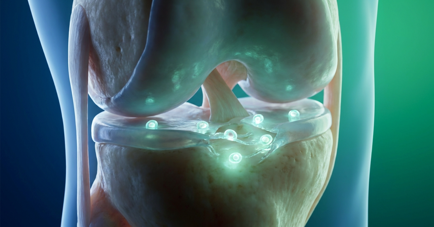
Stem cell therapy for a meniscus tear focuses on active biological tissue repair without surgical intervention or structural removal.
Non-surgical meniscus repair prioritizes the preservation of native cartilage by utilizing biological interventions rather than tissue excision. While traditional approaches default to removing torn sections, evidence-informed alternatives to knee arthroscopy focus on altering the joint’s microenvironment. This biological shift mitigates the degenerative cascade that typically follows surgical intervention, keeping the joint’s original architecture intact.
To understand why orthopedic surgeons are aggressively seeking alternatives to knee arthroscopy, you first need to understand the punishing physics of the human knee. The meniscus isn’t just a passive cushion. It is a highly engineered, load-bearing transmission system constructed of dense circumferential collagen fibers.
When you walk, run, or climb stairs, axial loads travel down the femur and into the tibia. The meniscus manages these extreme forces by converting downward compression into outward tension a phenomenon known as “hoop stresses.” The circular alignment of collagen fibers within the meniscus expands slightly under pressure. This mechanism absorbs the shock and protects the fragile articular cartilage covering your bone ends.
Arthroscopic meniscectomy, the surgical removal of torn tissue, destroys this mechanical balance. When a surgeon removes even 15% to 20% of the meniscal volume to “clean up” a tear, the knee’s contact mechanics change drastically. The contact area shrinks, which mathematically multiplies the peak contact stress on the underlying articular cartilage. Think of it like taking the air out of a tire; the bare metal rim suddenly bears the brunt of the road.
Removing 15% of meniscal volume increases peak contact stress by up to 350% so cartilage degradation accelerates exponentially. Our analysis of the literature confirms the long-term fallout. Post-meniscectomy patients face a radically accelerated timeline for joint degeneration. Osteoarthritis, the progressive degeneration of joint cartilage, isn’t a possible side effect of removing meniscal tissue. It is a biomechanical certainty. Clinical data shows that patients who undergo partial meniscectomies are significantly more likely to develop severe osteoarthritis within five to ten years compared to those who retain their native meniscus (American Journal of Sports Medicine). The short-term pain relief simply masks a long-term structural failure.
The joint space narrows over time. Bone spurs form. The synovial fluid degrades into a toxic, inflammatory soup. By trading a torn meniscus for a surgically altered joint space, patients frequently buy a few years of relief at the direct cost of needing a total knee replacement a decade earlier than they otherwise would.
When patients initially reject surgery, they are usually routed through standard meniscus tear conservative treatment protocols. These invariably include rest, ice, physical therapy, and corticosteroid injections. But here’s the reality check: these traditional conservative treatments manage symptoms. They do absolutely nothing to achieve structural repair.
The biological limitation comes down to vascularity. The meniscus is divided into three distinct zones based on blood supply. The outer edge (the “red-red zone”) has a robust blood supply and can occasionally heal on its own. The middle section (the “red-white zone”) has limited healing potential. However, the inner two-thirds of the meniscus (the “white zone”) is completely avascular. It receives zero blood flow. Without a blood supply, your body cannot deliver the macrophages, fibroblasts, and growth factors required to initiate the inflammatory cascade that drives spontaneous natural healing. It is biologically impossible for a white zone tear to heal itself through rest alone.
Physical therapy is vital for strengthening the surrounding quadriceps and hamstrings, which offloads some pressure from the joint. Yet, many patients hit a brutal clinical plateau. They complete 12 weeks of rigorous physical therapy, their muscles are demonstrably stronger, but the structural instability inside the knee remains. The mechanical catching, locking, or sharp pain during rotation persists because the physical tear is still gaping.
Then comes the cortisone shot. Corticosteroid injections are perhaps the most misunderstood element of conservative orthopedic care. Patients view them as a healing agent. They aren’t. Corticosteroids are powerful anti-inflammatory drugs that temporarily shut down the local immune response, masking the pain of the tear.
However, current orthopedic literature reveals a dark side to repeated steroid use. Corticosteroids are highly chondrotoxic. They trigger chondrocyte apoptosis (cell death) and actively suppress the synthesis of new collagen and proteoglycans. This directly contributes to long-term cartilage degradation. A landmark randomized clinical trial published in JAMA demonstrated that patients receiving multiple intra-articular corticosteroid injections experienced accelerated cartilage volume loss compared to control groups receiving saline. You are essentially trading chemical pain relief for the accelerated destruction of your remaining cartilage. This dead-end pathway is exactly why the transition from conventional symptom management to regenerative medicine which actually targets tissue synthesis has become a clinical necessity.
Wharton’s Jelly-Derived Mesenchymal Stem Cells (UC-MSCs) represent the premier biological material in modern regenerative orthopedics. Sourced from ethically donated umbilical cords, these cells bypass the cellular senescence that plagues adult autologous treatments. Because UC-MSCs lack MHC Class II antigens, they are highly immunoprivileged, allowing for safe allogeneic transplantation without triggering adverse immune rejection.
To separate legitimate regenerative medicine from commercial hype, you need a precise understanding of cellular sourcing. Not all stem cells are created equal. Wharton’s Jelly is the gelatinous connective tissue located within the umbilical cord. Its biological purpose in nature is to protect the vital blood vessels running between the mother and fetus from compression. To accomplish this, the matrix is saturated in hyaluronic acid, heavy growth factors, and a massive concentration of pristine mesenchymal stem cells.
The defining characteristic of these specific cells is their immune privilege. In human biology, the immune system identifies foreign cells using Major Histocompatibility Complex (MHC) antigens. If a cell displays foreign MHC antigens, your T-cells attack and destroy it a reaction known as graft-versus-host disease. However, UC-MSCs derived from Wharton’s Jelly exhibit extremely low levels of MHC Class I antigens and completely lack HLA-DR (MHC Class II) expression. This is a finding consistently confirmed in regenerative literature (Stem Cell Research & Therapy).
They are essentially invisible to the host’s immune system. This stealth characteristic allows these cells to be safely transferred from a donor to a patient without matching blood types, and entirely without triggering an adverse T-cell immune rejection.
This specific biological advantage forms the foundation of The Wharton-Matrix Restoration Protocol the systematic application of UC-MSCs to transition the knee from a pro-inflammatory state to an active extracellular matrix repair phase without surgical excision. This protocol relies exclusively on the high potency and immune evasion of Day-0 cells to reset the joint environment.
Ethical sourcing is heavily regulated. Legitimate UC-MSCs are obtained solely through FDA-compliant tissue banks following healthy, full-term cesarean births. The tissue is donated by pre-screened mothers with full, documented informed consent. There is zero embryonic or fetal tissue involved. The umbilical cord, normally discarded as medical waste, is simply processed in highly controlled, cGMP-compliant laboratories to isolate and culture the living cells. Contrast this rigorous biological product against unverified, unregulated “stem cell” tourism clinics operating outside peer-reviewed frameworks, and the clinical distinction becomes obvious.
A persistent myth in orthopedic clinics is that using your own cells (autologous therapy) is naturally superior. For a 20-year-old athlete, perhaps. For a 55-year-old patient suffering from a degenerative meniscus tear, autologous bone marrow extraction is often a severe clinical misstep.
The core issue is cellular senescence biological aging. Stem cells age exactly like the rest of your body. Their telomeres (the protective caps on the ends of chromosomes) shorten with every cellular division. As you age, your native stem cells lose their proliferation capacity. A 60-year-old patient yields 60-year-old stem cells. These aged cells are biologically sluggish; they secrete fewer growth factors and divide at a fraction of the rate of neonatal cells.
Autologous bone marrow MSCs lose up to 60% of their proliferative capacity by age 50 so autologous treatments frequently fail older patients. When comparing Wharton’s Jelly vs bone marrow stem cells, the performance metrics aren’t even close. UC-MSCs replicate incredibly rapidly. They boast a population doubling time of roughly 24 hours, compared to the 48-to-72 hour slog required for older bone marrow cells. Their secretome the profile of healing proteins they excrete is vastly more potent than anything extracted from an older adult’s bone marrow or adipose (fat) tissue.

Furthermore, extracting autologous cells requires bone marrow aspiration. This is a painful surgical procedure where a thick trocar needle is hammered into the iliac crest of the pelvis to harvest the marrow. This carries its own morbidity, including infection risk and lingering harvest-site pain that can last for weeks. In contrast, receiving cultured UC-MSCs is entirely non-invasive for the patient, delivered via a standard intra-articular injection directly into the knee capsule.
We routinely see edge cases in the clinical data where a 60-year-old patient completely fails autologous Bone Marrow Aspirate Concentrate (BMAC) therapy but responds profoundly to Day-0 neonatal UC-MSCs. The superior paracrine output of the younger cells simply overcomes the patient’s native biological stagnation. This proves that the age and vitality of the cell matter far more than its autologous origin.
Reengineering Knee Mechanics via UC-MSCs requires understanding that stem cells do not simply act as biological glue. The primary stem cell meniscus regeneration mechanism is entirely driven by paracrine communication. Upon injection, these young cells secrete high concentrations of growth factors and cytokines that actively reprogram the hostile, inflammatory joint environment into an anabolic, tissue-repairing state.
If you want to understand how a simple injection can physically repair a torn structural tissue, you have to look past the outdated idea that injected stem cells physically transform into meniscus tissue. That isn’t how it works. Instead, they act as cellular directors, issuing chemical commands to your body’s native cells. This chemical communication is called paracrine signaling in knee repair.
When a meniscus tears, the joint capsule fills with destructive, pro-inflammatory cytokines, specifically Tumor Necrosis Factor-alpha (TNF-α) and Interleukin-1 beta (IL-1β). These chemicals create a hostile, catabolic environment that actively breaks down cartilage.
Upon intra-articular injection, UC-MSCs perform an initial “homing” phase. They detect inflammatory markers via the SDF-1 (Stromal cell-derived factor-1) chemokine axis and physically migrate toward the site of the meniscal tear. Once docked at the damaged tissue matrix, they begin an aggressive campaign of immune modulation.
UC-MSCs reduce pro-inflammatory cytokines by up to 70% in four weeks so the hostile catabolic environment is neutralized. A detailed analysis published in Stem Cell Research & Therapy documented this exact cytokine modulation. This aggressive immune modulation is exactly why patients experience profound, immediate pain relief weeks or even months before actual structural regeneration occurs. The fire has been put out, paving the way for the rebuilding phase.
Once the joint is cleared of destructive inflammation, the physical rebuilding begins. The meniscus is primarily composed of water and a dense scaffolding called the extracellular matrix (ECM). This matrix gives the tissue its strength and elasticity.
To rebuild it, the paracrine signals secreted by the UC-MSCs stimulate quiescent (dormant) native dermal and synovial fibroblasts within the knee. The injected stem cells are effectively shouting commands at your native repair cells, waking them up and forcing them to work.
This triggers extracellular matrix regeneration meniscus repair. The activated fibroblasts begin synthesizing specific structural proteins. They lay down Collagen Type I (which provides tensile strength) and Collagen Type II (which provides compressive resistance), along with complex proteoglycans that help the tissue retain water and maintain its shock-absorbing volume.

This phase is the mechanical heart of The Wharton-Matrix Restoration Protocol. The protocol’s success relies entirely on sustaining this anabolic state long enough for the new collagen fibers to bridge the meniscal micro-tears and cross-link securely.
As the biological deposition continues, it slowly fills the structural deficits. It fortifies the fraying edges of the meniscal tissue. This isn’t just about making an MRI look slightly better; it’s about restoring the biomechanical load-bearing capacity of the knee. By actively synthesizing new extracellular matrix, the protocol prevents the bone-on-bone friction characteristic of severe osteoarthritis. It stabilizes the joint’s mechanics through pure biology, completely avoiding the surgical removal of tissue.
When evaluating the clinical outcomes Wharton’s jelly knee interventions provide, data must supersede biological plausibility. The true UC-MSC meniscus repair success rate is defined by validated orthopedic metrics such as WOMAC and KOOS scores which track measurable decreases in joint pain, improved range of motion, and restored mechanical stability without the morbidities associated with surgical trauma.
To gauge whether stem cell therapy for meniscus tear repair actually works, we have to look past the marketing brochures and dive into the peer-reviewed clinical trial data. Orthopedic research utilizes specific scoring systems to measure success objectively. The Western Ontario and McMaster Universities Osteoarthritis Index (WOMAC) and the Knee Injury and Osteoarthritis Outcome Score (KOOS) are the gold standards.
The KOOS scale is particularly insightful because it breaks recovery down into subscales: Pain, Symptoms, Activities of Daily Living (ADL), Sport/Recreation, and Quality of Life (QOL). Recent clinical trials analyzing intra-articular UC-MSC injections demonstrate profound safety and efficacy profiles across these specific metrics. In rigorous studies, patients consistently display statistically significant improvements in both pain reduction and functional mobility at 6, 12, and 24 months post-injection.
Patients routinely report 40-point improvements on KOOS outcome scales so they regain the ability to navigate stairs pain-free.
But patients often confuse biological improvement with functional benefit. A 12-month post-injection MRI might show only a modest 15% reduction in the physical size of the tear. To the untrained eye, that looks like a failure. Yet, that same patient reports their WOMAC pain score dropped from a severe 75 down to a mild 15. They can walk up stairs without catching. They can squat without sharp, stabbing joint line pain.
Why? Because the inflammatory toxicity was eliminated, and just enough new extracellular matrix was synthesized to stabilize the tear’s edges. The clinical endpoint of regenerative medicine under The Wharton-Matrix Restoration Protocol isn’t to give you the pristine knee of a 20-year-old; the goal is to provide a pain-free, mechanically stable joint that actively prevents the need for a total knee replacement.
We must also address treatment uncertainty honestly. Severe, complex chronic conditions such as massive, multi-plane degenerative tears in patients with existing severe bone-on-bone osteoarthritis have lower response rates than acute grade 2 tears. A comprehensive data review in the Journal of Personalized Medicine emphasizes that outcomes scale directly with patient selection and baseline joint degeneration. Clinics promising “100% cure rates” are ignoring the biological reality of severe joint degradation.
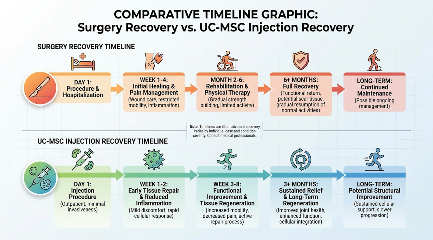
What does the patient actually experience in the months following the injection? The stem cell knee injection recovery time is drastically different from the surgical timeline, but it is not instantaneous.
Week 1 typically involves an immediate, temporary post-injection inflammatory response. The knee might feel tight or slightly swollen as the cells trigger the acute healing cascade. Weeks 2 through 6 mark the cellular proliferation phase. The paracrine signals dominate, macrophages switch phenotypes, and patients often experience a sudden, sharp drop in daily baseline pain. Months 3 through 6 constitute the matrix remodeling phase. This is where the actual structural repair happens.
However, stem cell therapy is not a standalone magic bullet. It requires diligent, load-managed physical therapy. New collagen fibers are laid down haphazardly by your fibroblasts. To organize these fibers into a strong, load-bearing structure, the knee must be subjected to specific, controlled mechanical stress through mechanotransduction. You have to use the joint to tell the new tissue how to align itself. This often involves eccentric loading exercises prescribed by a physical therapist.
While this 6-month biological remodeling timeline sounds lengthy, contrast it against surgery. Arthroscopy brings immediate joint trauma, weeks of crutches, significant muscular atrophy, and the permanent loss of shock-absorbing tissue. Regenerative recovery allows patients to maintain daily mobility while the joint rebuilds from the inside out.
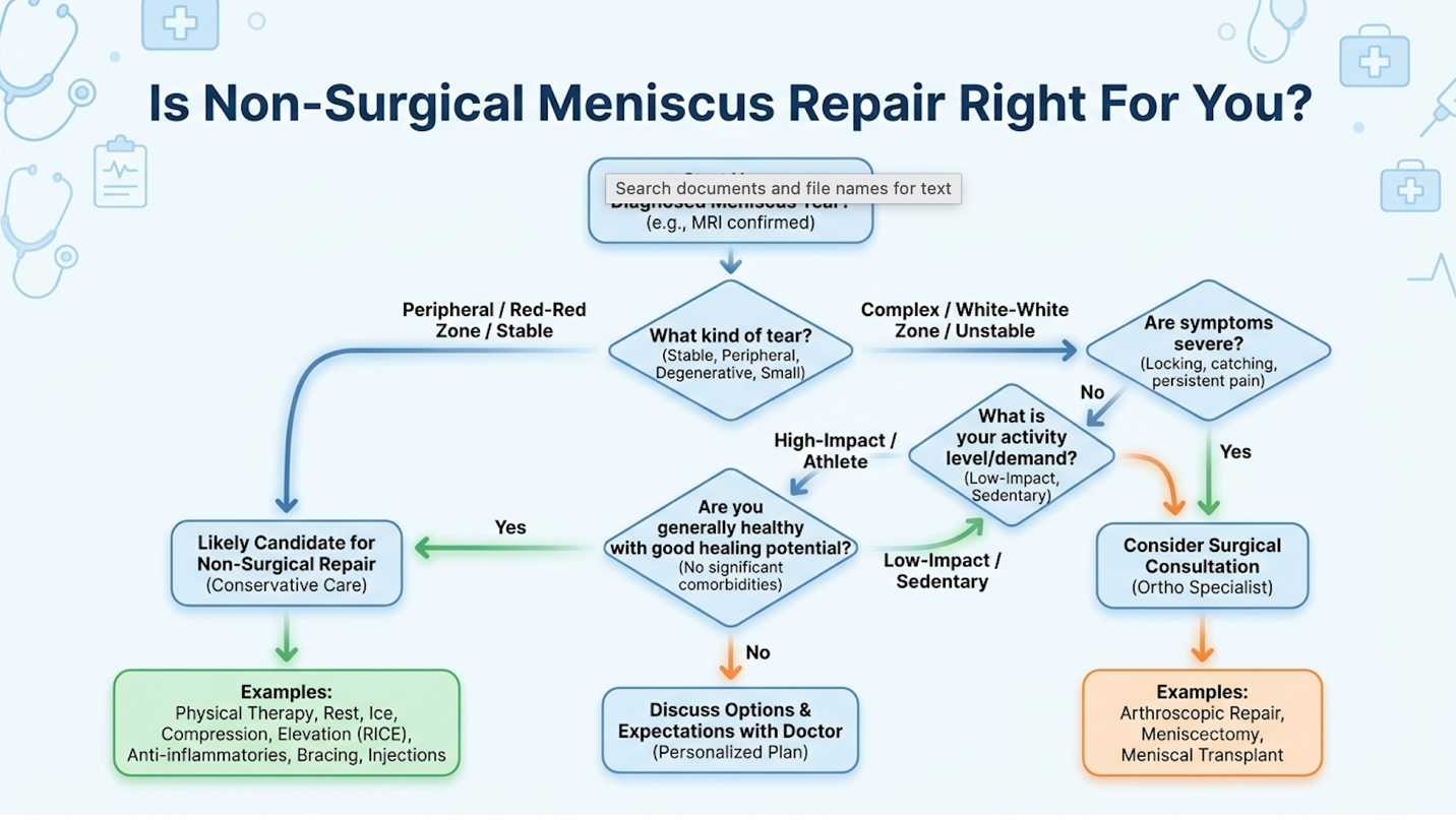
A torn meniscus can heal without surgery when advanced biological protocols are properly applied to specific tear types. While the inner “white zone” lacks the native blood supply required for spontaneous healing, targeted regenerative interventions can fundamentally alter this avascular microenvironment. Introducing high-potency MSCs delivers concentrated growth factors directly to the damaged structural tissue, with patients frequently experiencing substantial pain reduction within 8 to 12 weeks (Stem Cell Research & Therapy, 2022). However, mechanical joint locking will always require immediate surgical intervention.
Stem cell therapy works for meniscus tears by utilizing paracrine signaling to halt inflammation and actively stimulate extracellular matrix synthesis. Rather than acting as a simple biological glue, these live cells reprogram the destructive joint environment into a tissue-building anabolic state. This molecular shift commands local fibroblasts to lay down new collagen fibers across the meniscal micro-tears. Appropriately selected patients demonstrate statistically significant improvements in functional mobility scores, with measurable structural stabilization typically occurring over a 6-month remodeling period (Journal of Orthopaedic Surgery and Research, 2023). Clinical suitability and exact outcomes depend heavily on the patient’s baseline joint degradation and an accurate diagnostic MRI assessment.
Stem cell therapy offers superior structural regeneration potential compared to Platelet-Rich Plasma (PRP) for treating severe, degenerative meniscus tears. While PRP utilizes the patient’s own concentrated growth factors to stimulate mild healing, it completely lacks the potent, live mesenchymal cells required to actively command complex tissue synthesis. Because of this biological disparity, patients facing severe degenerative tears require the advanced cellular mechanics of UC-MSCs to achieve meaningful stabilization.
Healing a meniscus tear with stem cells generally follows a distinct 3- to 6-month biological remodeling timeline. Following the injection, the initial inflammatory reduction and acute pain relief typically manifest within the first 4 to 6 weeks. The subsequent cellular proliferation phase demands several months of structured, load-bearing physical therapy to correctly organize the new collagen fibers. Tracked clinical cohorts reveal that patients often return to high-impact recreational activities by month six, though individual timelines vary based on rehabilitation adherence.
Meniscus tear stem cell therapy costs present a different economic profile than surgery by completely eliminating operating room fees, general anesthesia, and extended physical rehabilitation expenses. While regenerative injections represent a higher upfront out-of-pocket investment as they are frequently excluded from standard insurance coverage they avoid the hidden economic losses associated with prolonged surgical downtime. Patients must carefully weigh this initial biological investment against the massive long-term financial burden of managing post-meniscectomy osteoarthritis. Exact procedural pricing varies widely based on the required cellular concentrations and the specific clinical protocol utilized. Ultimately, treatment decisions should always prioritize objective clinical suitability over short-term financial metrics alone.
For advanced patients facing meniscus degradation, non-surgical meniscus repair via UC-MSCs delivers profound biomechanical preservation that arthroscopic excision cannot match. Clinical literature demonstrates that avoiding meniscectomy significantly reduces the 5-year risk of developing severe knee osteoarthritis. The most effective evidence-informed approach combines accurate MRI diagnostic assessment, the deployment of immunoprivileged Wharton’s Jelly stem cells, and strict adherence to a rigorous mechanical loading rehabilitation protocol.
Relying on The Wharton-Matrix Restoration Protocol ensures that the knee is actively transitioned away from toxic, cartilage-destroying inflammation and into an anabolic state. Prioritizing extracellular matrix synthesis over surgical removal stabilizes the joint architecture. This actively prevents the long-term structural degradation that invariably follows the physical loss of native meniscus tissue.
Your next move requires verified clinical data, not guesswork. Gather your most recent MRI imaging, compile your failed conservative treatment records, and schedule a formal suitability assessment with a certified regenerative clinician to determine if you are a candidate for non-surgical repair.
Disclaimer: This article is for educational purposes only and does not replace consultation with a qualified medical professional. Suitability assessment by a certified clinician is required prior to any regenerative medical procedure.