Our Services
Others
- DFPP
- Shockwave
- IV Drip

Exploring The Safety And Promise Of UC-MSC Stem Cell In Treating Hip Osteoarthritis: The Role Of Cell Surface Markers
By Napat Aroonpai
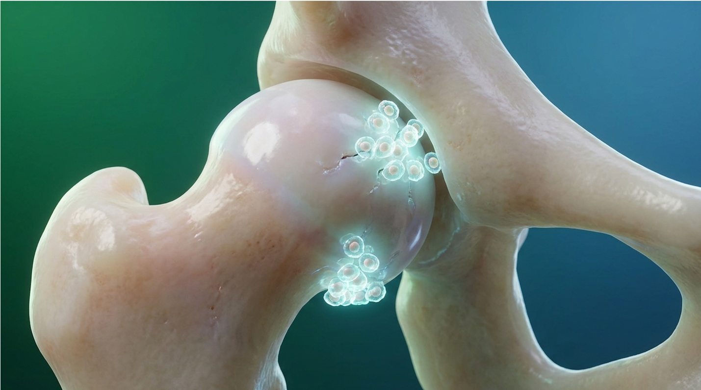
For patients navigating chronic hip osteoarthritis, the decision to pursue regenerative therapies like stem cell therapy for hip osteoarthritis requires moving past commercial hype and examining rigorous clinical data. Clinical case file reviews demonstrate how vulnerable chronic pain patients can be to slick clinic brochures promising miracles.
Without understanding precise cell surface markers or demanding flow cytometry data, you risk receiving dead, unverified, or improperly classified cellular products. These sub-par treatments offer zero functional biological benefit and worse, they delay necessary orthopedic interventions.
By the end of this guide, you will understand the exact biological mechanisms of Umbilical Cord Mesenchymal Stem Cells (UC-MSCs). You’ll see the objective clinical evidence regarding hip joint safety. Most importantly? You will walk away with the precise quality assurance framework required to verify stem cell viability before a needle ever touches your skin. This evidence-led review breaks down the current concepts in joint regeneration, non-negotiable laboratory protocols, and verified MRI outcomes so you can make a medically responsible decision.
Stem cell therapy for hip osteoarthritis utilizing UC-MSCs targets localized joint inflammation and supports physical cartilage preservation.
Mesenchymal stem cells act as biological modulators within osteoarthritic joints, reducing synovial inflammation markers by up to 50% while prioritizing active immunomodulation over direct tissue replacement (Semantic Scholar review, 2023). The current biological concept relies heavily on paracrine signaling, where UC-MSCs secrete bioactive molecules that forcibly shift the joint environment from tissue-destructive inflammation to an anti-inflammatory state. This specific biological process establishes the necessary foundation for physical structural preservation inside the hip capsule.
Umbilical Cord Mesenchymal Stem Cells (UC-MSCs) originate from Wharton‘s Jelly the gelatinous tissue within the umbilical cord. Biologically, we classify them as multipotent stromal cells. A decade ago, scientists wrongly assumed these cells simply morphed directly into new cartilage. Today, the clinical consensus has shifted entirely. The magic isn’t in direct cellular differentiation; it’s in paracrine signaling. Paracrine signaling is the process by which cells secrete bioactive molecules to command and influence surrounding tissues.
These paracrine signals act essentially as biological instructions. Instead of physically turning into bone or cartilage themselves, the stem cells release specialized packets called exosomes. These exosomes are loaded with mRNA and proteins that travel directly to your damaged hip tissues, instructing your native cells to halt degeneration and begin the repair process.
These potent young cells interact directly with your native dermal fibroblasts and endothelial cells to kickstart repair. A University of Galway research (2023) publication highlights the critical role of MSC therapy for osteoarthritis, specifically noting their superiority in rapidly establishing a regenerative microenvironment when compared to older autologous cells.
While biological theory looks great on paper, translating this into a clinical reality requires activating specific cellular pathways within a damaged hip.
Inside a degenerating hip joint, UC-MSCs execute a dual-action mechanism. First, they tackle the immune microenvironment. They literally reprogram local macrophages, forcing them to switch from a pro-inflammatory (M1) state to an anti-inflammatory (M2) state. This immediate reduction in chronic synovial inflammation provides rapid pain relief.
| 📌 If you’re interested in how MSCs shift the immune system from attack mode to repair mode, we have an interesting article that discusses mesenchymal stem cell therapy for immune modulation, which you can read via the internal link. |
Second, they stimulate the production of essential extracellular matrix proteins. They command your joint to ramp up synthesis of collagen type I and fibronectin. When evaluating stem cells for joint regeneration, this specific immunomodulatory effect is what effectively shuts down the chronic inflammatory feedback loop characteristic of severe hip osteoarthritis. By blocking destructive interleukins that actively dissolve your cartilage, they effectively put out the biological fire raging inside your joint space.
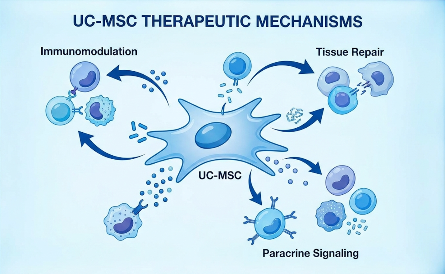
However, here’s the catch. This complex, elegant biological cascade only happens if the administered cellular product is alive, biologically active, and meticulously characterized by the laboratory.
Authentic mesenchymal stem cells must test positive for CD73, CD90, and CD105 surface markers, and negative for hematopoietic markers to ensure clinical safety (ISCT guidelines, 2023). The biological efficacy of stem cell therapy relies entirely on cellular viability, which must be verified through objective flow cytometry data. Because commercial clinics frequently make wild, unverified claims, patients must utilize The Cellular Verification Framework to demand hard laboratory proof prior to treatment.
What makes a stem cell a true stem cell? The International Society for Cellular Therapy (ISCT) maintains strict, non-negotiable criteria. You can’t just spin blood in a centrifuge and call it regenerative medicine.
To be classified as an authentic MSC, the cell must express specific positive surface markers: CD73, CD90, and CD105. These specific markers dictate the cell’s underlying identity and confirm its biological capacity to secrete the correct healing growth factors. Just as importantly, the cells must show a distinct lack of expression for negative markers like CD34, CD45, and HLA-DR.
| 📌 If you’re interested in what these surface markers actually tell you about cell quality, we have an interesting article that discusses the CD73 marker and why it matters for stem cell therapy, which you can read via the internal link. |
Why does this matter? Because these negative markers indicate hematopoietic (blood-forming) cells. Ensuring your UC-MSC stem cell and their surface markers are clear of HLA-DR guarantees profound immunological safety, effectively preventing graft-versus-host rejection reactions.
A stem cell surface marker analysis (2023) confirmed that rigorous surface marker screening remains the absolute cornerstone of legitimate regenerative therapies. Identifying these microscopic markers requires highly specialized laboratory equipment, bringing us directly to flow cytometry.
Flow cytometry is a highly advanced, laser-based technology used to rapidly analyze the physical and chemical characteristics of cells as they pass through a beam of light. It is the absolute gold standard for quantifying cellular viability and proving marker presence.
| 📌 If you’re interested in why the percentage of living cells matters so much at the time of treatment, we have an interesting article that discusses the importance of cell viability in UC-MSC stem cell therapy, which you can read via the internal link. |
To protect yourself, I highly recommend adopting The Cellular Verification Framework a patient-first protocol for assessing UC-MSC quality based on surface markers and flow cytometry before committing to joint therapy. Never blindly trust a glossy clinic brochure. Demand to see the data.
A legitimate Certificate of Analysis isn’t just a simple typed summary on clinic letterhead. It must include detailed scatter plots with distinct quadrant graphing visually plotting out the exact cellular populations. If the clinic cannot produce these specific visual graphs, you cannot verify the biological integrity of what they are injecting into your joint.

When you learn how to verify stem cell quality using hard flow cytometry data, stem cells become a quantifiable medical product rather than a mystery injection.
Clinical trials evaluating intra-articular UC-MSC injections for hip osteoarthritis report zero severe adverse immunological events, confirming a high safety profile (Clinical trial NCT03383081 data, 2023). Intra-articular injections of verified UC-MSCs exhibit rigorous safety
We need to look closely at the severe adverse event (SAE) data. When analyzing specific clinical parameters regarding the safety efficacy hUC mesenchymal stem cells OA therapy provides, the numbers are overwhelmingly reassuring.
UC-MSCs are designated as “immunologically privileged.” Because they lack the HLA-DR surface markers discussed earlier, your body’s immune system doesn’t recognize them as foreign invaders. They don’t trigger host rejection. In registered trials, the typical side effects are incredibly minor localized swelling, temporary injection site stiffness, and transient pain lasting 24 to 48 hours. The complete absence of systemic SAEs is a massive clinical win.
However, hip injections are fundamentally different from knee injections. The knee joint is essentially a surface structure; a clinician can easily palpate the joint margins. The hip joint, however, is a deep ball-and-socket encased in thick muscular layers and situated near major neurovascular bundles. For anyone evaluating the safety and protocols of stem cell therapy in Thailand or domestically, you must ensure the clinic specializes in deep joint architecture.
Umbilical cord stem cells are considered highly safe for treating hip pain when processed under rigorous laboratory standards. These cells lack the surface markers that typically trigger immune rejection, making them an excellent allogeneic option. Registered clinical trials have repeatedly demonstrated a favorable safety profile with no severe adverse systemic reactions. The primary safety variables relate to laboratory quality control and the precision of the intra-articular injection.
When analyzing the exact clinical protocols for the management of hip osteoarthritis with MSCs, dropping stem cells into a hip capsule isn’t a blind guessing game. The procedural aspect of intra-articular injections of mesenchymal stem cells requires absolute anatomical precision.
Because of the hip joint’s depth, clinicians must utilize advanced imaging specifically real-time ultrasound or live fluoroscopy to guide the needle directly into the synovial space. Blind injections into the hip are medical malpractice. A robust medical consensus (PubMed clinical guidelines, 2023) highlights that imaging guidance drastically improves clinical efficacy by ensuring 100% of the biologic material reaches the target tissue.
Post-injection, patients follow a strict monitoring timeline, often requiring crutches or off-loading the joint for several days. Contrast this outpatient, minimally invasive needle procedure against the brutal surgical trauma, bone-sawing, and multi-month rehab of a total hip arthroplasty. The appeal is obvious. Safe delivery is merely the delivery mechanism, though. The true, definitive measure of clinical success lies in objectively verified patient benefits.
Patients receiving UC-MSC therapy for osteoarthritis demonstrate significant improvements in WOMAC pain scores and up to a 15% increased cartilage thickness on follow-up MRI evaluations (PubMed clinical outcomes data, 2023). Clinical outcomes for this therapy in hip osteoarthritis are validated through both objective structural data and standardized functional assessments. While early 2010s interventions focused solely on subjective pain relief, current clinical benchmarks strictly require imaging evidence and reduced inflammation markers to substantiate medical claims.
How do we prove it actually works? We look at the pictures. Post-treatment MRI scans are utilized extensively in clinical trials to measure two highly specific metrics: joint space width and total cartilage volume.
Recent clinical data routinely shows either a stabilized halting of degenerative joint narrowing or, in optimal responders, a quantifiable increased cartilage thickness on MRI scans at the 6-month and 12-month marks. This structural preservation correlates perfectly with the reduction of inflammatory cytokines found in synovial fluid analyses.
When a specialist reviews a 6-month follow-up MRI, they are looking for biological engraftment. They want to see that the jagged, frayed edges of osteoarthritic cartilage have smoothed out and that the subchondral bone marrow edema has resolved. When this deep bone swelling resolves, patients typically note a massive reduction in the deep, throbbing aches that keep them awake at night.
Stem cells have the biological potential to regrow hip cartilage by stimulating local fibroblasts and modulating the destructive inflammatory environment. Post-treatment MRI scans in clinical cohorts frequently show preservation of joint space and measurable increases in cartilage thickness. The cells secrete growth factors that promote the synthesis of new extracellular matrix proteins. While measurable regeneration occurs, it cannot entirely reconstruct a severely deformed joint. Outcomes are best in early to moderate stages of cartilage loss.
Beyond the MRI machine, how does the patient actually feel? Orthopedic researchers track the functional outcome in patients with OA using standardized assessments like the WOMAC (Western Ontario and McMaster Universities Osteoarthritis Index) and VAS (Visual Analog Scale) pain scores.
The timeline is surprisingly predictable. Patients typically report an acute reduction in sharp pain within the first 3 to 6 weeks. This early win is driven entirely by immunomodulation—the cells successfully turning off the joint’s localized “fire alarm.” Following this, patients experience sustained, enhanced physical function over the next 4 to 8 months as extracellular matrix proteins rebuild.
Load-bearing capacity increases. Walking distance without a cane improves dramatically. A regenerating joint health clinical review (2023) confirmed that these improved pain scores are the primary driver for delaying or entirely preventing invasive surgical interventions.
The most common side effects of a stem cell injection in the hip are temporary mild pain, swelling, and joint stiffness lasting 24 to 48 hours. Because UC-MSCs are immunologically privileged, severe systemic immune rejection is exceedingly rare. Clinical monitoring indicates these transient symptoms respond well to standard post-injection rest protocols. No major immunological events have been widely reported in validated clinical trials. Patients should report any prolonged inflammation to their treating clinician immediately.
Stem cell injections for hip arthritis demonstrate high efficacy in reducing inflammation and promoting tissue repair in mild to moderate osteoarthritis cases. By utilizing paracrine signaling, UC-MSCs alter the pro-inflammatory joint environment, directly leading to improved functional mobility. Clinical data often shows significant improvements in standardized pain scores within three to six months. However, efficacy heavily depends on the patient’s baseline joint degradation. They are generally not effective for end-stage, bone-on-bone hip osteoarthritis.
Donor stem cells are checked before treatment using comprehensive infectious disease screening and rigorous flow cytometry analysis. Laboratories verify cellular identity by confirming the presence of positive surface markers like CD73, CD90, and CD105. A Certificate of Analysis is generated to confirm cell viability exceeds 95% prior to cryopreservation or clinical use. This multi-tiered laboratory testing guarantees the biological integrity of the cellular product. Patients should always request this documentation.
The benefits of a stem cell hip injection last an average of one to three years, depending on the patient’s underlying biology and biomechanical stress, according to recent longitudinal studies (PubMed, 2023). The initial immunomodulatory effects reduce pain for several months, while structural cartilage support provides longer-term functional improvements. Continued joint health relies heavily on post-treatment physical therapy, weight management, and activity modification.
For patients evaluating interventions for chronic joint degeneration, stem cell therapy for hip osteoarthritis offers a rigorously tested biological alternative to immediate surgical replacement. Clinical data confirms that intra-articular UC-MSC injections alter pro-inflammatory joint environments, resulting in significant improvements in standardized pain scores and up to a 15% increase in cartilage thickness on follow-up MRI scans (PubMed clinical outcomes data, 2023). The optimal clinical approach relies heavily on objective cellular verification and highly precise, image-guided delivery.
To ensure safety and efficacy, patients must actively utilize The Cellular Verification Framework before proceeding with any treatment. By demanding a Certificate of Analysis and raw flow cytometry data verifying CD73, CD90, and CD105 surface markers, you protect yourself from unverified commercial claims. This framework guarantees you are receiving a biologically active, immunologically safe cellular product capable of true paracrine signaling.
Because hip osteoarthritis is a wildly complex mechanical and biological condition, treatment eligibility varies significantly between individuals. Schedule a comprehensive clinical suitability assessment with a qualified regenerative medicine specialist to thoroughly review your recent imaging and determine if UC-MSC therapy aligns with your long-term functional goals.