Our Services
Others
- DFPP
- Shockwave
- IV Drip

For advanced osteoarthritis patients exploring offshore interventions, the contrast between domestic regulatory restrictions and Asian regenerative protocols requires objective clinical scrutiny. You’re likely reading this because you are staring down the barrel of a Total Hip Arthroplasty (THA) and wondering if there’s a legitimate biological alternative. Medical tourism is, quite frankly, fraught with marketing hyperbole. It leaves highly informed patients struggling to differentiate between verified mesenchymal stem cell (MSC) efficacy and unproven experimental procedures pushed by slick sales teams.
And let’s be honest when you’re dealing with chronic, mobility-stealing hip joint degeneration, the last thing you need is false hope. You need hard data.
By the end of this guide, you will possess a framework to objectively assess whether regenerative hip treatment with stem cells in Thailand aligns with your specific diagnosis and clinical needs. We aren’t going to feed you brochure copy. Instead, this analysis examines biological mechanisms, proven longevity timelines, FDA-regulated safety realities, and rigorous clinic credentialing. We will introduce and apply the Evidence-Led Cellular Assessment Framework (ECAF) a three-pillar evaluation model demanding you scrutinize the cellular source, the regulatory reality, and the biological longevity before booking a flight to Bangkok.
Navigating regenerative hip treatment with stem cells in Thailand requires applying the Evidence-Led Cellular Assessment Framework (ECAF) to verify clinical viability.
Cartilage regeneration stem cell therapy focuses on altering the joint’s pathological environment rather than simply depositing new structural tissue. Through advanced paracrine signaling, Mesenchymal Stem Cells (MSCs) multipotent stromal cells capable of profound immunomodulation secrete growth factors that suppress severe inflammation and stimulate native dermal fibroblasts. This biological intervention offers a mechanism for patients attempting to delay surgical joint replacement by actively changing how their joint manages friction and degradation. Make no mistake cellular medicine is not an overnight cure, but rather a profound biological shift that fundamentally alters the trajectory of joint degeneration.
Before exploring the deep biomolecular pathways, we must address the primary patient query: does this actually work for hips? Mesenchymal stem cell therapy demonstrates measurable clinical efficacy for moderate hip osteoarthritis by modulating chronic inflammation and promoting localized extracellular matrix synthesis. Clinical data continuously highlights significant improvements in both joint pain scores and functional mobility metrics when patients are correctly screened.
However, efficacy is highly conditional. The procedure is overwhelmingly most effective for patients caught in the early to moderate stages of joint degeneration (Kellgren-Lawrence Grades 2 and 3). Severe, bone-on-bone osteoarthritis typically fails to respond to biological treatments because the underlying structural scaffold is entirely gone. You can’t plant seeds in concrete. Suitability must be rigorously verified through high-resolution MRI imaging by an orthopedic specialist to ensure a high probability of cellular success.
The fundamental misunderstanding most patients have about biological joint repair is that they picture tiny cellular bricklayers rebuilding their hip cartilage overnight. That isn’t how orthobiologics actually function. The remarkable regenerative ability of umbilical cord-derived mesenchymal stem cells (UC-MSCs) is a direct result of their paracrine signaling rather than direct cellular differentiation. When injected into an arthritic hip, these cells act more like general contractors—they arrive at the damage site, secrete specific chemical signals, and command your body’s native repair mechanisms to get to work.
| 📌 If you’re interested in why umbilical cord cells are often preferred over bone marrow or fat-derived cells, we have an interesting article that discusses why UC-MSCs are superior to other stem cell sources, which you can read via the internal link. |
Specifically, MSCs secrete a complex cocktail of bioactive molecules. These include vascular endothelial growth factor (VEGF), epidermal growth factor, and basic fibroblast growth factor. These precise secretions forcefully shift the joint’s localized microenvironment. They downregulate destructive pro-inflammatory cytokines like TNF-alpha and Interleukin-1 which are actively eating away at your remaining cartilage matrix by activating tissue-destroying matrix metalloproteinases (MMPs). By directly inhibiting these specific destructive enzymes, stem cells essentially apply a biological brake to the degenerative process that causes daily friction and pain.
Simultaneously, this cellular signaling cascade promotes a critical M1 to M2 macrophage transition. This effectively forces the joint’s localized immune response to stop attacking the tissue and start preserving it. Mesenchymal stem cells (MSCs) have demonstrated significant improvements in WOMAC scores for joint pain (NIH, 2021). NIH research on cartilage regeneration confirms this mechanism, noting that MSCs show immense potential in regenerating damaged cartilage structures in hip osteoarthritis.
These bioactive factors also stimulate your quiescent dermal fibroblasts. Suddenly, dormant local cells wake up and begin synthesizing collagen type I, fibronectin, and elastin to reinforce the extracellular matrix. Clinical trials indicate a substantial reduction in pro-inflammatory markers within the first 90 days meaning the joint environment successfully transitions from active tissue degradation to measurable structural preservation. Furthermore, the introduction of high-viability cells ensures that this paracrine signaling remains active over several weeks, continuously conditioning the joint fluid to become more viscous and less hostile to native tissue.
Contrast this active, disease-modifying immunomodulation with the passive masking of pain provided by corticosteroid injections. Steroids offer rapid relief but are heavily documented to accelerate cartilage loss and induce localized osteoporosis over time. Recent studies in the Journal of Orthopaedic Translation emphasize that biological approaches using MSCs prioritize the long-term preservation of the native joint architecture over mere symptom suppression.
When evaluating a Stem Cell Joint Repair Treatment Osteoarthritis protocol against Total Hip Arthroplasty (THA) the traditional surgical joint replacement standard patients are comparing two entirely different philosophies of medicine. THA is an invasive mechanical salvage operation. It requires amputating the native femoral head, hollowing out the acetabulum with intramedullary reamers, and hammering in a combination of titanium and highly cross-linked polyethylene implants. While incredibly effective for end-stage joint failure, this procedure permanently alters your native anatomy.
The recovery from THA is long, painful, and carries inherent surgical risks ranging from deep vein thrombosis to adverse immune reactions against the metal hardware, not to mention the severe risk of periprosthetic joint infection. Conversely, a stem cell injection for hip pain is a minimally invasive outpatient procedure. Under live ultrasound or fluoroscopic guidance, millions of high-viability cells are delivered directly into the intra-articular space of the hip via a specialized needle. There is no surgical trauma. There is no extensive tissue cutting or disruption of the joint capsule.
Instead of triggering the massive fibrotic healing response typical of major surgery, MSCs deliver targeted anti-inflammatory macrophage reprogramming. THA provides excellent, immediate mechanical stability, but stem cells aim to preserve the native biomechanics and crucial bone stock of the hip. Consider a highly active 52-year-old patient suffering from early-stage hip joint degeneration.
| 📌 If you’re interested in how MSCs shift the immune system from attack mode to repair mode, we have an interesting article that discusses mesenchymal stem cell therapy for immune modulation, which you can read via the internal link. |
A THA at this age almost guarantees they will outlive the tribological lifespan of their mechanical hardware, requiring a complex, highly risky revision surgery in their 70s once osteolysis sets in. A successful biological intervention might delay that primary joint replacement by 5 to 10 years, pushing any potential revision surgery entirely out of their lifespan. This delay preserves essential quality of life during a patient’s most active decades, allowing them to continue low-impact sports
This brings us to the first pillar of the Evidence-Led Cellular Assessment Framework (ECAF): identifying patients with appropriate remaining joint space. The harsh reality of regenerative medicine is that it cannot fix everything. If a clinic tells you they can regrow a completely destroyed joint, grab your wallet and walk away immediately.
Bone-on-bone osteoarthritis classified clinically as Kellgren-Lawrence Grade 4—represents a complete structural collapse of the joint space. These patients are generally poor candidates for biological regeneration because the microenvironment is too severely compromised. The sheer mechanical grinding of a collapsed joint will instantly destroy newly introduced stem cells before they can even initiate paracrine signaling.
The ideal candidate for a non-surgical hip treatment looks completely different. We are looking for moderate (Grade 2 or 3) osteoarthritis where a viable cartilage scaffold still exists, even if it is actively fraying. We are looking for avascular necrosis candidates caught in the early stages (Ficat Stage I or II) before the femoral head collapses structurally. We are looking for patients with hip labral tears who haven’t yet developed end-stage, diffuse arthritis.
This is exactly why a rigorous suitability assessment utilizing current, high-resolution MRI data is a non-negotiable step in responsible medical tourism. A world-class stem cell clinic won’t even discuss travel dates until their orthopedic team has reviewed your imaging for subchondral sclerosis, osteophyte formation, and precise joint space narrowing. If you don’t meet the biological criteria, you must be rejected.
Determining how many years stem cell therapy lasts requires distinguishing between immediate anti-inflammatory relief and long-term structural stabilization. Unlike corticosteroids that mask symptoms temporarily, biological interventions initiate a cellular healing cascade that takes weeks to activate and months to fully mature within the hip joint. Patients who view stem cell injections
Patients frequently confuse different orthobiologics. They mistakenly group Platelet-Rich Plasma (PRP) an autologous concentration of human platelets in the exact same category as laboratory-expanded mesenchymal stem cells. This is a profound error when calculating clinical timelines. PRP relies on the rapid degranulation of alpha granules within the platelets to release growth factors. It creates a localized healing spike, but those factors dissipate quickly within a matter of days.
Because of this rapid dissipation, PRP treatments for severe hip osteoarthritis often necessitate booster injections within 6 to 12 months just to maintain baseline pain relief. MSCs operate on an entirely different, vastly prolonged biological timeline. Because they embed themselves in the synovial lining and continually secrete exosomes and growth factors, their mechanism of action is sustained over multiple cellular generations. Clinical follow-ups at 24 to 36 months indicate that MSC injections provide more durable symptom relief than temporary PRP treatments (NIH, 2019).
Long-term follow-up studies comparing MSCs to PRP have decisively shifted clinical consensus. Clinical follow-ups at 24 to 36 months indicate that MSC injections provide more durable symptom relief and structural cartilage preservation compared to the temporary effects of Platelet-Rich Plasma (PRP) (2019). Patients tracking functional mobility via WOMAC scores typically maintain over 60% of their peak improvements at the three-year mark proving biological durability significantly outlasts mechanical placebos.
I won’t offer you false guarantees, and neither should any clinician. However, evidence-informed tracking consistently shows that the disease-modifying potential of high-viability umbilical cord MSCs outpaces any PRP or stem-cell injections temporary relief. Patients frequently maintain their functional mobility improvements well past the three-year mark, provided they don’t actively sabotage the healing process. Data published in the Journal of Translational Medicine indicates that higher cell viability at the time of injection correlates directly with extended clinical longevity.
| 📌 If you’re interested in why the percentage of living cells matters so much at the time of treatment, we have an interesting article that discusses the importance of cell viability in UC-MSC stem cell therapy, which you can read via the internal link. |
The immediate logistical recovery time for a stem cell hip injection is relatively short, typically requiring 3 to 7 days of strict localized rest. During this initial window, patients are advised to utilize crutches to minimize compressive weight-bearing loads on the freshly injected joint capsule. While the injection itself is minimally invasive and requires only local anesthesia, the biological healing cascade takes weeks to initiate within the joint.
Patients often report noticeable reductions in acute joint inflammation and pain within the first 4 to 6 weeks as the paracrine signaling takes effect. However, true structural recovery is a marathon. Maximum functional improvement generally matures between the 3-month and 6-month marks as the newly synthesized extracellular matrix stabilizes. Strict adherence to progressive physical therapy guidelines after the initial rest period is vital to ensure the new collagen fibers align correctly along the lines of mechanical stress.
You cannot inject millions of premium stem cells into a toxic, inflamed host body and expect miracle longevity. Biological longevity is heavily dependent on the patient’s commitment to post-procedure rehabilitation. The newly forming extracellular matrix is incredibly fragile during the first 12 weeks. If you immediately return to high-impact distance running, you will mechanically shear the new tissue before it stabilizes.
Strict post-procedure joint offloading is mandatory. Top clinics recommend aquatic therapy and low-resistance stationary cycling to encourage mechanotransduction how cells sense and respond to physical loads which requires gentle, progressive stimulation to encourage proper tissue alignment. Systemic factors play an equally massive role in determining treatment lifespan. A patient’s biological age, baseline systemic inflammation, BMI, and overall metabolic health directly impact how long the regenerative effects last.
High blood sugar and chronic stress generate reactive oxygen species (ROS), which aggressively attack and degrade newly formed cartilage. Conversely, mitigating oxidative stress often through activating the SIRT1 longevity pathway via an anti-inflammatory diet and targeted supplementation extends cellular viability. Think of the joint like a garden. The stem cells are the
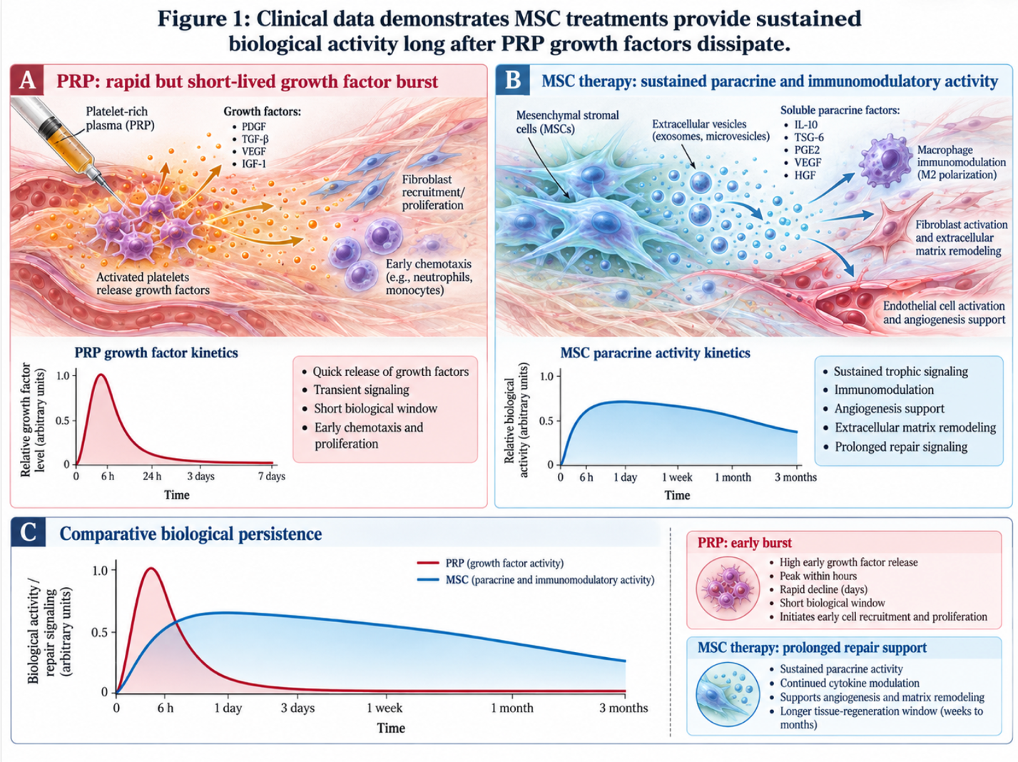
Figure 1: Clinical data demonstrates MSC treatments provide sustained biological activity long after PRP growth factors dissipate.
Understanding why the U.S. Food and Drug Administration (FDA)—the regulatory body overseeing biological products doesn’t approve stem cell therapy for orthopedic uses requires analyzing specific international regulatory frameworks. In the United States, cellular expansion is strictly classified as drug manufacturing, prompting patients seeking advanced regenerative orthopedics to evaluate the rigorous safety protocols maintained in Southeast Asian medical hubs. The regulatory environment dictates access, not necessarily biological validity.
You’ve likely wondered: if this technology is so effective, why is my local orthopedist only offering cortisone or surgery? The answer lies explicitly in the FDA’s dual-pathway regulatory system for human cells and tissues, outlined in 21 CFR Part 1271. The FDA divides biologics into the 361 pathway and the 351 pathway based on the concept of “minimal manipulation” and “homologous use.”
Under the 361 pathway, domestic doctors can extract your own cells (like bone marrow aspirate or adipose fat) and reinject them, provided they are minimally manipulated and used during the exact same surgical procedure. They cannot multiply them in a lab. The moment a laboratory takes those cells and expands (cultures) them over a three-week period to reach the optimal therapeutic dose of 50 to 100 million cells, the FDA instantly reclassifies the product under the 351 pathway as a biological drug.
Creating and approving a new 351 biological drug requires IND (Investigational New Drug) applications and a decade of Phase I-III clinical trials costing billions of dollars. Because no single company can patent naturally occurring umbilical cord stem cells to recoup those trial costs, the trials simply aren’t funded for joint repair. The FDA has not approved any stem cell-based products for use in orthopedic conditions like osteoarthritis (FDA, 2023). Official FDA warnings on unapproved treatments warn of significant safety risks from unproven domestic treatments trying to skirt these rules.
FDA regulations cap autologous bone marrow extractions at roughly 10% of the cellular volume found in expanded Thai protocols resulting in vastly inferior clinical outcomes for older patients relying on domestic US treatments. This is the exact regulatory reality pushing patients to Thailand. The Thai government framework allows for controlled, laboratory-expanded UC-MSCs within accredited clinical settings, utilizing the high-dose cell counts necessary for severe osteoarthritis that are legally restricted in the West.
Let’s address the most common, fear-driven query directly: can stem cells turn cancerous? This pervasive myth stems from a fundamental public misunderstanding of cellular biology, specifically pluripotent and multipotent cells. Not all stem cells are created equal, and their safety profiles diverge radically based on their specific lineage programming.
Pluripotent cells like embryonic stem cells or induced pluripotent stem cells (iPSCs) have the capacity to differentiate into literally any tissue type in the human body. Because their differentiation pathways are wild and unguided, injecting them carries a massive, well-documented oncology risk. The Mayo Clinic’s explanation of stem cell tumor risks notes that unproven therapies using unguided pluripotent stem cells have been associated with a severe risk of growing into irregular tissue types or tumors, known as teratomas.
However, reputable orthopedic clinics do not use pluripotent cells. They utilize adult, multipotent Mesenchymal Stem Cells. Multipotent cells are strictly lineage-restricted. An umbilical cord MSC can only differentiate into bone, cartilage, muscle, or fat. It literally lacks the biomolecular programming required to form a spontaneous teratoma.
Furthermore, normal multipotent MSCs are subject to the Hayflick limit they can only divide a certain number of times before naturally undergoing apoptosis (programmed cell death). They do not possess the immortal telomerase replication traits that characterize malignant cancer cells. Rigorous laboratory standards in top Thai clinics utilize exclusively these specialized multipotent UC-MSCs, completely negating the oncology risk associated with primitive cell types.
When discussing what are the potential downsides of stem cell injections, we have to separate the biological risks of the cells from the logistical risks of international medical tourism. The cellular therapy itself, when utilizing verified, lab-tested UC-MSCs, has an incredibly low adverse event profile. The real, catastrophic dangers stem entirely from unregulated “stem cell tourism” clinics operating in the shadows of the medical ecosystem.
These black-market operations offer dubious biological products at steep discounts without stringent QA/QC protocols. They skip expensive endotoxin screening. They avoid flow cytometry viability checks. Injecting dead, senescent, or contaminated cells into a major joint will cause severe septic arthritis, potentially destroying the joint completely. Moreover, the act of flying long-haul shortly after a medical procedure introduces a real, physiological risk of deep vein thrombosis (DVT). You must remain mobile and hydrated during your 15-hour flight home.
To protect yourself, you must verify hospital accreditation before making a deposit. Do they hold Joint Commission International (JCI) accreditation? Are their partner cellular laboratories ISO-certified? You can learn more about the safety of stem cell therapy in Thailand by demanding total transparency. If a clinic refuses to provide their independent laboratory certifications outlining specific CD surface markers (CD73, CD90, CD105), walk away immediately.
Mesenchymal stem cell therapy demonstrates measurable efficacy for moderate hip osteoarthritis by modulating inflammation and promoting extracellular matrix synthesis. Clinical data indicates improvements in joint pain and functional mobility scores. The procedure is most effective for patients in early to moderate stages of joint degeneration. Severe, bone-on-bone osteoarthritis typically requires surgical intervention rather than biological treatment.
Stem cell therapy restores function through advanced paracrine signaling and immunomodulation rather than direct mechanical replacement. Injected multipotent cells secrete bioactive molecules, including vascular endothelial growth factors, into the joint space. These factors instruct native dermal fibroblasts to synthesize new collagen and elastin while inhibiting destructive inflammatory pathways. This biological reprogramming enhances local microcirculation and tissue health. Results depend heavily on cellular viability and proper post-injection rehabilitation protocols. Without disciplined offloading, these newly formed structures remain highly vulnerable to shear forces.
The initial recovery period for a stem cell hip injection is typically 3 to 7 days of localized rest, followed by progressive physical therapy. While the injection itself is minimally invasive, maximum functional improvement generally matures between the 3-month and 6-month marks.
Umbilical cord-derived mesenchymal stem cells (UC-MSCs) are highly safe and widely utilized in accredited regenerative orthopedics. Unlike pluripotent embryonic stem cells, adult multipotent MSCs do not carry the risk of spontaneous teratoma or tumor formation. Furthermore, UC-MSCs exhibit immune-evasive properties, significantly reducing the risk of adverse immune rejection when properly processed. Safety is entirely contingent upon utilizing GMP-certified laboratories that rigorously screen for infectious diseases.
Ideal candidates are individuals with mild to moderate osteoarthritis, early-stage avascular necrosis, or specific labral tears who wish to delay surgery. These patients maintain adequate joint space and possess the systemic health necessary to support a cellular healing cascade. Individuals with complete cartilage loss (bone-on-bone arthritis) or severe structural deformities are generally poor candidates. Furthermore, patients with active cancers or acute systemic infections are medically disqualified. A comprehensive clinical review of recent medical imaging is mandatory to confirm eligibility.
Regenerative hip treatment in Thailand is highly safe when conducted within JCI-accredited hospitals and GMP-certified cellular laboratories. The Thai medical infrastructure strictly regulates the expansion and administration of biological products, maintaining clinical standards comparable to top Western institutions. However, risks increase exponentially if patients engage with unverified clinics operating outside the established medical tourism ecosystem. Thoroughly credentialing the attending orthopedic specialist and the partner biorepository eliminates the primary risks associated with offshore treatment. Patients must also prioritize safe travel protocols to prevent post-operative complications like deep vein thrombosis.
For patients facing progressive joint degeneration, regenerative hip treatment with stem cells in Thailand offers a viable biological alternative to invasive surgery. Clinical literature evaluating MSC efficacy indicates that appropriately selected candidates experience durable symptom relief lasting 24 to 36 months, significantly outperforming temporary orthobiologics (NIH, 2019). The most successful approach combines rigorous patient selection, high-viability umbilical cord cells, and adherence to strict post-injection physical therapy protocols.
You can protect your physical and financial health by rigorously applying the Evidence-Led Cellular Assessment Framework (ECAF). By demanding transparency regarding cellular sourcing, understanding the regulatory reality driving offshore access, and accepting realistic timelines for biological longevity, you eliminate the emotional vulnerability that medical marketers prey upon. Objective evaluation prevents poor decision-making and aligns your expectations with biological reality.
Act now by acquiring an updated, high-resolution MRI of your affected hip and compiling your current orthopedic records. Do not book travel or accept a generic consultation without verifying clinical benchmarks. Visit the website of an accredited, GMP-certified regenerative specialist in Thailand to submit your imaging directly for an objective, evidence-based suitability assessment before committing to any protocol.