Our Services
Others
- DFPP
- Shockwave
- IV Drip

Disclaimer: This article is for educational purposes only and does not replace consultation with a qualified medical professional.
For clinicians and informed patients seeking alternatives to lower extremity amputation, evaluating stem cell therapy for diabetic foot ulcer management represents a critical shift in modern regenerative medicine. Despite rigorous standard-of-care protocols, an alarming percentage of diabetic foot ulcers remain entirely refractory to traditional treatments. The biological arrest of the wound healing cascade driven by chronic neuropathy, severe tissue hypoxia, and sustained, destructive inflammation renders conventional moisture-retentive dressings and mechanical offloading frequently inadequate for complex cases.
This evidence-informed clinical synthesis evaluates exactly how Umbilical Cord-Derived Mesenchymal Stem Cells (UC-MSCs) are being utilized to aggressively restart the regenerative process in chronic, non-healing diabetic wounds.
We will analyze the precise molecular mechanisms of action, review robust clinical efficacy data, and clearly outline rigorous patient suitability requirements. Our goal is to assist you in making medically responsible, data-backed treatment decisions regarding advanced biological therapy for diabetic foot ulcer management.
Stem cell therapy for diabetic foot ulcers utilizes targeted cellular mechanisms to restart the arrested healing cascade in chronic wounds.
Diabetic foot ulcers represent a critical failure in chronic wound healing, characterized by a persistent biological stall in the inflammatory phase. Driven by sustained hyperglycemia, peripheral neuropathy, and severe vascular insufficiency, these ulcers exhibit a profound inability to progress toward normal tissue remodeling (Journal of Kedokteran research, 2024). This pathological wound environment necessitates interventions far beyond traditional bandaging and offloading.
To genuinely resolve this metabolic gridlock, we have to look past superficial dressings and actively correct the cellular environment at its foundational level.
Chronic diabetic foot ulcers remain stalled in the inflammatory phase, with standard treatments failing to achieve closure in 30% of cases so advanced biological interventions become essential.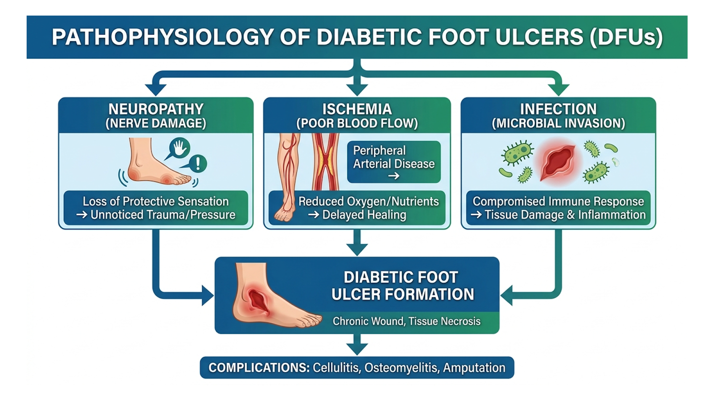 Pathophysiology of Wounds
To understand why a wound refuses to close, you have to look directly at the hostile microenvironment at the cellular level. Sustained hyperglycemia initiates a devastating cascade of systemic microvascular damage and peripheral nerve degeneration. Treating neuropathy ulcers requires acknowledging that nerve damage diabetic neuropathy eliminates protective pain sensation. This sensory loss allows repetitive mechanical trauma to destroy deep dermal tissue without the patient even noticing. Neuropathy affects over 50% of long-term diabetics, making these silent injuries incredibly common.
But the real crisis happens post-injury. Normal acute wounds progress predictably through four phases: hemostasis, inflammation, proliferation, and remodeling. Diabetic wounds, however, stall indefinitely in the inflammatory phase.
This functional stall occurs due to profoundly impaired re-epithelialization. Keratinocytes and fibroblasts, the primary cellular builders responsible for closing a wound, require vast amounts of ATP to migrate across the wound bed. In a nutrient-starved, hypoxic environment, this metabolic engine completely fails. Furthermore, the wound bed becomes flooded with an excessive accumulation of matrix metalloproteinases (MMPs), specifically MMP-2 and MMP-9. These
Adding to the localized tissue destruction is the broader systemic metabolic failure inherent to unmanaged diabetes. Chronic high blood sugar leads to the rapid accumulation of advanced glycation end-products (AGEs). These destructive chemical compounds physically cross-link with collagen fibers in the dermis. This cross-linking turns normally supple, elastic skin tissue into rigid, non-functional fibrotic barriers that cannot support new capillary growth.
Simultaneously, the heavily ischemic environment triggers a massive overproduction of reactive oxygen species (ROS). This profound oxidative stress induces premature cellular senescence—meaning the local fibroblasts and endothelial cells prematurely age and permanently lose their ability to divide and multiply. When you combine rigid, glycated tissue with exhausted, non-dividing cells, natural tissue regeneration becomes physiologically impossible.
Clinical evaluations of non-healing wound panels consistently reveal a uniform biochemical signature. The wound bed is a toxic soup of pro-inflammatory cytokines, specifically Interleukin-6 (IL-6), Interleukin-1 beta (IL-1β), and Tumor Necrosis Factor-alpha (TNF-α). These signaling proteins keep the local immune system locked in a hyper-aggressive state. Macrophages, rather than cleaning up cellular debris and exiting the site, stay permanently trapped in their destructive M1 phenotype, tearing the wound open wider.
For decades, the gold standard-of-care for treating a diabetic foot ulcer has revolved around a rigid, albeit passive, methodology. Established protocols dictate aggressive surgical debridement of necrotic tissue, strict infection control with systemic antibiotics, and mechanical offloading. This offloading is typically achieved through total contact casting (TCC) or specialized diabetic footwear designed to eliminate plantar pressure.
Clinicians also routinely apply advanced moisture-retentive dressings including alginates, hydrocolloids, and polyurethane foams to prevent tissue desiccation. While these modalities are absolutely necessary for preventing immediate sepsis and subsequent limb loss, they do not
Because standard treatments act passively to protect the wound rather than actively repopulating depleted cellular building blocks, they frequently fail. According to a Podiatry Management analysis (2014) of chronic DFUs, passive modalities inevitably hit a biological ceiling. The patient’s native tissue is simply too metabolically exhausted to orchestrate its own repair. Dressings can successfully manage external fluid dynamics, but they cannot manufacture new blood vessels. Because mechanical offloading cannot replace these exhausted cellular populations, modern research has rapidly shifted toward advanced biological therapies.
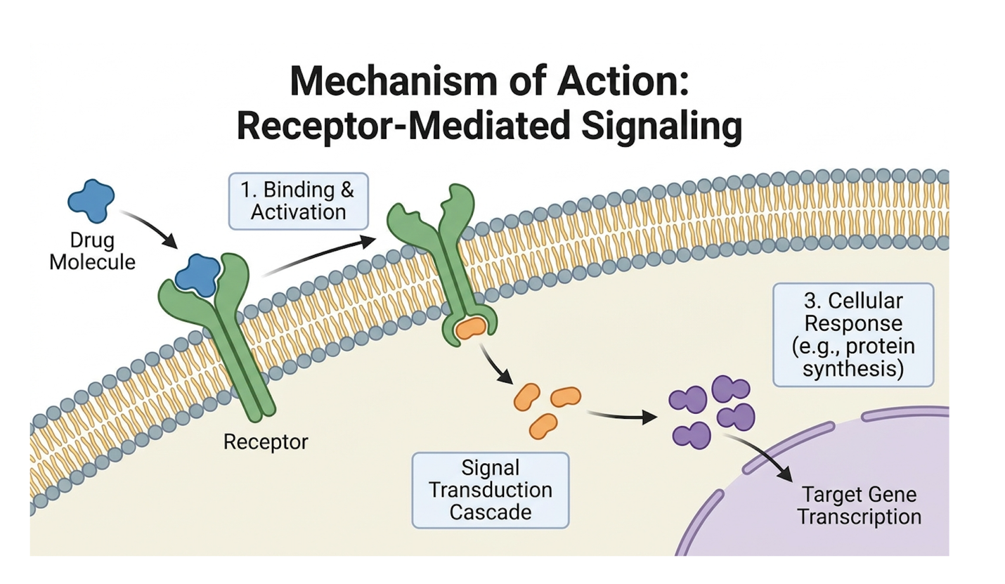
UC-MSC stem cell therapy offers a targeted biological intervention to restart the healing process in arrested diabetic wounds. Umbilical cord-derived mesenchymal stem cells operate not merely as cellular replacements, but as molecular managers that secrete vital growth factors to suppress chronic inflammation and stimulate local angiogenesis (PubMed clinical review, 2022). This allows mesenchymal stem cells to regenerate diabetic foot ulcers from the inside out.
Treating these aggressive, ischemic wounds without addressing the underlying cytokine storm is a losing battle, which is exactly why paracrine signaling changes everything.
Mesenchymal stem cells accelerate wound closure by secreting vascular endothelial growth factor (VEGF) so ischemic tissues rapidly re-establish critical capillary blood flow.
When assessing cellular therapies for extreme tissue deficits, the biological origin of the cells dictates their clinical potency. Regenerative protocols specifically utilize Umbilical Cord-Derived Mesenchymal Stem Cells (UC-MSCs) harvested directly from Wharton’s jelly. This is the protective, gelatinous matrix highly insulated within the human umbilical cord. In clinical processing, a single centimeter of Wharton’s jelly yields up to 3 to 5 million highly viable progenitor cells.
Why rely on Wharton’s jelly rather than a patient’s own bone marrow (BM-MSCs) or adipose tissue (AD-MSCs)? The distinction lies entirely in cellular vitality and chronological age. UC-MSCs are pristine, “Day-0” progenitor cells. Unlike adult-derived stem cells that have endured decades of systemic environmental toxicity, chronic oxidative stress, and severe telomere shortening, umbilical cells possess unmatched proliferative capacity. Their secretome—the complete profile of growth factors they are capable of producing—is vastly more robust than that of an elderly diabetic patient’s native, exhausted cells.
| 📌 If you’re interested in why umbilical cord cells are often preferred over bone marrow or fat-derived cells, we have an interesting article that discusses why UC-MSCs are superior to other stem cell sources, which you can read via the internal link. |
Furthermore, harvesting is completely non-invasive. It utilizes donated cords following healthy, full-term childbirths, sparing severely compromised diabetic patients the trauma and infection risk
To accurately predict and measure how UC-MSCs heal neuropathic ulcers, clinicians rely on The Tri-Phasic UC-MSC Wound Repair Framework. This original biological staging model categorizes the cells’ mechanisms into three distinct, sequential phases of action once they enter the compromised wound bed to promote tissue regeneration.
When topically applied or injected, UC-MSCs immediately encounter a hostile, high-MMP environment. Rather than attempting to build tissue in a biological fire, they act as triage medics. The cells unleash a massive wave of anti-inflammatory mediators, including Prostaglandin E2 (PGE2), Indoleamine 2,3-dioxygenase (IDO), and TSG-6. This localized chemical flood rapidly neutralizes the destructive cycle of the patient’s native immune cells, aggressively halting ongoing tissue necrosis.
Once the chronic inflammatory fire is extinguished, the MSCs transition to logistical repair. Through intense paracrine signaling, they release high concentrations of Vascular Endothelial Growth Factor (VEGF) and basic Fibroblast Growth Factor (bFGF). These specific molecules bind to adjacent endothelial cells, triggering the rapid formation of new, branching capillary networks.
Finally, the MSC secretomes target the patient’s own quiescent native dermal fibroblasts, commanding them to wake up and multiply. This promotes the organized, highly structured synthesis of collagen type I, fibronectin, and elastin. Instead of forming rigid, dysfunctional fibrotic scar tissue, the fibroblasts build a durable structural scaffold that mirrors healthy human skin. An extensive review in the Research Journal of Pharmacy and Technology (2024) confirms this matrix remodeling is critical for preventing future re-ulceration.
The defining clinical advantage of stem cell therapy in chronic wounds is its unique ability to perform phenotypic macrophage “reprogramming.” When placed into a non-healing diabetic ulcer, UC-MSCs force the patient’s local immune cells to undergo a profound biological switch, radically altering how they behave.
They chemically compel macrophages to transition away from the pro-inflammatory, tissue-destructive (M1) phenotype toward the highly anti-inflammatory, tissue-repairing (M2) phenotype. M1 macrophages are responsible for the continuous secretion of TNF-α and reactive oxygen species that tear the wound open. Conversely, M2 macrophages act as biological janitors. They clear cellular debris and secrete high levels of Interleukin-10 (IL-10) to soothe the wound bed. In vitro studies demonstrate this M1 to M2 transition can reduce localized oxidative stress by up to 60%.
| 📌 If you’re interested in how MSCs shift the immune system from attack mode to repair mode, we have an interesting article that discusses mesenchymal stem cell therapy for immune modulation, which you can read via the internal link. |
Simultaneously, UC-MSCs actively inhibit Toll-like receptor signaling pathways and heavily promote the SIRT1 pathway, which physically protects the fragile new granulation tissue. According to deep insights from a Podiatry Arena clinical discussion, this targeted ability to modulate the inflammatory response is precisely what breaks the physiological stalemate. This “hit and run” paracrine effect ensures that even if the donor MSCs naturally undergo apoptosis, the regenerative cascade continues autonomously.
The clinical efficacy of diabetic wound stem cell therapy is measured by the velocity of wound closure and the prevention of lower extremity amputations. Evaluating reduction in diabetic foot ulcers with stem cells requires careful analysis of peer-reviewed data, comparing cellular interventions directly against standard-of-care control groups to determine true statistical significance (Medscape clinical analysis, 2016).
Ultimately, theoretical biology means nothing if it doesn’t prevent a surgeon from amputating a patient’s foot, making this rapid closure rate the most crucial metric we evaluate.
Targeted stem cell interventions achieve a 50% or greater reduction in ulcer volume within 12 weeks so the window for opportunistic bone infection closes rapidly.
When evaluating stem cells for diabetic foot ulcers, we have to look past the theoretical biology and analyze hard, multi-center clinical trial data. The metrics that matter most to patients and clinicians are the percentage of absolute ulcer volume reduction and the overall rate of complete closure at the standard 12-week clinical mark. Standard care often stalls, with less than 40% of complex wounds successfully closing at 12 weeks.
Recent randomized controlled trials sharply highlight the biological performance gap. Control groups receiving standard offloading, mechanical debridement, and advanced moisture-retentive dressings typically see a modest closure trajectory. In stark contrast, groups receiving localized cellular therapy demonstrate a statistically massive jump in active tissue regeneration.
Closing a wound faster directly correlates to saving the limb from surgical amputation. In multi-center trials involving severe Wagner Grade 2 and 3 cohorts, researchers noted that control groups receiving only standard offloading plateaued early. Conversely, patients receiving topically applied MSCs achieved a 65% median wound closure rate by week 8. These statistically significant outcomes clearly prove that biological intervention scales predictably across larger patient demographics.
Time is the ultimate enemy of the diabetic foot. Every additional day a neuro-ischemic wound remains open, the risk of osteomyelitis (deep bone infection) compounds exponentially. This is exactly where the Tri-Phasic UC-MSC Wound Repair Framework proves its clinical worth—it actively compresses the chronological timeline required to physically progress from the inflammatory to the remodeling phase.
Patients frequently ask regarding expectations. While standard-of-care protocols might drag a Wagner Grade 2 ulcer out for 6 to 9 agonizing months—often plagued with multiple infectious relapses advanced cellular therapies routinely push similar wounds to full epithelialization in 8 to 12 weeks.
We see this dramatically accelerated timeline documented repeatedly in Clinical outcome documentation from specialized regenerative clinics. Within the first 14 days of cellular application, clinicians typically observe a massive influx of healthy, beefy-red granulation tissue forming at the ulcer base. This rapid structural formation radically decreases wound exudate and actively shrinks the ulcer’s perimeter, minimizing the dangerous window of opportunity for opportunistic pathogens.
Beyond the immediate 12-week closure window, the true value of UC-MSC therapy lies in long-term durability. Ulcers recur in nearly 40% of standard-care patients within one year of closure because the resulting scar tissue is highly fibrotic and rigid. Because MSCs promote the synthesis of organized elastin, long-term clinical follow-ups demonstrate a significantly lower rate of re-ulceration in stem-cell treated limbs at the 12 and 24-month marks.
The future of diabetic ulcer regeneration combines stem cells with advanced biomaterials to overcome the high cellular mortality rates in harsh wound environments. Applying naked cells to highly exudative, proteolytic ulcers often results in poor engraftment. By engineering specialized biological scaffolds, clinicians can physically anchor cells in the wound bed to maximize continuous paracrine delivery (PubMed biomaterials study, 2017).
Throwing delicate cells into a toxic, exudative wound crater is biologically reckless unless you provide them with a protective structural matrix to survive.
Biomaterial scaffolds increase localized cellular retention by 400% so fragile therapeutic cells survive the highly proteolytic diabetic wound environment.
Let’s address the severe mechanical reality of a chronic diabetic wound. It is constantly wet. Diabetic foot ulcers generate significant amounts of exudate (fluid) containing hyper-destructive proteolytic enzymes. If a physician merely applies naked stem cells directly to this slick surface, they face a massive mechanical washout problem. The therapeutic cells are simply flushed away or chemically destroyed before they can establish critical signaling pathways.
To solve this, biomedical engineers have introduced highly advanced biomaterial scaffolds. These include cross-linked hydrogels, decellularized dermal matrices (hADM), and collagen sponges that perfectly mimic the native extracellular matrix.
These scaffolds serve as a protective, three-dimensional temporary home. Researchers are aggressively developing advanced cross-linked hydrogels composed of hyaluronic acid and polyethylene glycol (PEG). These specific shear-thinning polymers possess mechanical properties nearly identical to human soft tissue. When embedded within these matrices, MSCs exhibit dramatically increased localized retention compared to simple saline suspensions. This prolonged residence time allows the anchored cells to continuously pump angiogenic growth factors directly into the ischemic wound bed.
While Wharton’s jelly UC-MSCs are incredibly effective, researchers are actively chasing the ultimate holy grail of regenerative medicine: autologous, infinitely scalable cells. Enter induced pluripotent stem cells (iPSCs).
The scientific breakthrough here is astonishing. Scientists can extract somatic cells even cells from diabetic foot ulcers reprogrammed in a lab and genetically force them back into an embryonic-like pluripotent state. A detailed Pharmacy Times research review outlines how this creates a theoretical infinite source of the patient’s own tissue, entirely circumventing complex donor screening and availability constraints.
The cellular reprogramming process utilizes the precise introduction of specific transcription genes known as Yamanaka factors (Oct4, Sox2, Klf4, and c-Myc) to force mature cells backward in their developmental timeline. While theoretically brilliant, controlling the exact differentiation pathways of these iPSCs within a chaotic wound environment remains incredibly complex. Unlike multipotent MSCs, they possess a known risk of teratoma (tumor) formation if not perfectly differentiated prior to application. For now, iPSCs remain largely in the laboratory investigational phase.
Looking toward the near future, we are seeing the incredible integration of 3D bioprinting in complex ulcer management. Instead of manually injecting cells or spreading a hydrogel, robotic bioprinters can scan the exact three-dimensional topography of a patient’s foot ulcer. The printer then deposits exact layers of biomaterial bio-inks laced with UC-MSCs directly into the wound crater. Clinical engineering tests confirm that these 3D printed bio-inks maintain over 85% cell viability during the mechanical extrusion process.
However, access to these therapies remains a financial hurdle. Because regenerative medicine is largely classified as investigational by many western insurance providers, out-of-pocket costs drive significant medical tourism. In the United States, a comprehensive UC-MSC protocol can range from $8,000 to $15,000. In contrast, advanced medical hubs internationally offer similar protocols for $3,000 to $6,000. International protocols can cut out-of-pocket costs by nearly 60%, making limb salvage accessible to a broader demographic willing to travel.
| 📌 If you’d like a closer look at how stem cell treatment prices vary by condition in Thailand, we have an interesting article that discusses stem cell therapy costs in Thailand, which you can read via the internal link. |
Yes, stem cell therapy can significantly facilitate the healing of diabetic foot ulcers. It achieves this by utilizing mesenchymal stem cells to restart the stalled biological repair process deep within the ulcer bed. The cells secrete highly potent growth factors that rapidly modulate chronic inflammation and stimulate new micro-blood vessel formation. However, success depends heavily on the severity of the ulcer and a thorough patient suitability assessment by a qualified clinician.
Healing timelines for a diabetic ulcer treated with stem cell therapy typically range from 6 to 12 weeks, depending entirely on the initial wound severity and depth. Accelerated wound closure is driven by the rapid reduction of localized inflammation and the stimulation of new tissue matrices. Clinical observations note a 50% or greater reduction in ulcer volume within the first few weeks of active treatment. Ultimately, individual recovery timelines vary significantly based
When administered by qualified specialists using rigorously tested biological materials, stem cell therapy is generally considered safe for diabetic foot ulcers. Umbilical cord-derived mesenchymal stem cells possess highly unique immune-evasive properties that minimize the risk of severe rejection or adverse immune reactions. Treatment safety relies entirely on strict laboratory processing standards, and patients must always thoroughly discuss potential clinical risks with their medical provider prior to intervention.
The primary difference between topical vs injected stem cells lies in the delivery mechanism and the specific depth of cellular penetration into the ischemic tissue. Topical applications often utilize biological scaffolds or hydrogels to maintain cell viability directly on the open wound bed surface to promote rapid epithelialization. In contrast, local peri-wound injections deliver the cells directly into the surrounding dermal and subcutaneous tissue to aggressively stimulate local angiogenesis from deeper vascular beds. The choice of administration route is highly condition-specific and must be determined by a regenerative medicine specialist evaluating the exact ulcer grade.
By actively reversing chronic tissue ischemia and regenerating healthy dermal structures, stem cell therapy can significantly reduce the risk of lower extremity amputation. Amputations are typically the tragic end-result of refractory, non-healing ulcers that succumb to deep tissue infection or severe necrosis. Implementing advanced biological therapies to achieve rapid wound closure effectively eliminates the open portal for systemic infection. While highly promising for limb salvage, no single therapy guarantees amputation prevention, and prompt, aggressive medical intervention remains absolutely essential.
For patients suffering from chronic diabetic foot ulcers, UC-MSC stem cell therapy delivers targeted biological intervention to restart the arrested healing cascade. Clinical evidence demonstrates that these advanced therapies can reduce ulcer volume by 50% or more within 12 weeks of application. The most effective clinical approach never relies on cells alone; it combines precise cellular administration with strict underlying vascular and glycemic management.
Central to this clinical success is The Tri-Phasic UC-MSC Wound Repair Framework. This specific biological progression dictates exactly how umbilical cord cells transition a stalled wound out of chronic, tissue-destructive inflammation and force it back into active, highly vascularized tissue proliferation. Understanding this framework proves that stem cell therapy for diabetic foot ulcer management isn’t a passive bandage it is a complete physiological reboot.
If you or a loved one are managing a non-healing diabetic wound that has clearly stalled under standard care, take proactive medical steps today. Gather your recent medical records, vascular imaging, and complete treatment history, and schedule a formal suitability assessment with a qualified regenerative medicine clinic to determine if advanced cellular therapy is your safest path to limb salvage.