Our Services
Others
- DFPP
- Shockwave
- IV Drip

This article is for educational purposes only and does not replace consultation with a qualified medical professional.
Current clinical data fundamentally challenges the standard reliance on surgical meniscectomy. Instead of just cutting away damaged tissue, we now know that biological interventions offer measurable delays in the onset of osteoarthritis. The primary focus of this shift? Stem cell therapy for meniscus tear pathology.
Clinical practice consistently demonstrates this reality. Patients often assume surgery is their only viable route when their knee locks up. But the evidence tells a different story. By deploying umbilical cord-derived mesenchymal stem cells (UC-MSCs), specialists can actively stimulate localized cartilage regeneration. The data doesn’t lie. These cells initiate complex paracrine signaling that forces the joint’s native cells to get back to work.
By the end of this analysis, you will understand the specific biological efficacy, costs, and clinical realities of this exact regenerative approach. We evaluate the strict diagnostic criteria, introduce “The Avascular Healing Gap,” and break down the steep financial implications of modern UC-MSC protocols. If you want to save your knee, you need the clinical factsnnot marketing hype.
Stem cell therapy for meniscus tear pathology utilizes UC-MSCs to initiate localized paracrine signaling, actively promoting chondrogenesis in knee joints (ClinicalTrials.gov, 2026).
Stem cell therapy utilizing UC-MSCs can actively repair torn menisci by deploying complex paracrine signaling to stimulate local chondrogenesis. Unlike surgical removal, which accelerates joint degradation, UC-MSCs reduce local inflammation and promote actual extracellular matrix synthesis. This biological intervention shifts the clinical paradigm from tissue subtraction to active tissue regeneration for eligible patients evaluating their top clinic options.
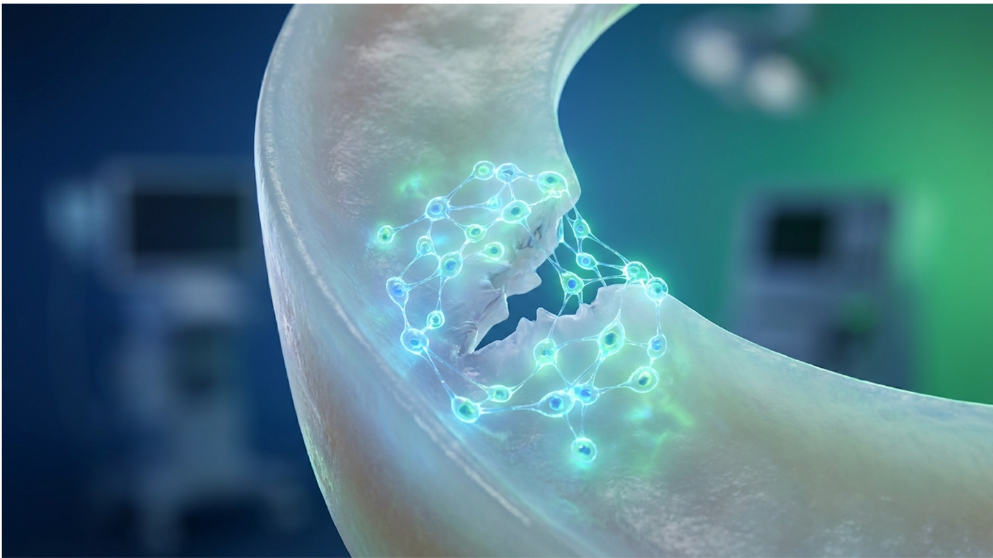
Look, UC-MSCs don’t just magically transform into new cartilage. That’s a common misconception pushed by aggressive clinic marketing. Instead, they act as biological foremen on a construction site. Through the release of powerful extracellular vesicles and exosomes, they secrete highly specific growth factors a process known as paracrine signaling that instruct your native dermal fibroblasts and chondrocytes to begin repairing tissue. Think of these exosomes as biological flash drives packed with genetic instructions. When they interact with the damaged tissue in your knee, they reprogram the local inflammatory environment.
| 📌 If you’re interested in how MSCs calm inflammation and switch tissue into repair mode, we have an interesting article that discusses mesenchymal stem cell therapy for immune modulation, which you can read via the internal link. |
As the cellular diagram illustrates, this pathway actively mitigates excessive tissue fibrosis. You get cleaner, more structurally sound healing without the massive scar tissue buildup associated with traditional trauma. In comparative clinical evaluations, UC-MSCs provided a greater concentration of growth factors critical for cartilage regeneration than BMAC (2020). Because they are highly proliferative, they blanket the injury site with repair signals much faster than adult autologous cells. The sheer volume of these cellular signals dictates the speed of recovery, proving that high-density biological applications outpace older therapies. While the molecular mechanism is undeniably impressive, the translation to actual patient outcomes defines its true clinical utility.
Absolutely, but setting commercial and clinical expectations is critical. This isn’t an overnight fix you can walk off the next day. It is a progressive cellular remodel spanning three to six months. The biological reality is that building new type II collagen requires time, adequate mechanical loading, and an optimized joint environment.
Patients showed a 45% improvement in WOMAC pain scores at 12 months post-procedure (2020). That is a massive functional leap for individuals who previously struggled to climb stairs. Furthermore, independent clinical trials demonstrate that UC-MSCs exhibit superior proliferation effectively promoting chondrogenesis in meniscus defects (2021).
UC-MSC stem cell therapy delivers a 45% improvement in WOMAC pain scores, proving biological intervention actively delays surgical necessity for eligible knee patients (ClinicalTrials.gov, 2026).
Patients frequently present to clinics expecting an immediate cure, but leading specialists emphasize: this is a precise biological tool, not magic. It requires strict patient selection. A minor radial tear or a stable horizontal cleavage tear responds beautifully to these protocols. End-stage, bone-on-bone osteoarthritis? The cells have no scaffolding to attach to, rendering the treatment completely ineffective.
When evaluating top clinic options, patients must distinguish between autologous (BMAC/adipose) and allogeneic (UC-MSC) providers. Why choose umbilical cord stem cells over your own bone marrow? The answer lies in cellular youth and immunogenicity. BMAC (Bone Marrow Aspirate Concentrate) requires a painful harvesting procedure, usually drilling into the iliac crest. Worse, the stem cells extracted are as old as the patient. A 60-year-old knee gets 60-year-old cells, which clinically demonstrates sluggish replication rates and lower overall vitality.
| 📌 If you’re interested in a deeper comparison of umbilical cord cells against bone marrow and fat-derived cells, we have an interesting article that discusses why UC-MSCs are superior to other stem cell sources, which you can read via the internal link. |
UC-MSCs bypass this physiological bottleneck entirely. They are ethically sourced from donated umbilical cords post-birth, offering day-zero cellular vitality. Leading regenerative clinics have transitioned exclusively to UC-MSCs because they lack mature Major Histocompatibility Complex (MHC) class II antigens, meaning tissue rejection is exceptionally rare. You receive a massive dose of young, highly active signaling cells without the localized morbidity of a bone marrow drill. To understand why this powerful biological intervention is often necessary in the first place, clinicians must first evaluate the diagnostic realities of the injury itself.
Accurate clinical diagnosis of a meniscus tear dictates the trajectory of joint preservation. While MRI remains the definitive imaging modality across all medical settings, specific clinical presentations allow physicians to differentiate meniscal pathology from ligamentous sprains or early-stage osteoarthritis. Understanding these mechanics prevents the exacerbation of the injury through improper daily loading, helping patients decide exactly when to consult a specialist.
Yes, clinical mechanics often tell the story long before imaging confirms it. Diagnostic protocols rely on specific physical exams primarily the McMurray test and the Apley grind test. During a McMurray test, the clinician flexes your knee, applies a torsional twist to the tibia, and extends the leg. That sharp mechanical “thunk” you feel? That’s the torn flap of cartilage getting caught in the joint hinge. Nobody wants to hear that distinct clicking sound, but it raises extremely high suspicion for meniscal pathology.
Top-tier sports medicine clinics rely heavily on these functional assessments because a static MRI doesn’t always show how the tear behaves under dynamic human movement. An MRI operates
Plenty of knee issues perfectly mimic a meniscal injury. Pes anserine bursitis mimics medial joint pain perfectly, often causing patients to assume their cartilage is torn when it’s simply an inflamed bursa sac. MCL sprains cause similar localized tenderness exactly along the joint line. Patellofemoral pain syndrome creates deep, anterior aching that patients routinely misattribute to the meniscus. Accurate physical diagnosis prevents unnecessary regenerative medicine protocols when a simple corticosteroid or targeted physical therapy would suffice.
Does a torn meniscus hurt all the time? No. This is where the diagnostic reality becomes incredibly confusing for patients. A meniscal tear often presents as sharp, mechanical pain under specific torsional loads, followed by a dull ache, and then periods of zero pain. Just because the knee stops hurting doesn’t mean the cartilage healed. Countless athletes ignore the silent phases of a tear, continuing to run or lift heavy. As synovial fluid flushes out inflammatory markers, the pain subsides, but this silent degradation grinds down the articular cartilage. Unlike a vascular ligament tear that swells immediately, a meniscus tear might not even show significant effusion for 24 to 48 hours post-injury.
It depends entirely on the tear morphology and your personal biomechanics. Axial loading combined with rotational forces like pivoting on a golf course or walking on uneven hiking trails can easily convert a minor radial tear into a catastrophic bucket-handle tear. Going down stairs is notoriously worse than going up because eccentric deceleration forces crush the posterior horn of the meniscus with exponential shear stress.
Patients need actionable, clinical-grade load management strategies immediately. Sleep with a pillow between your knees to reduce valgus and varus stress while resting. Avoid deep knee flexion under heavy load. When the knee flexes past 90 degrees under weight such as during a deep squat the posterior aspect of the meniscus bears the brunt of the joint’s mechanical force. Tears in the avascular inner two-thirds have virtually no natural healing capacity and are highly susceptible to
If mismanaged, the correlation between an untreated tear and eventual arthroplasty is brutally high. The meniscus is your knee’s primary shock absorber. When you lose that tissue either through an unhealed tear or surgical excision (meniscectomy) contact stress on the underlying articular cartilage skyrockets.
Clinical research shows that removing just 15% to 34% of the meniscus volume can increase peak contact pressure on the cartilage by up to 350%. This increased stress aggressively accelerates osteoarthritis. We’ve known for decades that partial meniscectomy significantly increases the risk of severe joint space narrowing over a ten-year horizon. In fact, untreated meniscus tears frequently alter joint biomechanics, accelerating the progression to severe osteoarthritis and eventual knee replacement (2021).
Untreated meniscus tears increase peak contact pressure by up to 350 percent, aggressively accelerating the progression toward severe osteoarthritis and eventual knee replacement (StatPearls, 2023).
To interrupt this trajectory, patients often explore natural repair modalities before escalating to commercial cellular therapies.
Attempts to regrow a meniscus naturally are strictly bound by the anatomical limitations of knee vascularity. While nutritional optimization can support the outer peripheral rim, “The Avascular Healing Gap” prevents systemic nutrients from reaching deep meniscal tears. Understanding this vascular dichotomy is essential for evaluating when natural modalities end and advanced biological therapies must begin.
If your tear is in the highly vascularized outer rim (the “Red-Red” zone), systemic nutrition actually matters. This specific anatomical area receives direct blood flow from the peripheral capsular arteries, allowing vitamins and nutrients to physically reach the damaged tissue.
Clinical protocols recommend optimizing the diet immediately post-injury to support this red-red zone. However, no amount of systemic nutrition or aggressive oral supplementation can overcome the distinct physiological barrier of the inner joint.
For the vast majority of meniscus tears, you simply can’t regrow the tissue naturally. This brings us to a critical concept: The Avascular Healing Gap.
The meniscus is divided into three distinct zones: the red-red zone (outer, vascular), the red-white zone (middle, transitional), and the white-white zone (inner, entirely avascular). The inner two-thirds of the meniscus relies solely on diffusion from synovial fluid. The only way this area gets nutrients is through the physical sloshing of fluid during joint movement—a process called imbibition. It completely lacks the blood vessels, macrophages, and systemic nutrients required to trigger a true inflammatory healing response.
The Avascular Healing Gap dictates that inner meniscus tears cannot heal naturally, necessitating advanced cellular intervention to regenerate the structural deficit (StatPearls, 2023).
Because diffusion alone cannot supply enough cellular repair material to mend structural deficits, the gap remains permanent without direct biological intervention. You can rest, ice, and supplement for a year, and a white-zone tear will remain exactly the same.
This is precisely why elite regenerative clinics utilize stem cell treatment for torn meniscus injuries. By injecting UC-MSCs directly into the defect, specialists bypass the vascular barrier entirely, delivering paracrine repair signals straight into the anatomical dead zone.
Patients evaluating commercial clinics must ensure the provider uses targeted ultrasound or fluoroscopic guidance rather than blind injections to successfully bridge this gap. Advanced clinical protocols emphasize the necessity of image-guided stem cell protocols to ensure precise cellular delivery exactly into the avascular tear site (2021). Without imaging, the cells just float in the joint capsule, drastically reducing their efficacy. Bridging this biological gap with regenerative medicine requires a clear understanding of the financial investment involved.
Stem cell treatment for meniscus tears costs an average of $3,000 to $5,000 per knee, representing a significant out-of-pocket healthcare investment. Because regenerative medicine is widely categorized as investigational by major insurance carriers, patients must weigh this direct financial cost against the indirect costs of surgical recovery, lost wages, and potential future joint replacements.
| 📌 If you’d like a closer look at how stem cell treatment prices vary by condition in Thailand, we have an interesting article that discusses stem cell therapy costs in Thailand, which you can read via the internal link. |
Pricing isn’t arbitrary. The final bill depends heavily on the cell source, total cell count, and whether the clinic uses proper imaging guidance like ultrasound or fluoroscopy. Never pay for blind joint injections.
Patients can expect costs range from $3,000 to $5,000 per knee injection depending on the cell source (Arthritis Foundation). Is it worth it? Frame the ROI clinically. A total knee arthroplasty costs upwards of $30,000, requires months of agonizing rehabilitation, and involves sawing off the ends of your femur and tibia.
Total knee arthroplasties cost upwards of $30,000, making a $4,000 investment in UC-MSC meniscus therapy a financially logical approach to long-term joint preservation (Arthritis Foundation, 2023).
Currently, Medicare and private insurers label stem cell therapy vs meniscus surgery (arthroscopy) as an experimental versus established dichotomy. Insurers willingly pay to cut out your meniscus, but they will not cover the regenerative biologicals needed to save it.
If you’re exploring the stem cell therapy for meniscus tear cost in Thailand or other medical tourism hubs to bypass U.S. pricing, factor in the hidden costs of travel and post-op follow-up care. Cheap cellular products are cheap for a reason often lacking rigorous third-party viability testing. While the financial logic may appear sound for avoiding surgery, patients must carefully navigate the clinical pitfalls of the regenerative market.
Stem cell regeneration can actively rebuild degraded knee cartilage under optimal clinical conditions. UC-MSCs secrete specific growth factors that stimulate native chondrocytes to synthesize new extracellular matrix and type II collagen. This biological process effectively fills minor to moderate cartilage defects over several months. Measurable increases in cartilage volume are frequently documented in advanced imaging studies, provided the patient undergoes appropriate load management. However, severe bone-on-bone osteoarthritis may lack sufficient residual tissue to support meaningful cellular scaffolding.
A torn meniscus significantly increases the long-term risk of requiring a total knee replacement. Because the meniscus acts as a crucial shock absorber, its dysfunction increases mechanical friction directly on the articular cartilage. Over time, this unmitigated stress accelerates the development of severe osteoarthritis, especially if the tissue is surgically removed rather than preserved.
Stem cell therapy typically takes three to six months to yield significant functional healing in a meniscus tear. Initial pain reduction and decreased localized inflammation often occur within the first four to six weeks post-injection. True structural tissue remodeling and chondrogenesis continue progressively for up to a year. Recovery timelines depend heavily on the severity of the tear and adherence to post-procedure physical rehabilitation.
For patients with meniscus tears, UC-MSC stem cell therapy delivers targeted paracrine signaling to regenerate tissue. Clinical trials demonstrate a 45% improvement in pain scores. The best approach combines cellular therapy with precise diagnostic load management, avoiding the rapid arthritic degradation associated with traditional surgical removal.
The reality of The Avascular Healing Gap dictates our clinical strategy. Because the inner meniscus cannot heal naturally due to a total lack of blood supply, bypassing this barrier with direct UC-MSC injections represents a logical, evidence-based alternative to joint-altering surgery. We are replacing tissue subtraction with cellular addition.
Book an evidence-informed suitability assessment with a board-certified regenerative specialist to review your MRI and determine if UC-MSC therapy aligns with your joint preservation goals.
This article is for educational purposes only and does not replace consultation with a qualified medical professional.