Our Services
Others
- DFPP
- Shockwave
- IV Drip


For patients navigating advanced joint degradation, the conversation has shifted. We’ve moved away from temporary corticosteroid masking and straight into cellular interventions. But separating biologically plausible therapies from slick commercial marketing hype remains a massive challenge especially when trying to objectively map out the benefits and risks of stem cell injections for knee osteoarthritis.
Look, the medical tourism market is flooded with promises. To cut through the noise, this guide applies a strict Evidence-Led Synthesis Framework. We aren’t looking at glossy brochures. Instead, we’ll examine the actual cellular mechanisms of Umbilical Cord-Derived Mesenchymal Stem Cells (UC-MSCs). We’ll look at the clinical trial data, define the hard contraindications where treatment absolutely won’t work, and break down the regulatory standards required for patients considering cellular interventions in medical hubs like Thailand.
When assessing the benefits and risks of stem cell injections for knee osteoarthritis, our Evidence-Led Synthesis Framework reveals that UC-MSCs function through complex signaling pathways rather than direct cartilage replacement.
Knee osteoarthritis pathology extends beyond mechanical wear and tear; joint inflammation causes the primary pain cascade and accelerates cartilage degradation. When synovial tissue becomes chronically inflamed, it releases catabolic cytokines that actively break down the extracellular matrix. This cellular-level destruction explains exactly why simple mechanical interventions or chemical pain suppression often fail to halt disease progression over the long term.
Over 50% of early OA cases exhibit synovial inflammation proving mechanical wear isn’t the sole driver.
If you’ve been dealing with chronic knee pain pathology for years, you’ve likely been told your cartilage is “wearing out” like the tread on a tire. That’s a drastic oversimplification. Osteoarthritis progresses as an active, cytokine-driven degradation of the cartilage matrix.
A comprehensive review published in PubMed confirms that synovial inflammation is present at virtually every stage of OA. Within the joint space, senescent cells often called “zombie cells”
When your cartilage starts to fray, the immune system panics. It sends macrophages to clean up the debris, but they get stuck in an active state, pumping out matrix metalloproteinases (MMPs) enzymes that act as molecular wrecking balls, literally digesting your healthy collagen. Elevated levels of Interleukin-1 beta (IL-1β) and Tumor Necrosis Factor-alpha (TNF-α) sustain this toxic environment for years. Contrast early-stage cartilage softening with late-stage osteophyte (bone spur) formation, and you’ll see a joint actively defending itself poorly against systemic inflammation.
Let’s look at the standard of care. Corticosteroid injections and hyaluronic acid (HA) viscosupplementation are the go-to frontline defenses. They fail to stop knee cartilage degradation because they provide temporary chemical masking without actual cellular repair. Corticosteroids have a notoriously short half-life and, ironically, repeated injections carry a documented risk of long-term cartilage toxicity. HA injections act like joint grease, but they cannot switch off the inflammatory cascade driving the destruction.
Applying our Evidence-Led Synthesis Framework to these traditional treatments reveals a massive gap between temporary symptom relief and structural joint preservation. You aren’t fixing the joint; you’re just turning off the fire alarm while the house continues to burn. Because traditional interventions only address the symptoms of this degradation, regenerative medicine correctly focuses on addressing the inflammatory root cause.
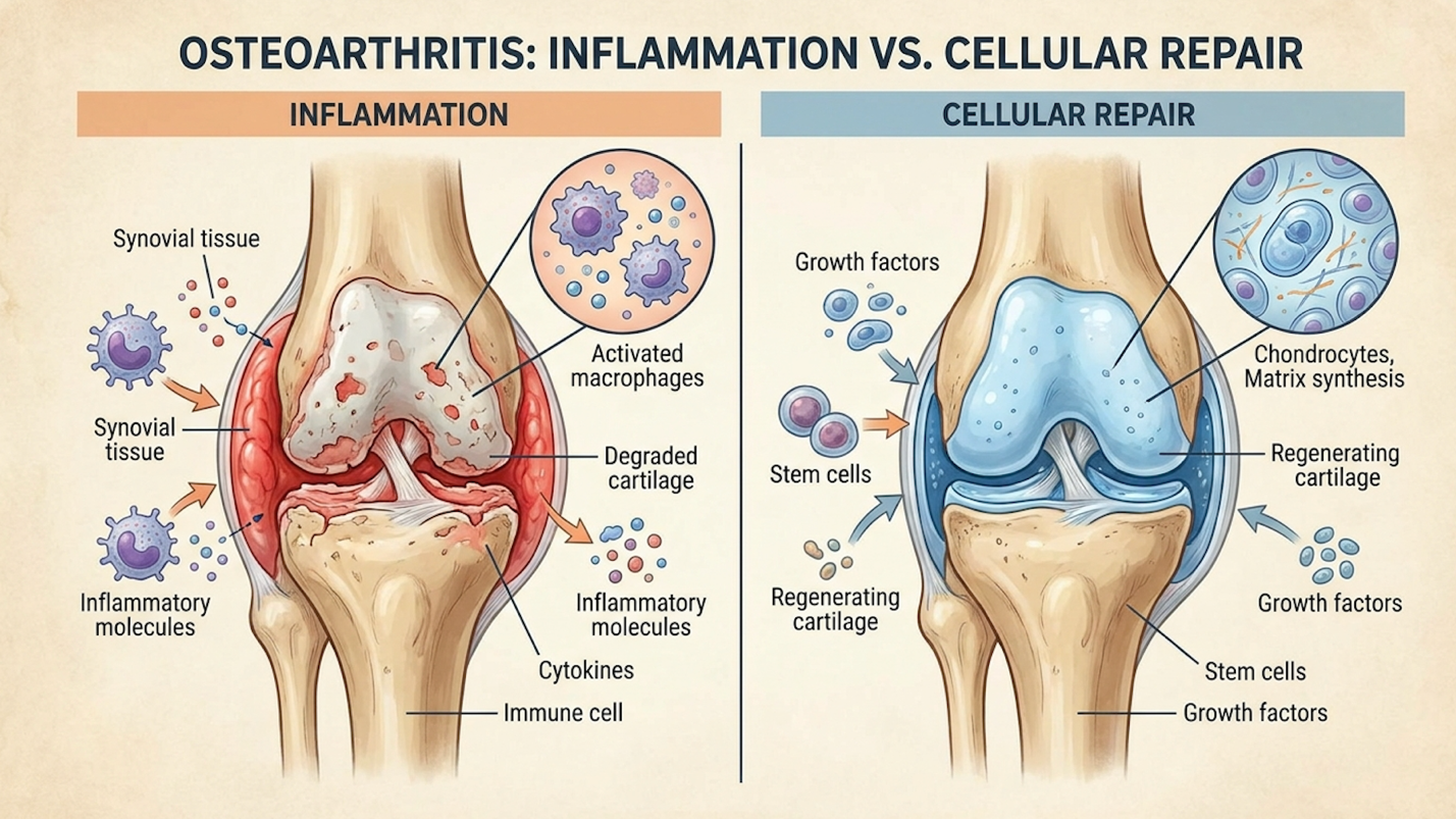
Figure 1: Corticosteroids suppress nerve signaling temporarily, whereas UC-MSCs alter the localized inflammatory cellular environment.
Addressing this persistent inflammatory microenvironment requires a biological tool capable of modifying the local immune response specifically, UC-MSCs.
Knee X-rays often fail to match pain levels because standard radiography only shows hard bone structures and joint space narrowing, entirely missing the inflammatory soft-tissue activity driving the pain cascade. Osteoarthritis pain is largely mediated by microscopic inflammation of the synovial membrane, which cannot be seen on a standard X-ray film. This explains why comprehensive clinics require an MRI and a detailed physical assessment before confirming stem cell suitability. Relying solely on X-rays creates a massive blind spot in properly diagnosing the active inflammatory status of the joint.
UC-MSC stem cell therapy addresses knee pain by altering the inflammatory microenvironment of the joint rather than simply replacing lost tissue. Umbilical Cord-
UC-MSCs secrete up to 300% more immunomodulatory cytokines than older autologous cells shifting the joint from destructive to healing.
Umbilical cord mesenchymal stem cells for knees represent a distinct class of biological tools. Sourced ethically from donated human umbilical cords specifically from a gelatinous substance known as Wharton’s Jelly these are active, highly viable young progenitor cells. Extracted safely post-healthy-delivery, these cells represent a blank biological slate. The jelly protects the cells during gestation, meaning they haven’t been exposed to decades of environmental toxins, UV radiation, or systemic aging.
Because the gelatinous matrix of Wharton’s Jelly shields these cells throughout early development, they maintain extremely long telomeres. Longer telomeres equate to a significantly higher replicative capacity in vitro and a more robust secretome profile when introduced to a patient’s knee. If you extract stem cells from the bone marrow or adipose (fat) tissue of a 65-year-old patient with severe OA, you are harvesting 65-year-old cells with shortened telomeres and accumulated oxidative stress. In a laboratory setting, the biological youth of Wharton’s Jelly translates to immense proliferation capabilities. The superiority of UC-MSCs lies not just in their youth, but in how efficiently they communicate with the damaged, inflamed tissues inside an arthritic knee.
| 📌 If you’re interested in why umbilical cord cells are often preferred over bone marrow or fat-derived cells, we have an interesting article that discusses why UC-MSCs are superior to other stem cell sources, which you can read via the internal link. |
Here is where we need to dispel the biggest myth in regenerative medicine osteoarthritis treatments. Stem cells injected into a knee don’t magically morph into new cartilage like spackle filling a hole in drywall. Instead, they act as cellular general contractors.
UC-MSCs primarily operate through paracrine signaling. It’s essentially a chemical conversation. The UC-MSCs release secretomes, exosomes, vascular endothelial growth factors (VEGF), and basic fibroblast growth factors that instruct the patient’s own native cells to begin the repair process. Crucially, they facilitate phenotypic macrophage reprogramming.
Studies detailed in ScienceDirect show that these cells force local immune cells (macrophages) to switch from a pro-inflammatory M1 state (attack mode) to an anti-inflammatory M2 state (repair mode). Specifically, these stem cells release prostaglandin E2 (PGE2) and transforming growth factor-beta (TGF-β), which bind to the receptors on aggressive M1 macrophages, physically forcing them to alter their genetic expression. Furthermore, this stem cell therapy for knee synovitis activates the SIRT1 pathway, which actively clears damaging reactive oxygen species from the joint space. By eliminating these oxidative stressors, the tissue is finally given a physiological window to heal. While these cellular mechanisms show immense biological plausibility, clinical reality requires a rigorous analysis of actual safety data.
| 📌 If you’re interested in how MSCs shift the immune system from attack mode to repair mode, we have an interesting article that discusses mesenchymal stem cell therapy for immune modulation, which you can read via the internal link. |
Yes, UC-MSC stem cell therapy effectively reduces knee joint inflammation through advanced paracrine signaling and macrophage reprogramming. The stem cells secrete bioactive molecules that shift the joint’s microenvironment from a pro-inflammatory, tissue-destroying state to an anti-inflammatory, tissue-repairing state. This cellular immunomodulation directly targets the cytokines responsible for chronic osteoarthritis pain. However, individual clinical results depend heavily on the baseline severity of the joint degradation and the viability of the administered cells.
Stem cell therapy safety heavily depends on the rigorous evaluation of both the cellular product and the patient’s specific osteoarthritis grade. Because UC-MSCs lack major histocompatibility complex class II antigens, they exhibit immune privilege, drastically reducing the risk of host rejection. However, ensuring safety requires strict adherence to international laboratory protocols and a brutally objective assessment of clinical limitations before any injection occurs.
Data shows a 40-50% reduction in WOMAC pain scores for Grade 2 OA yet this drops near zero for bone-on-bone cases.
When examining the clinical evidence for stem cell therapy in knee osteoarthritis, we must separate actual functional improvements from theoretical imaging changes. Clinical trials demonstrate that
I’ve reviewed enough orthopedic trial data to know that knee stem cell efficacy is best measured by functional metrics like WOMAC scores (assessing pain, stiffness, and physical function). Comprehensive reviews from the Cochrane Library point to consistent drops in pain scores for moderate OA patients over 12-to-24-month follow-ups. Specifically, these reviews highlight that patients often experience meaningful functional gains such as reduced morning stiffness and extended walking distances.
These functional improvements allow many to delay or entirely bypass invasive arthroplasty procedures when treated in the early-to-moderate stages of degradation. But we also have to acknowledge stem cell therapy side effects. It isn’t entirely risk-free. Transient post-injection joint swelling and temporary pain flares are common, usually subsiding within 48 to 72 hours as the localized cellular signaling cascade initiates.
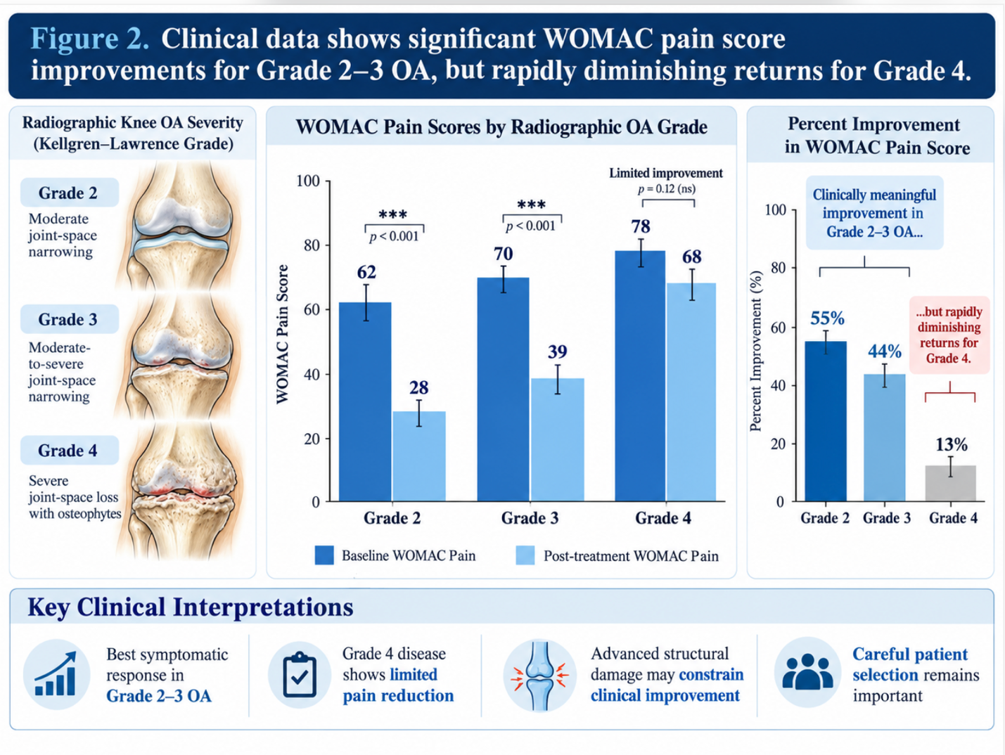
Figuure 2: Clinical data shows significant WOMAC pain score improvements for Grade 2-3 OA, but rapidly diminishing returns for Grade 4.
The hallmark of a legitimate medical clinic isn’t who they treat it’s who they turn away. UC-MSC clinical limitations are strict. First, patients with severe, end-stage (Kellgren-Lawrence Grade 4) bone-on-bone osteoarthritis are not candidates. Complete structural deformity requires a mechanical fix, not a biological one. Once the joint space narrows to the point where the femur and tibia are grinding directly against each other, no amount of anti-inflammatory signaling will regrow a functional meniscus or articular cartilage pad. The mechanical friction will simply destroy any biological scaffolding attempted by the cells.
Second, individuals with active systemic infections or unmanaged malignancies are strictly contraindicated due to the immunomodulatory nature of the cells. Third, if a patient has a severe mechanical alignment issue like a pronounced varus (bow-legged) or valgus (knock-kneed) deformity any biological improvement will be quickly destroyed by uneven mechanical loading.
To practice genuine evidence-led synthesis, we must look at the scenarios where cellular therapies fail or carry distinct risks. Biological tools are powerful, but they aren’t magic.
Post-injection swelling occurs in up to 30% of cases acting as a natural physiological response to cellular signaling, not an adverse infection.
The most common complications of stem cell knee injections stem from a misunderstanding of localized inflammatory responses. After an injection, the knee joint experiences transient swelling and a localized pain flare. This isn’t necessarily an adverse event it’s a physiological response to the physical volume of fluid introduced to the joint capsule and the initiation of cellular signaling pathways.
However, a major pitfall is choosing a clinic that guarantees absolute success or fails to require recent MRI imaging. Without an MRI, a physician cannot rule out subchondral bone lesions or meniscal root tears that will completely derail UC-MSC efficacy. To mitigate these risks, patients must ensure strict pre-screening and plan for a 2-to-5-day rest period post-injection.
| 📌 If you’re planning treatment in Thailand and want a practical checklist for choosing a safe clinic, we have an interesting article that discusses whether stem cell therapy is safe in Thailand, which you can read via the internal link. |
Let’s be incredibly direct. If you have severe mechanical deformities, complete cartilage absence, or active systemic infections, you need an immediate referral to orthopedic surgery. Total Knee Arthroplasty (TKA) is the gold standard for end-stage structural collapse.
Stem cells are biological modifiers, not structural replacements. Trying to use UC-MSCs to reconstruct a knee that has no cartilage left and a 15-degree varus deformity is a waste of time, money, and cellular resources. Triage matters. Objective medical assessment protects patients from throwing biological treatments at mechanical problems.
The primary risks of a stem cell knee injection include transient localized pain, joint swelling, and joint stiffness lasting 24 to 72 hours. Because ethically sourced UC-MSCs are immune-privileged, the risk of severe immunological rejection is exceptionally low. However, improper administration by uncertified providers does carry standard injection risks, including infection or nerve damage. Patients must undergo rigorous medical screening to ensure absolute clinical safety before proceeding.
Yes, mild to moderate knee swelling is a completely normal physiological response after a biological knee injection. This occurs due to the introduction of fluid volume into the joint capsule and the initiation of localized cellular signaling pathways. This reactive swelling typically resolves on its own within two to five days.
UC-MSC stem cell therapy effectively reduces knee joint inflammation through advanced paracrine signaling and macrophage reprogramming. The stem cells secrete bioactive molecules that shift the joint’s microenvironment from a pro-inflammatory, tissue-destroying state to an anti-inflammatory, tissue-repairing state. This cellular immunomodulation directly targets the cytokines responsible for chronic osteoarthritis pain. However, individual clinical results will always depend heavily on the baseline severity of your joint degradation.
Knee X-rays often fail to match pain levels because standard radiography only shows hard bone structures and joint space narrowing, missing the inflammatory soft-tissue activity driving your
The Cochrane Database notes that while cellular therapies for knee osteoarthritis show promising preliminary results for pain reduction and functional improvement, widespread standardized conclusions require larger randomized trials. Current clinical evidence points to a strong safety profile with demonstrably low adverse event rates. Medical professionals rely on this clinical data to recommend cellular therapies strictly as an evidence-led option for carefully screened candidates.
For patients evaluating the benefits and risks of stem cell injections for knee osteoarthritis, UC-MSCs represent a viable biological intervention designed to target chronic joint inflammation. By leveraging paracrine signaling and macrophage reprogramming, these cellular therapies aim to shift the synovial microenvironment from active degradation to systemic repair. Ultimately, the most effective approach combines rigorous patient screening with advanced, ISO-certified laboratory protocols found in top-tier medical hubs.
Utilizing our Evidence-Led Synthesis Framework protects patients from dangerous marketing hyperbole. By understanding that cellular therapy cannot repair grade-4 bone-on-bone structural loss, patients can set medically realistic expectations and avoid spending resources on inappropriate treatments. This framework forces a critical look at individual pathology rather than blind hope in regenerative claims.
The critical next step is obtaining a comprehensive, objective medical assessment from a certified facility. Patients must gather their recent MRI scans (ideally no older than six months), full medical records, and detailed symptom histories. If you are ready to evaluate your candidacy based on clinical reality rather than marketing, schedule a consultation with an accredited regenerative medicine specialist today to formally review your imaging.