Our Services
Others
- DFPP
- Shockwave
- IV Drip


“This comprehensive literature review explores the interaction between developmental pathways in early embryonic neurulation and post-natal central canal remodeling, and acquired hydrodynamic and cellular drivers of pathological central canal dilation in syringomyelia.”
Syringomyelia is a disabling neurological disease in which fluid-filled cavities form in the parenchyma of the spinal cord or in the central canal. Previously these entities were grouped under broad descriptive names such as hydromyelia and hydrosyringomyelia. Current neurobiological research suggests a multifactorial continuum of congenital embryonic susceptibility and acquired biophysical triggers. This literature review summarizes the present scientific knowledge on the development of the central canal from embryogenesis to pathological dilatation in adult life. In primary neurulation the primitive neural tube is formed. This process involves key signaling molecules such as transforming growth factor beta and fibroblast growth factors. The primitive neural tube has a neural canal which is lined with ependymal cells. After birth, the central canal progressively stenoses and involutes, especially in the thoracic segments, because of ependymal cell turnover and intracanalicular gliosis. Altered cerebrospinal fluid flow dynamics when combined with secondary mechanical or structural disruptions, such as Chiari malformations, spinal trauma or arachnoid scarring, result in destructive pressure gradients across the spinal cord tissue. Recent experimental evidence suggests that occlusion of the central canal and ependymal ciliary impairment are important physiological prerequisites for syrinx development, while cellular signaling cascades such as the transforming growth factor beta receptor and Smad3 pathway are implicated in ependymal cell migration and tissue expansion. This review integrates embryological development, physical fluid mechanics, and cellular pathways to create a unified framework for understanding central canal dilation and identifies emerging therapeutic targets for restoring cerebrospinal fluid homeostasis.
Syringomyelia is a complex neurological disorder that is characterized by the abnormal formation of fluid-filled cavities, called syringes, within the spinal cord(Sumei Liu et al., 2023). The fluid contained in these cavities is generally the same or very similar to cerebrospinal fluid (CSF) or extracellular fluid(Fadila et al., 2025). In clinical practice, a growing syrinx mechanically compresses the crossing spinothalamic tracts and anterior horn neurons, resulting in the classic neurological signs including dissociated sensory loss, muscle atrophy, spasticity, and neuropathic pain. The disease was first described in the historical literature by Charles Prosper Ollivier d’Angers in 1827 and has been a diagnostic and therapeutic problem for neurologists and neurosurgeons for a long time(Demetriades, 2012).
The precise anatomical relationship between the central canal and the pathological cavity is a major topic in spinal pathology. Historically, the term hydromyelia was used to describe a uniform dilation of the ependymal-lined central canal, whereas true syringomyelia pertained to eccentric, glial-lined cavities that interrupted the surrounding spinal parenchyma(Hagizawa, Moriguchi, Yamada, & Kashii, 2024). However, current imaging and histopathological analyzes demonstrate that these entities frequently coexist along a continuous morphological spectrum. The central canal itself is the central axial lumen of the spinal cord and plays an important role in the initiation and propagation of fluid retention(Saker et al., 2016).
Understanding the reasons and mechanisms for central canal dilation requires a dual perspective linking embryonic development with acquired post-natal pathology. The central canal is embryologically a direct remnant of the lumen of the primitive neural tube formed early in neurulation. The human central canal is functional in the fetus but is gradually occluded and compromised structurally after birth creating focal anatomical weak spots(Jiang et al., 2023). Later acquired insults such as Chiari malformations, trauma, arachnoiditis, or intramedullary tumors disrupt normal craniospinal CSF circulation. This disturbance displaces hydrostatic and hydrodynamic forces and forces fluid into the spinal cord parenchyma or central canal lumen. Recent molecular insights have further emphasized that the ependymal lining is not merely a passive barrier, but an active player in tissue remodeling through ciliary movement, cellular migration and signaling cascades. This review consolidates these various origins to elucidate the mechanisms of central canal dilatation in syringomyelia(Martinelli & Massimi, 2026).
Early embryonic development lays the structural basis of the central canal of the spinal cord. In the fourth week of human gestation, the ectoderm above is stimulated by pre-existing mesodermal structures and notochord to thicken into the neural plate. This inductive cascade is mediated by the major morphogenetic signaling families of transforming growth factor-beta (TGF-β) activins and fibroblast growth factors (FGFs), prominently(Saker et al., 2016). The lateral edges of the neural plate elevate to form neural folds that gradually approximate and fuse in a craniocaudal direction along the dorsal midline, transforming the flat plate into a hollow neural tube. The neural canal, the internal cavity within this tube, is open at its rostral and caudal ends to the amniotic cavity, the neuropores(Saker et al., 2016).
The continuous lumen becomes an enclosed fluid system with the closure of the rostral neuropore at approximately embryonic day 25 and the caudal neuropore at approximately day 27. The anterior part expands to form the cerebral ventricular system. The caudal part narrows to form the embryonic central canal of the spinal cord. During this period, the cells of the walls of the neural tube become neuroepithelial and proliferate rapidly(Gray & Lewis, 1918). The lateral walls thicken substantially, creating the mantle and marginal zones. The dorsal roof plate and the ventral floor plate remain relatively thin. By the ninth to tenth week of gestation this surrounding parenchymal expansion narrows the wide neural canal to a tiny uniform central canal(Moore, Persaud, & Torchia, 2013).
The neuroepithelium surrounding the lumen differentiates into specialized columnar ependymal cells which form a continuous monociliated and multiciliated epithelial layer lining the canal, as shown in figure 1. During early embryogenesis, these ependymal cells have long basal processes that extend to the pial surface and motile cilia on the apical surface that aid in fluid movement. Secondary neurulation forms the conus medullaris and filum terminale at the caudal end of the spinal cord. Within the conus the central canal is dilated into a small triangular cavity of fluid, called the ventriculus terminalis of Krause. Embryonic studies reveal that structural variations (e.g., canal duplication, localized forking, and microscopic outpouchings) are common in this terminal phase of redifferentiation. These developmental differences suggest that the central canal is inherently dynamic and structurally complex from its earliest development(Saker et al., 2016).
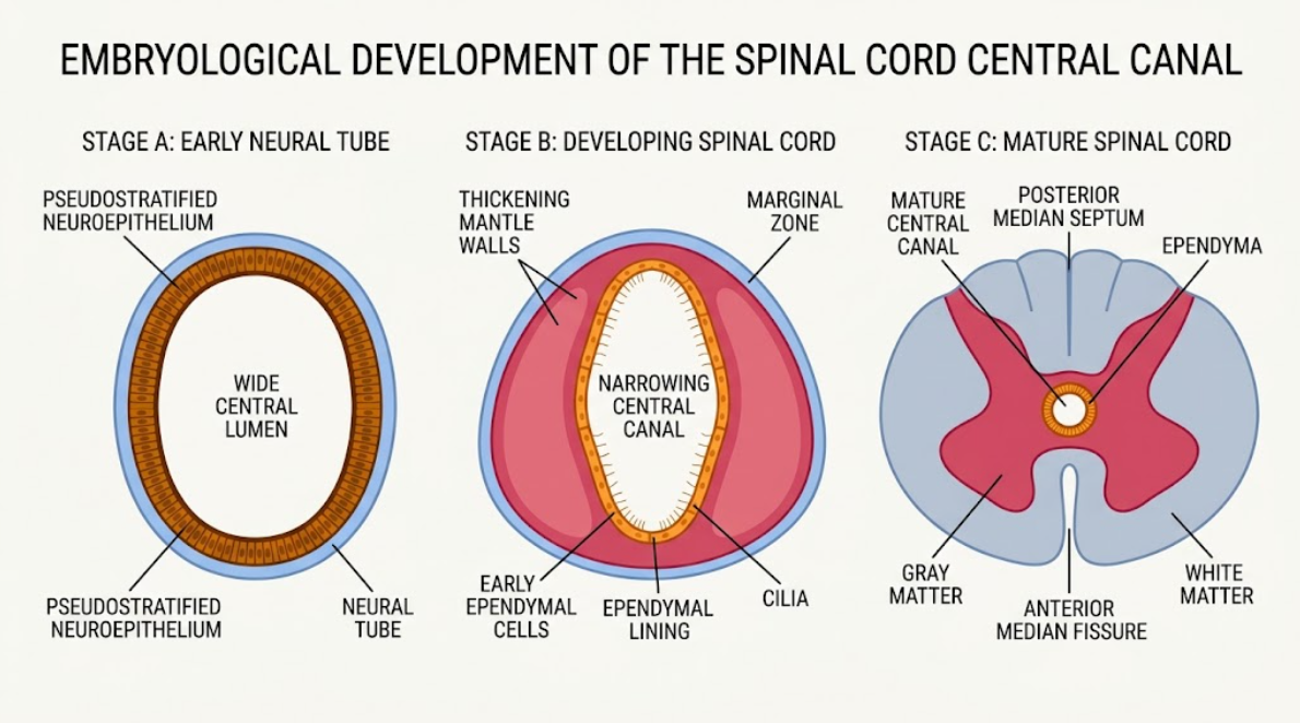
Figure 1. Development of human spinal cord and central canal. Diagrammatic cross-sections illustrating the transition from a wide embryonic neural tube with pseudostratified neuroepithelium (Stage A), to narrowing of the lumen due to mantle wall thickening and differentiation of ciliated ependymal cells (Stage B), to the fully formed mature spinal cord with a tiny central canal, gray matter commissure, and white matter (Stage C).
In many non-human mammalian species the central canal remains widely patent and functionally integrated into fluid circulation throughout life, but in humans the central canal undergoes marked post-natal structural alteration. In the infant and young child the central canal has a continuous, open lumen lined by a distinct layer of simple columnar ependymal cells. However, a physiological regressive process starting in late childhood and accelerating through young adulthood leads to progressive stenosis and closure of the canal lumen(Milhorat, Kotzen, & Anzil, 1994; Sakata, Yashika, & Hashimoto, 1993).
Autopsy and histologic studies indicate stenosis of the central canal in approximately 70% to 90% of normal adults. This process of occlusion is very region-specific, beginning and peaking typically in the mid-thoracic spinal cord (particularly between T2 and T8). Conversely, the cervical cord segments and the caudal conus medullaris frequently retain localized areas of patency into middle age(Hagizawa et al., 2024). Histologically closure occurs by progressive desquamation and breakdown of the lining ependymal cells. Loss of ependymal integrity is associated with proliferation of subependymal astrocytes that extend dense meshes of glial fibrillary acidic protein (GFAP)-positive processes into the lumen. This astrocytic proliferation with accumulation of cellular debris and formation of intracanalicular gliovascular buds converts the open canal into a fibrous, discontinuous glial cord(Jiang et al., 2023; Saker et al., 2016).
The precise trigger for this post-natal stenosis is still being investigated. While previously considered an inevitable degenerative or age-dependent process, other hypotheses suggest that the repetitive subclinical viral ependymitis during life leads to focal ependymal destruction. Influenza A, parainfluenza and reoviruses, among other viral agents, replicate selectively in ependymal cells. Localized desquamation and subsequent reactive gliosis ensue(Mims, 1960).
Whatever the etiology, the physiologic occlusion creates a structural environment with its own biomechanical characteristics. A totally patent canal provides a potential low-resistance route for fluid drainage, but a partially or segmentally occluded central canal creates closed fluid compartments. These isolated patent segments with fibrotic or gliotic margins are prone to focal distension and pressure build-up in the setting of abnormal fluid dynamics(Jiang et al., 2023).
Embryological factors and post-natal stenosis give a structural predisposition, but acquired pathophysiological factors are the main drivers of central canal dilation in adult syringomyelia(Martinelli & Massimi, 2026). The vast majority of acquired cases are the result of structural lesions that change normal CSF circulation in the spinal SAS. The most common of these is the Chiari Type I malformation, which is characterized by caudal herniation of cerebellar tonsils through the foramen magnum. Other acquired causes of note include spinal injury after trauma, postinfectious arachnoiditis, surgical scarring, intervertebral disk herniation, and extramedullary tumors(Wong et al., 2012).
In the last several decades, many physical and biomechanical theories have been developed to describe the process of obstruction in the subarachnoid space that results in fluid accumulation in the spinal cord(Rai & Rai, 2015). The classic “Water-Hammer Theory” of Gardner proposed that obstruction at the outlets of the fourth ventricle causes the pressure waves of systolic arterial CSF to be rerouted directly down the patent central canal via the obex(Sivakumaran, Ashwood, Kamal, & Jayakumar, 2023). In the design process of the role of the fluid pulse waves, Gardner’s model fell out of favor as modern imaging showed that there was no anatomical communication between the fourth ventricle and syrinxes in most clinical cases(Greitz, 2006).
Williams proposed the concept of “Craniospinal Pressure Dissociation” and the “Suck and Slosh” mechanism to explain the non-communicating cavities. Williams showed that brief increases in intra-abdominal and intra-thoracic pressure (as in coughing, sneezing or straining) fill the epidural venous plexus and rapidly compress the dural sac(Milhorat, Johnson, Milhorat, Capocelli, & Pevsner, 1995). In the normal individual, CSF flows freely upward into the cranial vault to equalize pressure. However, when a subarachnoid block occurs at the foramen magnum, a ball-valve effect prevents rapid dissipation of the fluid and a transient pressure gradient between the cranial and spinal compartments is established. The craniospinal pressure differential creates a suction force that pulls fluid into the spinal cord. Longitudinal “slosh” dynamics caused by patient motion cause the cavity to expand in rostral and caudal directions(Sivakumaran et al., 2023).
Later refinements led to Oldfield’s “Piston Theory” focusing on the dynamic movement of herniated cerebellar tonsils. In systole, the rapid influx of arterial blood distends the brain and forces the cerebellar tonsils downward as a solid piston into the blocked spinal subarachnoid space. This piston-like movement creates strong systolic CSF pressure waves in the spinal SAS and forces fluid into the spinal tissue through perivascular spaces (Virchow-Robin spaces)(Oldfield, Muraszko, Shawker, & Patronas, 1994).
For this purpose, Greitz suggested the “Intramedullary Pulse Pressure” model and the use of the Venturi effect. If a fluid is moving at a higher velocity through a narrowed channel in the subarachnoid space then there will be a localized drop in lateral pressure according to hydrodynamic principles(Martinelli & Massimi, 2026; Rai & Rai, 2015).
This drop in pressure causes a transmedullary pressure gradient and an outward suction force on the cord parenchyma (see Figure 2). This mechanical tension increases extracellular spaces and impairs normal interstitial fluid clearance resulting in local fluid pooling and central canal dilation(Heiss et al., 1999).
Elaborating on those mechanical perspectives, the “Volume Change Theory” by Rai and Rai addresses biomechanical changes in normal spinal motion. The bony spinal canal lengthens and expands during flexion of the spine and shortens during extension, so the internal volume of the canal varies dynamically over the course of the day(Rai & Rai, 2015). In healthy subjects these volume shifts are buffered by free exchange of CSF with the posterior cranial cavity. However, when the subarachnoid space is blocked, the spinal cord tissue undergoes severe mechanical deformation. The central canal dilation develops as a secondary mechanical adaptative response, the fastest physical mechanism to compensate for the cyclical volume changes during flexion and extension is the fluid accumulation within the central canal(Rai & Rai, 2015).
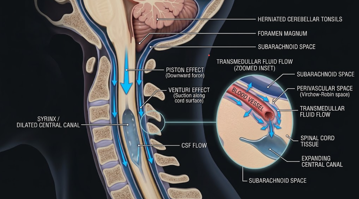
Figure 2. Fluid dynamics of central canal dilation in syringomyelia Forces of fluid dynamics driving syringomyelia. Herniated cerebellar tonsils protrude into the subarachnoid space at the foramen magnum, resulting in downward pressure (Piston effect) and suction from the surface (Venturi effect). The zoomed inset highlights the transmedullary movement of cerebrospinal fluid (CSF) through perivascular (Virchow-Robin) spaces that causes progressive dilation of the central canal (syrinx).
Physical hydrodynamics describe the external forces acting on the spinal cord, but recent scientific studies suggest that active cellular, molecular and ciliary responses in the central canal wall control syrinx expansion and tissue remodeling. The ependyma lines the central canal and contains a specialized niche of neural stem and progenitor cells that respond to mechanical stress and injury(S. Liu & Chen, 2019).
Recent experimental studies in animal models of spinal cord injury and of extradural compression have demonstrated that the mere occlusion of the subarachnoid space does not lead to syringomyelia. In controlled investigations it has been shown that in experimental animals with only subarachnoid compression, true syrinx cavities only develop when there is structural occlusion of the central canal and localized ciliary impairment(Jiang et al., 2023). The central canal is lined by ependymal cells, which possess motile cilia that beat in a coordinated, synchronized manner to provide continuous fluid movement and prevent fluid stasis. Physical trauma, such as prolonged mechanical pressure or direct trauma to the cord, leads to severe physical damage and desquamation of ependymal cilia. Scanning electron microscopy shows extensive loss of cilia in dilated regions of canals. Loss of ciliary motility interferes with directional fluid clearance, causing local fluid turbulence, micro-stasis and progressive mechanical weakening of the canal boundary(Cosan, Tel, Durmaz, Gülec, & Baycu, 2000).
On the cellular level, mechanical strain and fluid accumulation rapidly activate and proliferate ependymal cells of the central canal. Activated ependymal cells re-express neural stem cell markers like Nestin and Sox2 and proliferative markers like Ki67. The persistent intraluminal pressure stimulates proliferation of ependymal cells and formation of tunnel-like cellular protrusions along the central canal(Sumei Liu et al., 2023).
Ependymal cell lineage tracking shows these cells undergo active migration facilitated by a cadherin switch as shown in the figure 3. Ependymal cells express high levels of epithelial cadherin (E-cadherin) under normal physiological conditions, which maintains tight cell-to-cell adhesion(Lamouille, Xu, & Derynck, 2014). Mechanical injury and fluid distension activate the transforming growth factor beta receptor 1 (TGFβR1)-Smad3 signaling axis, which downregulates E-cadherin and upregulates neural cadherin (N-cadherin). This change, typical of epithelial-mesenchymal transition (EMT), decreases inter-cellular adhesion, allowing migration of ependymal cells along the wall of the canal and formation of irregular cellular protrusions that favor enlargement of the cavity. Experimental use of specific chemical inhibitors of the Smad3 pathway (e.g. SIS3) significantly reduces N-cadherin expression, maintains E-cadherin, inhibits abnormal ependymal migration, and prevents progressive central canal dilation(Sumei Liu et al., 2023).
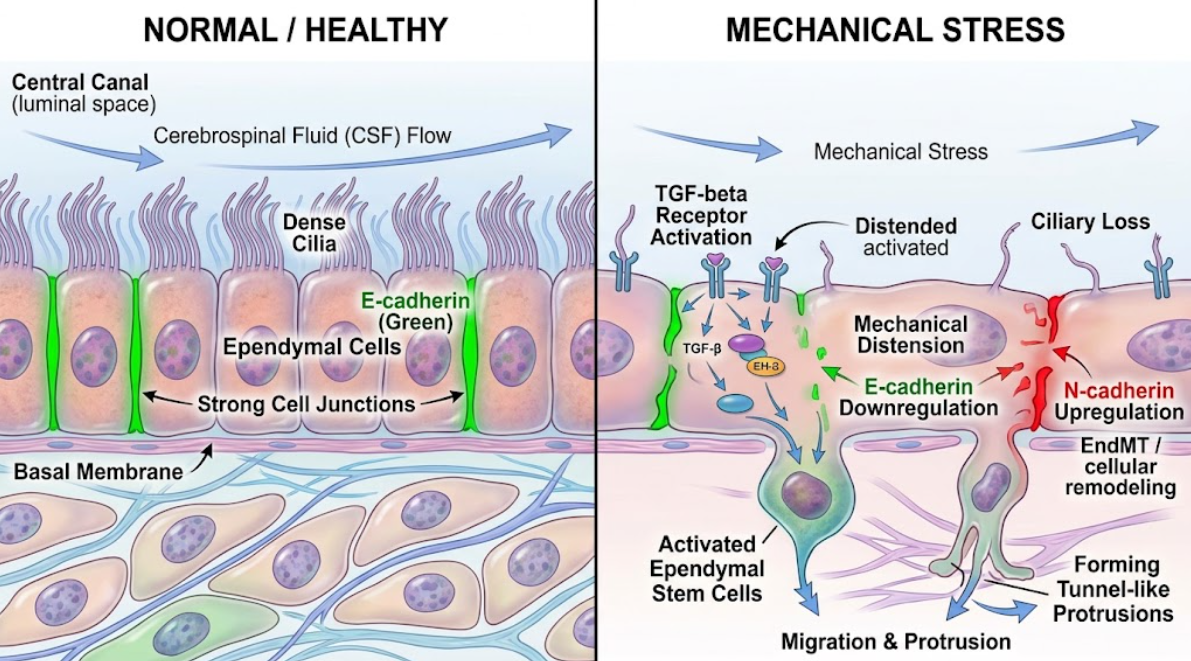
Figure 3. Morphological and Molecular Changes of Ependymal Cells of the Spinal Cord Under Mechanical Stress Left. Normal ependymal cells lining the central canal have dense cilia and stable E-cadherin junctions. Mechanical stress causes loss of cilia, activation of the TGF-β pathway, cadherin switching (E- to N-cadherin) and migration of activated ependymal stem cells through tunnel-like protrusions (Right).
The combination of embryological development, physical fluid mechanics, and cellular signaling shows that the central canal of syringomyelia is not an isolated congenital anomaly or a purely mechanical phenomenon. It is a complex multi-stage pathological process and not a single disease(Jiang et al., 2023).
Embryonic neurulation sets up the basic architecture of the central canal and the surrounding neuroepithelial niche. Regional differences in canal patency develop during post-natal development, with normal physiological stenosis of the thoracic cord forming isolated, potential fluid compartments delineated by fibrotic gliosis. When acquired pathologies such as Chiari I malformations, trauma or arachnoid webs cause obstruction of the subarachnoid space the normal compliance of the craniospinal fluid system is severely compromised(Jiang et al., 2023; Martinelli & Massimi, 2026).
These hydrostatic and hydrodynamic disturbances are explained by a combination of craniospinal pressure dissociation, piston-like tonsillar impaction, Venturi-like pressure and volume deformation caused by posture and produce ongoing transmedullary pressure gradients. These physical forces push fluid into the parenchyma of spine and the lumen of central canal(Fadila et al., 2025).
Importantly, the transition from temporary fluid accumulation to permanent, enlarging syrinx spaces is controlled by cellular and microstructural processes. Ciliary desquamation reduces local fluid clearance and mechanical strain activates the ependymal stem cell niche. Downstream activation of the TGFβR1-Smad3 signaling cascade promotes an epithelial-mesenchymal transition, characterized by an E-cadherin to N-cadherin switch(Sumei Liu et al., 2023). This molecular change favors ependymal migration and degradation of the structural wall resulting in irreversible cavity expansion. This broad framework has important clinical implications. In the past, neurosurgical treatment has been almost exclusively directed at mechanical decompression of the subarachnoid space or direct shunting of the syrinx cavity. Suboccipital decompression can restore gross CSF pathways in many Chiari patients, but clinical results are variable, especially in the presence of established parenchymal damage or isolated central canal occlusion(Jiang et al., 2023; Martinelli & Massimi, 2026). The recognition of central canal occlusion and ciliary destruction as primary prerequisites for syrinx formation suggests that future treatments will have to extend beyond subarachnoid decompression. Promising adjunct therapeutic approaches to stop cellular migration and prevent progressive destruction of the spinal cord include restoring central canal patency, maintaining or restoring ependymal ciliary function, and targeting molecular pathways (e.g., TGFβR1-Smad3 inhibition)(Jiang et al., 2023; Sumei Liu et al., 2023).
Central canal dilation in syringomyelia is due to a dynamic interplay between embryological structural determinants, physiological stenosis in the post-natal period, acquired hydrodynamic disturbances and active cellular remodeling. Early embryonic neurulation constructs the first ependymal lumen which ages and develops segmental occlusion in post-natal life. If acquired lesions prevent subarachnoid cerebrospinal fluid flow, the complex hydrodynamics create transmedullary pressure gradients that permit fluid to enter the vulnerable portions of the central canal. Mechanical stress leads to ciliary loss, fluid stasis, and ependymal activation, inducing TGFβR1-Smad3 mediated cellular migration and persistent cavity enlargement. A full explanation of these combined embryologic, mechanical, and biological origins provides a holistic paradigm that links basic anatomy to neurosurgical practice and opens the door for targeted cellular and biophysical therapies.
Cosan, T. E., Tel, E., Durmaz, R., Gülec, S., & Baycu, C. (2000). Non-hindbrain-related syringomyelia. Obstruction of the subarachnoid space and the central canal in rats. An experimental study. J Neurosurg Sci, 44(3), 123-127.
Demetriades, A. K. (2012). The first descriptions of syringomyelia. J R Soc Med, 105(7), 278. doi:10.1258/jrsm.2012.120082
Fadila, M., Sarrabia, G., Shapira, S., Yaacobi, E., Baruch, Y., Engel, I., & Ohana, N. (2025). Orthopedic Manifestations of Syringomyelia: A Comprehensive Review. J Clin Med, 14(9). doi:10.3390/jcm14093145
Gray, H., & Lewis, W. H. (1918). Anatomy of the human body (20th ed.). Philadelphia and New York,: Lea & Febiger.
Greitz, D. (2006). Unraveling the riddle of syringomyelia. Neurosurg Rev, 29(4), 251-263; discussion 264. doi:10.1007/s10143-006-0029-5
Hagizawa, H., Moriguchi, Y., Yamada, T., & Kashii, M. (2024). Syringomyelia with Central Canal Enlargement Caused by Postoperative Inflammation after Surgery for Intradural Lumbar Disc Herniation. NMC Case Rep J, 11, 421-425. doi:10.2176/jns-nmc.2024-0165
Heiss, J. D., Patronas, N., DeVroom, H. L., Shawker, T., Ennis, R., Kammerer, W., . . . Oldfield, E. H. (1999). Elucidating the pathophysiology of syringomyelia. J Neurosurg, 91(4), 553-562. doi:10.3171/jns.1999.91.4.0553
Jiang, C., Wang, X., Lu, C., Li, Q., Ma, L., Li, W., . . . Jian, F. (2023). The Physiological Occlusion of the Central Canal May Be a Prerequisite for Syringomyelia Formation. Neurospine, 20(4), 1346-1357. doi:10.14245/ns.2346834.417
Lamouille, S., Xu, J., & Derynck, R. (2014). Molecular mechanisms of epithelial-mesenchymal transition. Nat Rev Mol Cell Biol, 15(3), 178-196. doi:10.1038/nrm3758
Liu, S., & Chen, Z. (2019). Employing Endogenous NSCs to Promote Recovery of Spinal Cord Injury. Stem Cells Int, 2019, 1958631. doi:10.1155/2019/1958631
Liu, S., Ma, L., Qi, B., Li, Q., Chen, Z., & Jian, F. (2023). Suppression of TGFβR-Smad3 pathway alleviates the syrinx induced by syringomyelia. Cell & Bioscience, 13(1), 98. doi:10.1186/s13578-023-01048-w
Martinelli, R., & Massimi, L. (2026). Idiopathic Syringomyelia: A Systematic Scoping Review. J Clin Med, 15(16). doi:10.3390/jcm15166216
Milhorat, T. H., Johnson, R. W., Milhorat, R. H., Capocelli, A. L., Jr., & Pevsner, P. H. (1995). Clinicopathological correlations in syringomyelia using axial magnetic resonance imaging. Neurosurgery, 37(2), 206-213. doi:10.1227/00006123-199508000-00003
Milhorat, T. H., Kotzen, R. M., & Anzil, A. P. (1994). Stenosis of central canal of spinal cord in man: incidence and pathological findings in 232 autopsy cases. J Neurosurg, 80(4), 716-722. doi:10.3171/jns.1994.80.4.0716
Mims, C. A. (1960). Intracerebral injections and the growth of viruses in the mouse brain. Br J Exp Pathol, 41(1), 52-59.
Moore, K. L., Persaud, T. V. N., & Torchia, M. G. (2013). The Developing Human: Clinically Oriented Embryology: Saunders/Elsevier.
Oldfield, E. H., Muraszko, K., Shawker, T. H., & Patronas, N. J. (1994). Pathophysiology of syringomyelia associated with Chiari I malformation of the cerebellar tonsils. Implications for diagnosis and treatment. J Neurosurg, 80(1), 3-15. doi:10.3171/jns.1994.80.1.0003
Rai, S. K., & Rai, P. S. (2015). Volume change theory for syringomyelia: A new perspective. Asian J Neurosurg, 10(4), 245-251. doi:10.4103/1793-5482.162680
Sakata, M., Yashika, K., & Hashimoto, P. H. (1993). Caudal aperture of the central canal at the filum terminale in primates. Kaibogaku Zasshi, 68(2), 213-219.
Saker, E., Henry, B. M., Tomaszewski, K. A., Loukas, M., Iwanaga, J., Oskouian, R. J., & Tubbs, R. S. (2016). The Human Central Canal of the Spinal Cord: A Comprehensive Review of its Anatomy, Embryology, Molecular Development, Variants, and Pathology. Cureus, 8(12), e927. doi:10.7759/cureus.927
Sivakumaran, P., Ashwood, N., Kamal, M., & Jayakumar, N. (2023). The Pathogenesis of Chiari Malformation and Syringomyelia: A Case Report and Systematic Review of Current Theories. Cureus, 15(10), e47301. doi:10.7759/cureus.47301
Wong, J., Hemley, S., Jones, N., Cheng, S., Bilston, L., & Stoodley, M. (2012). Fluid outflow in a large-animal model of posttraumatic syringomyelia. Neurosurgery, 71(2), 474-480; discussion 480. doi:10.1227/NEU.0b013e31825927d6