Our Services
Others
- DFPP
- Shockwave
- IV Drip


“This literature review provides an in-depth mechanistic synthesis of how splanchnic arterial vasodilation results in systemic hyperdynamic circulation, vascular hypo-responsiveness and end-organ failure in decompensated liver cirrhosis.”
Decompensated liver cirrhosis is a major clinical watershed with severe systemic complications such as intractable ascites, variceal hemorrhage, hepatic encephalopathy and hepatorenal syndrome. Intrahepatic structural collapse and increased resistance to flow in sinusoids initiate the development of portal hypertension, but the progression to overt clinical decompensation is largely related to profound systemic and splanchnic hemodynamic derangements. Splanchnic arterial vasodilation is the main vascular driver of a hyperdynamic circulatory state, with increased cardiac output, resting tachycardia, reduced systemic vascular resistance, and arterial hypotension. Here, a comprehensive review summarizes the accumulating literature on cellular, molecular and neurogenic mechanisms of splanchnic vasodilation in decompensated cirrhosis. We discuss multifactorial overproduction of endogenous vasodilators, notably endothelial nitric oxide synthase-derived nitric oxide, carbon monoxide, prostacyclin, endocannabinoids and neuropeptides, and their intracellular signaling via cyclic guanosine monophosphate, cyclic adenosine monophosphate and hyperpolarizing potassium channels. We also discuss the cellular mechanisms of vascular hypo-responsiveness to endogenous pressor agents, which is mediated by G-protein-coupled receptor desensitization, regulator of G-protein signaling upregulation, and altered RhoA/Rho-kinase contractile signaling. Finally, we emphasize the synergistic effects of mesenteric sympathetic ganglionic atrophy, gut-derived bacterial translocation, and systemic immune activation in sustaining pathologically low splanchnic vascular tone. A detailed understanding of these integrated mechanisms is essential to improve the efficacy of targeted therapeutic approaches aimed at reducing circulatory failure in severe liver disease.
Cirrhosis of the liver is the final common pathway of progressive chronic liver injury from multiple etiologies including alcohol-related liver disease, metabolic dysfunction-associated steatotic liver disease and chronic viral hepatitis(Ververeli et al., 2025). Cirrhosis has long been defined as a static structural disease characterized by fixed fibrous septa and regenerative nodules, contrary to this view, hepatologists have come to recognize cirrhosis as a highly dynamic biological continuum, influenced by complex vascular, metabolic and neurohumoral interactions. The most important prognostic landmark on this spectrum is the transition from compensated to decompensated liver cirrhosis. Compensated disease is characterized by preserved functional reserve and minimal symptoms but decompensation heralds a dramatic reduction in median survival driven by the development of ascites, variceal bleeding, hepatic encephalopathy and functional renal impairment(Al-Hamoudi, 2010; Di Pascoli, Sacerdoti, Pontisso, Angeli, & Bolognesi, 2017).
Previously we have reviewed the pathophysiological progression from compensated to decompensated cirrhosis characterized by progressive loss of parenchymal metabolic redundancy, mitochondrial oxidative energy collapse and systemic detoxification failure. The fundamental ideas underlying the previous work on structural sinusoidal capillarization and intrahepatic resistance are now clearer that extrahepatic hemodynamic dysregulation is a major systemic driver that precipitates overt clinical decompensation(Kim, Baik, & Lee, 2010). The most important mechanical trigger of portal hypertension is the increased intrahepatic resistance due to structural tissue remodeling and sinusoidal endothelial dysfunction. However, increased intrahepatic resistance alone is not sufficient to sustain the severe extrahepatic manifestations of advanced liver disease(Bolognesi, Di Pascoli, Verardo, & Gatta, 2014; Colle, Geerts, Van Steenkiste, & Van Vlierberghe, 2008).
In fact, the clinical course of decompensated liver cirrhosis is characterized by a profound systemic hemodynamic rearrangement, the hyperdynamic circulatory syndrome(Iwakiri & Groszmann, 2006). The hallmark of this syndrome is marked and sustained arterial vasodilation, predominantly in the splanchnic vascular bed. The splanchnic circulation, consisting of the mesenteric, gastric and splenic arterial networks, shows a pronounced decrease in vascular tone(Geerts et al., 2006). This profound vasodilation results in massive splanchnic pooling of blood, effectively decreasing central arterial blood volume and triggering a compensatory neurohumoral cascade that paradoxically sustains and exacerbates portal hypertension(Arroyo & Fernández, 2013; Tsai, 2007).
The current literature review aims to provide a profound mechanistic synthesis of the hemodynamic alterations and splanchnic vasodilation in decompensated liver cirrhosis. In this paper, we describe how endothelial hyper-reactivity, receptor desensitization, autonomic nerve atrophy and gut-derived systemic inflammation act in concert to induce splanchnic hyperemia and multi-organ circulatory dysfunction, on the basis of evidence across the cellular, molecular, paracrine and neurogenic domains.
The splanchnic vascular system normally receives about one quarter to one third of the total resting cardiac output. In decompensated liver cirrhosis this extensive vascular network undergoes structural and functional remodeling to become a low-resistance high-flow reservoir(Mulvany & Aalkjaer, 1990). Vasodilation of splanchnic arteries is mainly observed at the precapillary resistance arterioles of the mesenteric and splenic circulations. This loss of arteriolar tone leads to an increase in blood flow into the portal venous system(Bolognesi et al., 2014). This excessive splanchnic inflow results in a pathophysiologic paradox: although the portosystemic collateral networks are extensive and attempt to decompress the high-pressure portal vein, the portal pressure remains persistently high or worsens because of the massive volume of incoming blood(Martell, Coll, Ezkurdia, Raurell, & Genescà, 2010).
The change in hemodynamics in the splanchnic bed starts a systemic chain reaction. Massive splanchnic arterial dilatation leads to relative unfilling of the systemic arterial tree, a condition termed effective arterial hypovolemia(Henriksen & Moller, 2006). In the early compensated phase of cirrhosis, the body copes with this relative hypovolemia by increasing plasma volume and cardiac output. However, as cirrhosis progresses to decompensation, splanchnic pooling becomes so severe that arterial baroreceptors and volume receptors in the carotid sinus and aortic arch are continuously stimulated(Martell et al., 2010).
Figure 1 shows that the cascade starts with splanchnic arterial vasodilation, leading to central arterial hypovolemia, which in turn activates the neurohumoral reflex arches and generates the classical hyperdynamic circulatory state(Iwakiri & Groszmann, 2006).
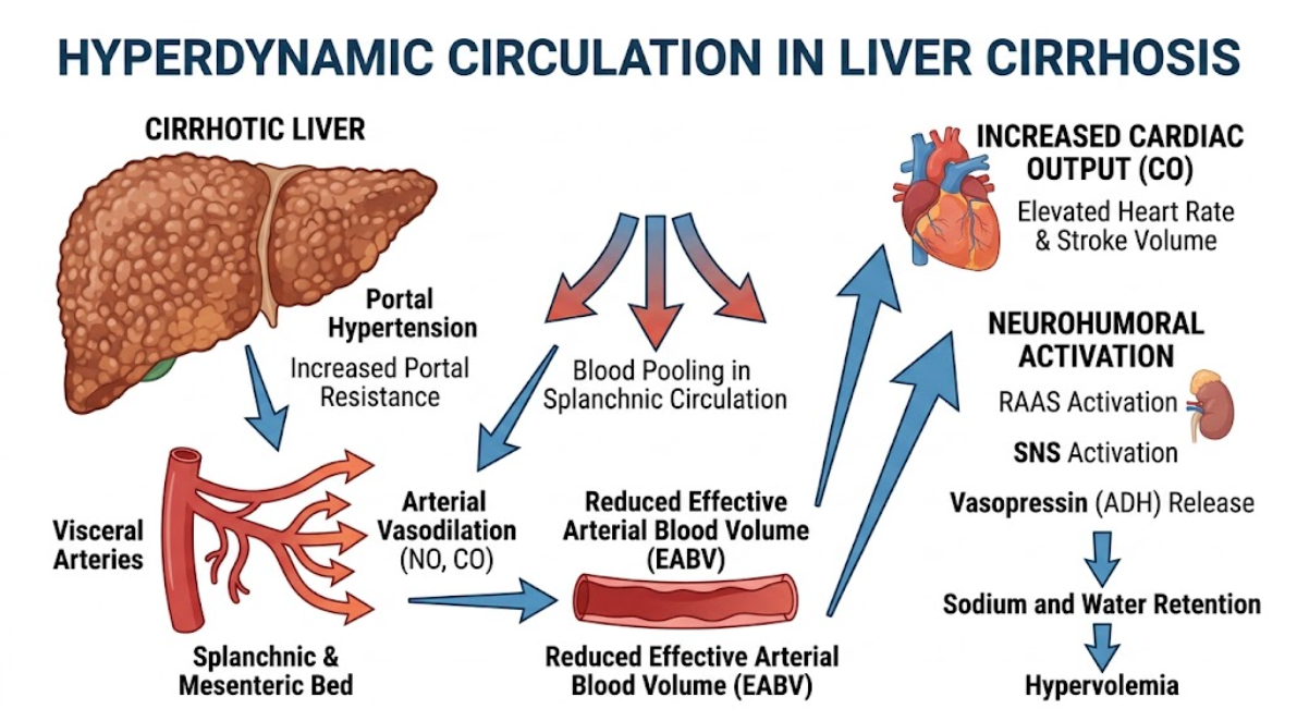
Figure 1. Pathophysiologic Sequence of Hyperdynamic Circulation in Cirrhosis of Liver. Schematic depiction of the hyperdynamic state caused by portal venous hypertension. Severe intrahepatic resistance leads to splanchnic and mesenteric arterial vasodilation with blood pooling and a decrease in effective arterial blood volume (EABV). In response compensatory neurohumoral pathways (RAAS, SNS and ADH) are activated to retain sodium and water and cardiac output and heart rate are increased culminating in hyperdynamic circulation.
The neurohumoral response is characterized by a pronounced compensatory activation of the renin-angiotensin-aldosterone system, the sympathetic nervous system and non-osmotic arginine vasopressin release(Al-Hamoudi, 2010). These systems stimulate avid renal tubular reabsorption of sodium and water resulting in total plasma volume expansion. The total blood volume is increased but the fluid is pathologically sequestered in the dilated splanchnic basin and the peritoneal cavity as ascites(Arroyo & Fernández, 2013). At the same time there is a baseline increase in cardiac output by increase in heart rate and stroke volume to establish the hyperdynamic circulatory state with low mean arterial pressure, bounding pulses, low systemic vascular resistance and high cardiac index(Kim et al., 2010).
Structural remodeling and neoangiogenesis of splanchnic stroma in addition to arteriolar vasodilatation are important for low vascular resistance. Mesenteric vasculature undergoes mechanical stretch and shear stress due to chronic portal hypertension(Colle et al., 2008). This mechanical stimuli triggers the release of pro-angiogenic factors, primarily vascular endothelial growth factor, placental growth factor, and platelet-derived growth factor. These factors favor sprouting of new, immature microvessels and dilatation of pre-existing portosystemic collaterals(Bolognesi et al., 2014). The newly formed vessels have defective smooth muscle coverage and altered junctions of endothelial cells. This leads to decreased resistance in the regional resistance arteries and hyperdynamic splanchnic inflow(Geerts et al., 2006).
The cellular mechanisms of splanchnic vasodilation in decompensated liver cirrhosis are complex and involve a complex network of local paracrine molecules, circulating humoral factors and altered intracellular signaling cascades within endothelial and vascular smooth muscle cells(Genesca et al., 1999).
Among the many vasoactive mediators, endothelial nitric oxide synthase-derived nitric oxide has been identified as the principal mediator of splanchnic arterial vasodilation(Whittle & Moncada, 1992). While endothelial dysfunction drastically decreases intrahepatic sinusoidal bioavailability of nitric oxide as described in the previous article, splanchnic and systemic arterial beds have a diametrically opposite abnormality: a significant and continuous overproduction of nitric oxide(Genesca et al., 1999).
Nitric oxide is a soluble free radical gas that is generated from L-arginine by the enzymes nitric oxide synthase. Endothelial nitric oxide synthase activity is markedly increased in the splanchnic endothelium. In the early phase of portal hypertension, the initial activation of endothelial nitric oxide synthase is caused by post-translational modifications induced by mechanical shear stress along the mesenteric arterial walls(Iwakiri et al., 2002). Specifically, shear stress and angiogenic signaling by vascular endothelial growth factor activate the phosphoinositide 3-kinase/Akt pathway, resulting in phosphorylation of endothelial nitric oxide synthase at the Serine-1177 residue(Colle et al., 2008). This phosphorylation event greatly increases the catalytic efficiency of the enzyme, allowing sustained nitric oxide generation even at basal cytosolic calcium levels(Dimmeler et al., 1999).
As portal hypertension approaches decompensation, molecular chaperones and cofactors continue to sustain the hyperactivation of endothelial nitric oxide synthase. Heat shock protein 90 is a chaperone that directly binds to phosphorylated endothelial nitric oxide synthase and stabilizes the active conformation, thus increasing nitric oxide production(Schrier et al., 1988). In addition, bacterial translocation from the gut induces the rate-limiting enzyme in the tetrahydrobiopterin biosynthetic pathway, endothelial GTP-cyclohydrolase I. Tetrahydrobiopterin is an essential cofactor that prevents the uncoupling of endothelial nitric oxide synthase, resulting in the robust and sustained production of nitric oxide rather than superoxide(Di Pascoli et al., 2017). This mechanism is a multi-step molecular cascade of Akt-mediated phosphorylation, coupling with heat shock protein 90 and stabilization of tetrahydrobiopterin, resulting in excessive release of nitric oxide into vascular smooth muscle cells (Figure 2)(Martell et al., 2010).
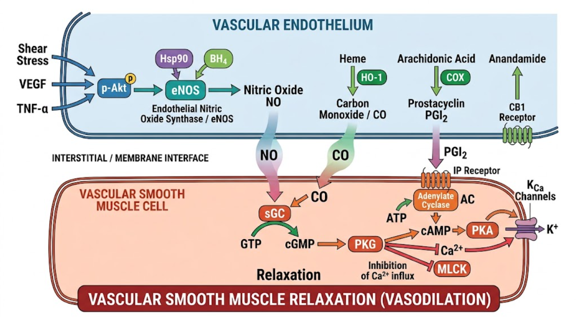
Figure 2. Splanchnic Vasodilation: Molecular Signaling Pathways. Schematic description of molecular mechanisms involved in splanchnic vasodilation at the interface of the vascular endothelium and smooth muscle cells. Shear stress, VEGF and TNF-α activate eNOS through phosphorylation of Akt and coupling of Hsp90/BH4 to generate nitric oxide (NO). Companion pathways include heme oxygenase-1 (HO-1) producing carbon monoxide (CO), cyclooxygenase (COX) producing prostacyclin (PGI2), and anandamide activating CB1 receptors. NO and CO are diffusible, activate soluble guanylyl cyclase (sGC), increasing cGMP and activating PKG. PGI2 activates adenylate cyclase (AC) to increase cAMP and activate PKA. Both pathways converge to inhibit Ca2+ influx and myosin light chain kinase (MLCK) and activate calcium-activated potassium (KCa) channels, inducing relaxation of vascular smooth muscle (vasodilation) together.
After its synthesis, nitric oxide diffuses rapidly across the endothelial membrane and into adjacent vascular smooth muscle cells. There it binds to the heme moiety of soluble guanylyl cyclase, catalyzing the conversion of guanosine triphosphate to cyclic guanosine monophosphate(Bolognesi et al., 2014). Cyclic guanosine monophosphate accumulation activates protein kinase G, which leads to smooth muscle relaxation via numerous downstream targets: increased intracellular calcium sequestration into the sarcoplasmic reticulum, blockade of voltage gated calcium entry, and calcium dependent potassium channel activation inducing membrane hyperpolarization(Ververeli et al., 2025).
Besides the endothelial constitutive isoform, there is growing evidence for contributions from neuronal nitric oxide synthase and inducible nitric oxide synthase in advanced cirrhosis(Di Pascoli et al., 2017). Increased neuronal nitric oxide synthase expression in perivascular adventitial nerves and mesenteric arterial walls contributes to neurogenic vasodilation. Inducible nitric oxide synthase is expressed in adventitial macrophages and vascular smooth muscle cells, in response to circulating endotoxins and pro-inflammatory cytokines, and generates high-capacity, long-lasting pools of nitric oxide that exacerbate splanchnic vasorelaxation during acute inflammatory decompensation(Genesca et al., 1999).
Carbon monoxide is an important synergistic vasodilator of nitric oxide and is produced during heme degradation by heme oxygenase enzymes. The inducible isoform heme oxygenase-1 is significantly upregulated in splanchnic arterial beds of cirrhotic models in response to oxidative stress, mechanical stretch and circulating inflammatory mediators(Bolognesi et al., 2005).
Like nitric oxide, CO diffuses into vascular smooth muscle cells and activates soluble guanylyl cyclase, increasing levels of cyclic guanosine monophosphate. Carbon monoxide is a weaker activator of soluble guanylyl cyclase than nitric oxide, but it has a distinctive, high-affinity interaction with large-conductance, calcium-activated potassium channels on the smooth muscle cell membrane. L-type voltage-dependent calcium channels are subsequently closed(Bolognesi et al., 2014). Carbon monoxide directly increases the open-state probability of these potassium channels, producing an outward efflux of potassium ions with marked cell membrane hyperpolarization. In advanced decompensated cirrhosis with ascites, mesenteric resistance arteries show increased sensitivity to carbon monoxide that contributes to vascular hyporeactivity to pressor agents(Di Pascoli et al., 2017).
In vascular endothelial cells, arachidonic acid is metabolized via the cyclooxygenase pathway to the potent vasodilator lipid prostacyclin. Circulating and splanchnic prostacyclin and its stable urinary metabolites are markedly increased in patients with decompensated cirrhosis(Martell et al., 2010).
Altered hemodynamics and increased portal pressure increase endothelial cyclooxygenase-1 expression in mesenteric resistance arteries. Prostacyclin acts locally at smooth muscle prostacyclin receptors that are coupled to G-s proteins that activate adenylyl cyclase(Guarner et al., 1992). This results in an intracellular accumulation of cyclic adenosine monophosphate and activation of protein kinase A. Protein kinase A promotes smooth muscle relaxation by phosphorylating myosin light chain kinase, thereby decreasing its affinity for calcium-calmodulin, and by stimulating sarcoplasmic/endoplasmic reticulum calcium ATPase pumps. Pharmacological inhibition of cyclooxygenase by non-steroidal anti-inflammatory agents partially reverses splanchnic arterial hyperemia and decreases portal pressure in cirrhotic models, thus confirming the functional role of prostacyclin in the maintenance of splanchnic vasodilation(Guarner et al., 1992).
The endogenous cannabinoid system is a key non-classical pathway involved in splanchnic vasodilation. Endocannabinoids are bioactive lipid mediators that are synthesized on demand from membrane phospholipids. One endocannabinoid is anandamide (N-arachidonoylethanolamine). In decompensated cirrhosis, circulating monocytes, vascular endothelial cells and mesenteric tissue produce high concentrations of anandamide, stimulated by gut-derived endotoxemia and systemic inflammation(Martell et al., 2010).
Anandamide is a ligand for vascular cannabinoid type-1 receptors on endothelial cells, vascular smooth muscle cells and perivascular nerve terminals in the splanchnic bed. Endothelial expression of cannabinoid type-1 receptor activates endothelial nitric oxide synthase resulting in paracrine release of nitric oxide(Bátkai et al., 2001). At the same time, anandamide directly interacts with smooth muscle transient receptor potential vanilloid-1 channels on adventitial sensory nerves and induces neurogenic release of powerful peptide vasodilators. Pharmacological blockade of the cannabinoid type-1 receptor reversed mesenteric arterial vasodilatation, increased systemic vascular resistance and reduced portal pressure, suggesting that endocannabinoids are major players in vascular collapse in advanced cirrhosis(Di Pascoli et al., 2017).
Endothelium-derived hyperpolarizing factors mediate sustained arterial relaxation in resistance arterioles with impaired nitric oxide synthase or cyclooxygenase signaling. The endothelium-derived hyperpolarizing factors are a heterogeneous group of signaling molecules including epoxyeicosatrienoic acids (from cytochrome P450 epoxygenases), hydrogen sulfide and potassium ions(Martell et al., 2010).
These factors are mainly responsible for opening of small- and intermediate-conductance calcium-activated potassium channels on endothelial cells leading to a hyperpolarization wave that is directly propagated to adjacent smooth muscle cells through myoendothelial gap junctions(Nishimatsu et al., 2001). The hyperpolarization of the membrane inhibits depolarization of smooth muscle cells and the influx of voltage gated calcium, and thus maintains vasodilation in small precapillary resistance vessels. Furthermore, the increased production and decreased hepatic clearance by the diseased liver of humoral vasodilators such as glucagon, adrenomedullin, calcitonin gene-related peptide, substance P, vasoactive intestinal peptide and atrial natriuretic peptide, which are present in increased circulating levels, act in concert to suppress vascular tone(Henriksen & Moller, 2006).
Vascular hyporeactivity to endogenous vasoconstrictors is a pathological characteristic of splanchnic hemodynamics in decompensated liver cirrhosis. The splanchnic arterial bed remains paradoxically dilated, despite severe, compensatory systemic activation of pressor pathways, as evidenced by markedly increased circulating levels of norepinephrine, angiotensin II, endothelin-1 and vasopressin. This loss of contractile responsiveness prevents the body from restoring effective arterial blood volume and perpetuates splanchnic hyperdynamic inflow(Martell et al., 2010).
Vascular hyporesponsiveness has several cellular mechanisms, including receptor desensitization, abnormalities in post-receptor signal transduction, and altered regulation of the contractile apparatus in vascular smooth muscle cells(Ververeli et al., 2025).
Vasoconstrictors signaling through G-protein-coupled receptors include norepinephrine on α1 adrenergic receptors and angiotensin II on angiotensin II type-1 receptors. In normal vascular smooth muscle, agonists binding to these receptors activate G-q proteins which stimulate phospholipase C to cleave phosphatidylinositol 4,5-bisphosphate into inositol trisphosphate and diacylglycerol. Inositol trisphosphate mediates calcium release from sarcoplasmic reticulum and diacylglycerol activates protein kinase C, together causing myosin light chain phosphorylation and vascular contraction(Martell et al., 2010).
In decompensated cirrhosis, sustained exposure to high circulating levels of catecholamines and angiotensin II results in desensitization of homologous and heterologous receptors. Agonist-induced up-regulation and membrane translocation of G protein-coupled receptor kinase-2 in mesenteric arterial smooth muscle cells(Du et al., 2013). G-protein-coupled receptor kinase-2 phosphorylates serine and threonine residues on the occupied vasoconstrictor receptor intracellular loops. Phosphorylation enhances the binding of β-arrestin-2, which physically uncouples the receptor from its downstream G-q protein and targets the receptor for clathrin-mediated endocytosis and lysosomal degradation. This leads to a significant reduction in the functional receptor density on the surface of the smooth muscle cell(Ferguson, 2001; Henriksen & Ring-Larsen, 1994).
In addition, regulators of G-protein signaling, such as regulator of G-protein signaling-2 and regulator of G-protein signaling-4, are overexpressed in cirrhotic mesenteric arteries. These proteins enhance the intrinsic GTPase activity of G-α-q subunits, thus quickly terminating G-protein signaling, and preventing intracellular calcium mobilization even if vasoconstrictors successfully bind to remaining surface receptors(Di Pascoli et al., 2017).
Vascular smooth muscle contraction depends not only on cytosolic calcium elevation, but also on calcium sensitization regulated by the small GTPase RhoA and its downstream effector Rho-kinase (ROCK). Under physiological conditions, however, vasoconstrictors activate RhoA which subsequently activates Rho-kinase. Active Rho-kinase phosphorylates and inactivates myosin light chain phosphatase, preventing myosin light chain dephosphorylation, and thus sustaining smooth muscle contraction even as intracellular calcium levels decline(Martell et al., 2010).
The RhoA/Rho-kinase signaling cascade is severely blunted in mesenteric arteries of cirrhotic animals and patients. Excessive production of nitric oxide and carbon monoxide leads to continuous activation of protein kinase G and protein kinase A(Martell et al., 2010). These cyclic nucleotide–dependent kinases directly phosphorylate RhoA at serine-188, sequestering active RhoA to the cytosol and preventing translocation to the cell membrane(Colle et al., 2008). Inhibition of the RhoA/Rho-kinase pathway allows myosin light chain phosphatase to dominate and promotes rapid dephosphorylation of myosin light chains and prevents stable cross-bridge cycling between actin and myosin filaments(Martell et al., 2010).
The adrenergic neurotransmission in mesenteric vasculature is mediated by the co-release of norepinephrine and neuropeptide Y from sympathetic nerve terminals. Neuropeptide Y produces direct vasoconstriction and potentiates α-1 adrenergic vasoconstriction by acting on post-junctional Y1 receptors(Colle et al., 2008).
In portal hypertension the impaired responsiveness to norepinephrine is due to reduced availability of neuropeptide Y in the vascular wall. High systemic catecholamine levels activate presynaptic α-2 adrenergic auto-receptors which inhibit synthesis and release of neuropeptide Y from the nerve endings. Similarly, activity of dipeptidyl-peptidase IV, an enzyme that degrades neuropeptide Y into inactive fragments, is significantly increased in the adventitia of cirrhotic mesenteric arteries(Di Pascoli et al., 2017). The removal of a key endogenous vasopressor brake by the loss of neuropeptide Y co-transmission facilitates mesenteric vascular relaxation(Martell et al., 2010).
In addition to local endothelial hyper-reactivity and smooth muscle hyporesponsiveness, central and peripheral neurogenic remodeling is crucial in the maintenance of splanchnic arterial vasodilation in advanced cirrhosis(Coll et al., 2008).
The autonomic nervous system controls vascular tone via a complex loop of sensory afferent nerves, brainstem cardiovascular integration centers, and efferent sympathetic post-ganglionic fibers. Long-term portal venous pressure elevation in decompensated liver cirrhosis leads to continuous mechanical stretch of mesenteric vessels and portal radicles(Gibbins, Jobling, & Morris, 2003). Mechanical distortion drives primary mechanosensitive afferent sensory nerves that project to cardiovascular regulatory centers in the brainstem, such as the nucleus tractus solitarius and the rostral ventrolateral medulla(Al-Hamoudi, 2010).
The sensory afferent signals from the high-pressure portal and mesenteric vessels to the brainstem elicit compensatory central autonomic reflexes that result in postganglionic sympathetic nerve regression and mesenteric adrenergic atrophy (figure 3)(Martell et al., 2010).
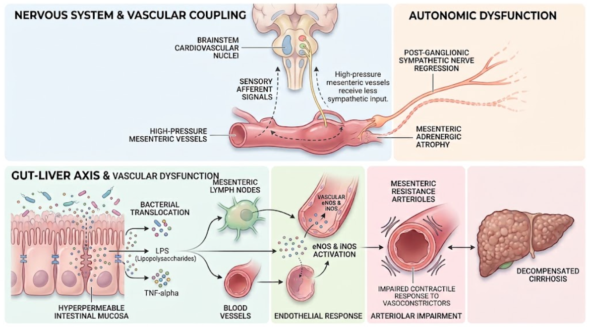
Figure 3. Pathophysiology of the gut-liver-brain axis in decompensated cirrhosis. The diagram shows that high pressure in mesenteric vessels causes autonomic dysfunction, which results in the regression of sympathetic nerves and mesenteric adrenergic atrophy. Bacteria, LPS and TNF- α translocate through the hyperpermeable intestinal mucosa to lymph nodes and blood vessels. This stimulates eNOS and iNOS and impairs the contractile response of mesenteric resistance arterioles to vasoconstrictors.
Chronic non-physiological stimulation of this afferent sensory reflex arc leads to a severe neuro-structural degeneration, named mesenteric sympathetic ganglionic atrophy. Neurons in the celiac and superior mesenteric ganglia withdraw axons, trim dendrites and die by apoptosis. This local sympathetic regression results in loss of adrenergic nerve fibers innervating precapillary mesenteric resistance arterioles(Coll et al., 2008).
There is a localized neurogenic denervation, with a marked decrease in the local release of norepinephrine from the sympathetic ganglionic atrophy within the mesenteric vascular wall. Systemic sympathetic activity remains highly elevated, with high plasma concentrations of circulating norepinephrine causing renal and cardiac vasoconstriction, but the mesenteric arterial tree is subject to localized adrenergic deficiency. The absence of sympathetic vasoconstrictor tone means that the local paracrine vasodilators like nitric oxide and prostacyclin are unopposed in their action, leading to further splanchnic vasodilation(Coll et al., 2008).
At the same time, activated capsaicin-sensitive primary sensory afferent nerves release powerful vasodilator neuropeptides from their peripheral terminals in the vascular adventitia. Perivascular release of calcitonin gene-related peptide and substance P evokes potent endothelium-independent vasodilation and inhibits sympathetic nerve-mediated vasoconstriction(Song, Sharkey, Breitman, Zhang, & Lee, 2001). Capsaicin-induced ablative destruction of primary sensory afferent nerves attenuates mesenteric vasodilation and hyperdynamic circulatory development. These studies emphasize the functional significance of neurogenic pathways in liver disease(Martell et al., 2010).
The gut-liver axis is a close bidirectional functional unit that catastrophically breaks down during the transition into decompensated liver cirrhosis. Portal hypertension, splanchnic venous congestion, and intestinal ischemia-reperfusion injury structurally disrupt the intestinal mucosal barrier and set off a cascade of inflammatory vasodilation(Ververeli et al., 2025).
As discussed in the previous review, decompensated cirrhosis is characterized by failure of hepatic clearance pathways and systemic detoxification. Chronic venous congestion of the gastrointestinal tract causes edema of the intestinal mucosa, disruption of epithelial tight junction proteins such as occludin and zonula occludens-1, and pathological hyperpermeability, a phenomenon clinically known as the leaky gut. Concurrently, small intestinal bacterial overgrowth and dysbiosis alter the luminal microbial composition by increasing the abundance of Gram-negative pathobionts(Dirchwolf & Ruf, 2015).
Disruption of the intestinal barrier allows pathologic translocation of viable gut bacteria, bacterial fragments, and pathogen-associated molecular patterns, particularly lipopolysaccharide (endotoxin), from the intestinal lumen to the mesenteric lymph nodes and splanchnic circulation. Lipopolysaccharide binds to lipopolysaccharide-binding protein and is presented to Toll-like receptor 4 expressed on vascular endothelial cells, adventitial resident macrophages and circulating monocytes(Colle et al., 2008).
Toll-like receptor 4 activates downstream intracellular signaling pathways such as nuclear factor kappa B and mitogen-activated protein kinases. This activation of the transcription factor results in robust gene expression and the release of pro-inflammatory cytokines, such as tumor necrosis factor- α, interleukin-1β, and interleukin-6(Dirchwolf & Ruf, 2015).
These inflammatory cytokines aggravate splanchnic vasodilation by various synergistic pathways:
Selective intestinal decontamination with non-absorbable or systemic antibiotics such as norfloxacin or rifaximin markedly decreases circulating endotoxin levels, reduces vascular nitric oxide synthase expression, improves vascular reactivity to vasoconstrictors and attenuates the hyperdynamic circulation in cirrhotic models and patients. These clinical findings confirm that gut-derived bacterial translocation and systemic immune activation are important extrinsic drivers of splanchnic vasodilation during clinical decompensation(Colle et al., 2008; Ververeli et al., 2025).
The hemodynamic derangements, which arise in the splanchnic circulation, propagate throughout the body and constitute the pathogenetic basis for the primary life-threatening complications of decompensated liver cirrhosis(Al-Hamoudi, 2010).
Splanchnic arterial vasodilation increases precapillary hydrostatic pressure in the intestinal and mesenteric microcirculation. In parallel, the increased portal venous pressure results in an increased postcapillary resistance. This shift in Starling’s forces with high capillary hydrostatic pressure and severe hypoalbuminemia secondary to hepatic synthetic failure causes an excessive transudation of fluid across the visceral peritoneum into the peritoneal cavity which is called ascites. Ongoing accumulation of fluid within the abdominal cavity progressively reduces effective arterial blood volume and stimulates the neurohumoral cascade of renal sodium and water retention(Iwakiri & Groszmann, 2006).
Hepatorenal syndrome is a severe, functional renal failure occurring in patients with advanced decompensated cirrhosis without primary structural renal disease. The pathogenesis of hepatorenal syndrome is directly related to extreme splanchnic arterial vasodilation(Al-Hamoudi, 2010).
As splanchnic vasodilation progresses, effective arterial blood volume falls below a critical level. The renin-angiotensin-aldosterone system, the sympathetic nervous system and the release of arginine vasopressin are activated in a compensatory manner and reach extreme levels(Martell et al., 2010). The splanchnic vascular bed is insensitive to these vasoconstrictors because of local nitric oxide overproduction and receptor desensitization. The renal vasculature is not protected by local vasodilator activity and is subject to intense, sustained vasoconstriction(Al-Hamoudi, 2010).
Severe renal cortical ischemia causes a marked reduction in glomerular filtration rate, oliguria, and progressive azotemia. In type 1 hepatorenal syndrome (now termed HRS-AKI), an acute precipitating event such as spontaneous bacterial peritonitis further exacerbates systemic inflammation and splanchnic vasodilation leading to rapid renal circulatory collapse(Al-Hamoudi, 2010).
For decades, the hyperdynamic circulation of cirrhosis was thought to be maintained by a healthy heart that responded correctly to the decreased afterload. However, more detailed cardiac evaluation has shown that the chronic hyperdynamic stress results in cirrhotic cardiomyopathy, a disease entity characterized by a baseline high cardiac output but blunted inotropic and chronotropic responses to physiological or pharmacological stress, diastolic dysfunction, and electrophysiological abnormalities including the prolongation of the QT interval(Al-Hamoudi, 2010).
Chronic sympathetic overactivation causes G-s proteins uncoupling and downregulation of cardiac β-adrenergic receptors and thus reduces cyclic adenosine monophosphate production in cardiomyocytes. Furthermore, circulating cardiodepressant mediators, such as nitric oxide, carbon monoxide, tumor necrosis factor-α and endocannabinoids, have direct negative inotropic effects on the myocardium(Al-Hamoudi, 2010). Acute stressors such as variceal bleeding, infection, or transjugular intrahepatic portosystemic shunt placement cannot be compensated by a further increase in cardiac output due to the failing heart and cannot meet systemic demands. This precipitous drop in cardiac output in the setting of severe splanchnic vasodilation precipitates acute multi-organ hypoperfusion and heralds acute on chronic liver failure(Arroyo & Fernández, 2013; Ververeli et al., 2025).
The literature synthesis performed in this review points to a fundamental conceptual evolution in hepatology: from the view of decompensated liver cirrhosis as a localized liver disease with structural scarring and intrahepatic mechanical obstruction, to a complex, systemic cardiovascular and inflammatory syndrome(Al-Hamoudi, 2010).
Increased intrahepatic vascular resistance is the fundamental inciting event in the development of portal hypertension. However, it is the gradual and unregulated vasodilation of the splanchnic arterial bed that converts stable compensated cirrhosis to a fragile decompensated state. The splanchnic circulation is a hemodynamic sink that sequesters a large proportion of the intravascular volume, leading to central arterial hypovolemia and a cascade of neurohumoral and inflammatory reflexes that ultimately damage distant organ systems, especially the kidneys, heart, and brain(Arroyo & Fernández, 2013; Colle et al., 2008; Martell et al., 2010).
A key lesson from recent mechanistic studies is the profound physiological compartmentalization of vascular signaling in cirrhosis. The bioavailability of nitric oxide represents a remarkable dichotomy being pathologically reduced in the intrahepatic sinusoidal microcirculation (endothelial cell capillarization and impaired Akt signaling) and excessively enriched in the extrahepatic splanchnic and systemic arterial beds (shear stress, vascular endothelial growth factor upregulation and bacterial translocation)(Genesca et al., 1999). This double abnormality poses a remarkable therapeutic challenge. Systemic administration of non-selective nitric oxide donors aggravates splanchnic vasodilation and arterial hypotension and systemic nitric oxide synthase inhibitors aggravate intrahepatic sinusoidal constriction and precipitate severe renal ischemia(Bolognesi et al., 2014; Martell et al., 2010).
The complex pathophysiology of portal hypertension is reflected in therapeutic interventions:
These therapeutic advances have not filled all knowledge gaps. The exact molecular switches that mediate the transition from local mesenteric sympathetic overactivity to sympathetic ganglionic atrophy remain to be fully elucidated. Additionally, the temporal relationship between gut microbial dysbiosis and specific vascular receptor desensitization needs to be longitudinally investigated(Hori et al., 2016). Filling these gaps in research will be important for the development of novel molecular therapies, such as liver-targeted nitric oxide donors, selective Rho-kinase activators or gut-barrier protective agents, which might prevent splanchnic vascular collapse and halt the progression to clinical decompensation(Kim et al., 2010).
To summarize, splanchnic arterial vasodilation is the main vascular mechanism responsible for hyperdynamic circulation, increased portal pressure and clinical decompensation in liver cirrhosis. This complex phenomenon stems from the complex convergence of endothelial vasodilator overproduction (mainly nitric oxide, carbon monoxide, prostacyclin and endocannabinoids), smooth muscle vasoconstrictor hyporesponsiveness mediated by receptor desensitization and RhoA/Rho-kinase uncoupling, mesenteric sympathetic ganglionic atrophy, and gut-derived inflammatory signaling due to bacterial translocation.
This results in a state of effective arterial hypovolaemia with intense neurohumoral activation, fluid retention, intractable ascites, hepatorenal failure and cirrhotic cardiomyopathy. A better understanding of the complex cellular and neurogenic mechanisms that control splanchnic hemodynamics is imperative for the development of novel pharmacological approaches and the identification of disease-modifying therapies that can improve the survival of patients with advanced liver disease.
Al-Hamoudi, W. K. (2010). Cardiovascular changes in cirrhosis: pathogenesis and clinical implications. Saudi J Gastroenterol, 16(3), 145-153. doi:10.4103/1319-3767.65181
Arroyo, V., & Fernández, J. (2013). Relationship between systemic hemodynamics, renal dysfunction, and fluid retention in cirrhosis. Clin Liver Dis (Hoboken), 2(3), 120-122. doi:10.1002/cld.185
Bátkai, S., Járai, Z., Wagner, J. A., Goparaju, S. K., Varga, K., Liu, J., . . . Kunos, G. (2001). Endocannabinoids acting at vascular CB1 receptors mediate the vasodilated state in advanced liver cirrhosis. Nat Med, 7(7), 827-832. doi:10.1038/89953
Bolognesi, M., Di Pascoli, M., Verardo, A., & Gatta, A. (2014). Splanchnic vasodilation and hyperdynamic circulatory syndrome in cirrhosis. World J Gastroenterol, 20(10), 2555-2563. doi:10.3748/wjg.v20.i10.2555
Bolognesi, M., Sacerdoti, D., Di Pascoli, M., Angeli, P., Quarta, S., Sticca, A., . . . Gatta, A. (2005). Haeme oxygenase mediates hyporeactivity to phenylephrine in the mesenteric vessels of cirrhotic rats with ascites. Gut, 54(11), 1630-1636. doi:10.1136/gut.2004.063735
Coll, M., Genescà, J., Raurell, I., Rodríguez-Vilarrupla, A., Mejías, M., Otero, T., . . . Martell, M. (2008). Down-regulation of genes related to the adrenergic system may contribute to splanchnic vasodilation in rat portal hypertension. J Hepatol, 49(1), 43-51. doi:10.1016/j.jhep.2008.03.015
Colle, I., Geerts, A. M., Van Steenkiste, C., & Van Vlierberghe, H. (2008). Hemodynamic Changes in Splanchnic Blood Vessels in Portal Hypertension. The Anatomical Record, 291(6), 699-713. doi:https://doi.org/10.1002/ar.20667
Di Pascoli, M., Sacerdoti, D., Pontisso, P., Angeli, P., & Bolognesi, M. (2017). Molecular Mechanisms Leading to Splanchnic Vasodilation in Liver Cirrhosis. Journal of Vascular Research, 54(2), 92-99. doi:10.1159/000462974
Dimmeler, S., Fleming, I., Fisslthaler, B., Hermann, C., Busse, R., & Zeiher, A. M. (1999). Activation of nitric oxide synthase in endothelial cells by Akt-dependent phosphorylation. Nature, 399(6736), 601-605. doi:10.1038/21224
Dirchwolf, M., & Ruf, A. E. (2015). Role of systemic inflammation in cirrhosis: From pathogenesis to prognosis. World J Hepatol, 7(16), 1974-1981. doi:10.4254/wjh.v7.i16.1974
Du, Q. H., Han, L., Jiang, J. J., Li, P. T., Wang, X. Y., & Jia, X. (2013). Increased endothelin receptor B and G protein coupled kinase-2 in the mesentery of portal hypertensive rats. World J Gastroenterol, 19(13), 2065-2072. doi:10.3748/wjg.v19.i13.2065
Ferguson, S. S. (2001). Evolving concepts in G protein-coupled receptor endocytosis: the role in receptor desensitization and signaling. Pharmacol Rev, 53(1), 1-24.
Geerts, A. M., De Vriese, A. S., Vanheule, E., Van Vlierberghe, H., Mortier, S., Cheung, K. J., . . . Colle, I. (2006). Increased angiogenesis and permeability in the mesenteric microvasculature of rats with cirrhosis and portal hypertension: an in vivo study. Liver Int, 26(7), 889-898. doi:10.1111/j.1478-3231.2006.01308.x
Genesca, J., Gonzalez, A., Segura, R., Catalan, R., Marti, R., Varela, E., . . . Guardia, J. (1999). Interleukin-6, nitric oxide, and the clinical and hemodynamic alterations of patients with liver cirrhosis. Am J Gastroenterol, 94(1), 169-177. doi:10.1111/j.1572-0241.1999.00790.x
Gibbins, I. L., Jobling, P., & Morris, J. L. (2003). Functional organization of peripheral vasomotor pathways. Acta Physiol Scand, 177(3), 237-245. doi:10.1046/j.1365-201X.2003.01079.x
Guarner, C., Soriano, G., Such, J., Teixidó, M., Ramis, I., Bulbena, O., . . . et al. (1992). Systemic prostacyclin in cirrhotic patients. Relationship with portal hypertension and changes after intestinal decontamination. Gastroenterology, 102(1), 303-309.
Henriksen, J. H., & Moller, S. (2006). Liver cirrhosis and arterial hypertension. World J Gastroenterol, 12(5), 678-685. doi:10.3748/wjg.v12.i5.678
Henriksen, J. H., & Ring-Larsen, H. (1994). Hepatorenal disorders: role of the sympathetic nervous system. Semin Liver Dis, 14(1), 35-43. doi:10.1055/s-2007-1007296
Hori, T., Ogura, Y., Onishi, Y., Kamei, H., Kurata, N., Kainuma, M., . . . Uemoto, S. (2016). Systemic hemodynamics in advanced cirrhosis: Concerns during perioperative period of liver transplantation. World J Hepatol, 8(25), 1047-1060. doi:10.4254/wjh.v8.i25.1047
Iwakiri, Y., & Groszmann, R. J. (2006). The hyperdynamic circulation of chronic liver diseases: from the patient to the molecule. Hepatology, 43(2 Suppl 1), S121-131. doi:10.1002/hep.20993
Iwakiri, Y., Tsai, M. H., McCabe, T. J., Gratton, J. P., Fulton, D., Groszmann, R. J., & Sessa, W. C. (2002). Phosphorylation of eNOS initiates excessive NO production in early phases of portal hypertension. Am J Physiol Heart Circ Physiol, 282(6), H2084-2090. doi:10.1152/ajpheart.00675.2001
Kim, M. Y., Baik, S. K., & Lee, S. S. (2010). Hemodynamic alterations in cirrhosis and portal hypertension. Korean J Hepatol, 16(4), 347-352. doi:10.3350/kjhep.2010.16.4.347
Martell, M., Coll, M., Ezkurdia, N., Raurell, I., & Genescà, J. (2010). Physiopathology of splanchnic vasodilation in portal hypertension. World J Hepatol, 2(6), 208-220. doi:10.4254/wjh.v2.i6.208
Mulvany, M. J., & Aalkjaer, C. (1990). Structure and function of small arteries. Physiol Rev, 70(4), 921-961. doi:10.1152/physrev.1990.70.4.921
Nishimatsu, H., Suzuki, E., Nagata, D., Moriyama, N., Satonaka, H., Walsh, K., . . . Hirata, Y. (2001). Adrenomedullin induces endothelium-dependent vasorelaxation via the phosphatidylinositol 3-kinase/Akt-dependent pathway in rat aorta. Circ Res, 89(1), 63-70. doi:10.1161/hh1301.092498
Schrier, R. W., Arroyo, V., Bernardi, M., Epstein, M., Henriksen, J. H., & Rodés, J. (1988). Peripheral arterial vasodilation hypothesis: a proposal for the initiation of renal sodium and water retention in cirrhosis. Hepatology, 8(5), 1151-1157. doi:10.1002/hep.1840080532
Song, D., Sharkey, K. A., Breitman, D. R., Zhang, Y., & Lee, S. S. (2001). Disordered central cardiovascular regulation in portal hypertensive and cirrhotic rats. Am J Physiol Gastrointest Liver Physiol, 280(3), G420-430. doi:10.1152/ajpgi.2001.280.3.G420
Tsai, M. H. (2007). Splanchnic and systemic vasodilatation: the patient. J Clin Gastroenterol, 41 Suppl 3, S266-271. doi:10.1097/MCG.0b013e3181576d1a
Ververeli, C.-L., Dimitroglou, Y., Soulaidopoulos, S., Cholongitas, E., Aggeli, C., Tsioufis, K., & Tousoulis, D. (2025). Cardiac Remodeling and Arrhythmic Burden in Pre-Transplant Cirrhotic Patients: Pathophysiological Mechanisms and Management Strategies. Biomedicines, 13(4), 812. Retrieved from doi:10.3390/biomedicines13040812
Whittle, B. J., & Moncada, S. (1992). Nitric oxide: the elusive mediator of the hyperdynamic circulation of cirrhosis? Hepatology, 16(4), 1089-1092. doi:10.1002/hep.1840160437